. 2019 Jul;73(6-7):365-374.
doi: 10.1007/s10858-019-00260-6. Epub 2019 Jun 26.

NMR and MD studies combined to elucidate inhibitor and water interactions of HIV-1 protease and their modulations with resistance mutations

Affiliations

NMR and MD studies combined to elucidate inhibitor and water interactions of HIV-1 protease and their modulations with resistance mutations

Rieko Ishima et al. J Biomol NMR. 2019 Jul.

Abstract

Over the last two decades, both the sensitivity of NMR and the time scale of molecular dynamics (MD) simulation have increased tremendously and have advanced the field of protein dynamics. HIV-1 protease has been extensively studied using these two methods, and has presented a framework for cross-evaluation of structural ensembles and internal dynamics by integrating the two methods. Here, we review studies from our laboratories over the last several years, to understand the mechanistic basis of protease drug-resistance mutations and inhibitor responses, using NMR and crystal structure-based parallel MD simulations. Our studies demonstrate that NMR relaxation experiments, together with crystal structures and MD simulations, significantly contributed to the current understanding of structural/dynamic changes due to HIV-1 protease drug resistance mutations.

Keywords: Crystal structures; Drug design; HIV-1; Inhibitor; MD; NMR; Protease.


Figures

Figure 1.
Figure 1.
HIV-1 protease structure, showing the locations of residues L10, G48, I54 and V82 (yellow spheres) that are mutated in Flap+. Two subunits, A and B, are depicted in green and light blue, respectively. The flap region (residues 45-55), the P1 loop (residues 79 to 84) and the α-helix (residues 85 to 94) are highlighted in pink, green and red. Key residue numbers are indicated in small font. The structure was generated using PDB: 1T3R (Surleraux et al. 2005b).
Figure 2.
Figure 2.
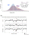
(a) Distribution of distances between the nitrogen atoms in the amino group of residue K55 and K55′ side chain, calculated 80 monomers during 100 ns MD trajectories for WT (blue) and Flap+ (purple) protease; (b) NMR relaxation data for WT (filled circles) and Flap+ protease (open triangles). The red dashed square indicates the flap region. The figures were adapted from (Cai et al. 2012) Reprinted with permission Cai Y, Yilmaz NK, Myint W, Ishima R & Schiffer CA (2012). Differential Flap Dynamics in Wild-type and a Drug Resistant Variant of HIV-1 Protease Revealed by Molecular Dynamics and NMR Relaxation. J Chem Theory Comput 8: 3452-3462. from Copyright 2012 American Chemical Society.
Figure 3.
Figure 3.
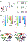
(a) Chemical structure of DRV and its analogs with substitutions at P1′ and P2′ moieties, (b) van der Waals contact energies for P1′ analogs (DRV, U1 and U6) and those for P2′ analogs (U1 – U5), (c) differences in ΔCSP between DRV and UX-bound forms (here, X = 2, 3, 7, 10; orange and red, ΔCSP >0.05 ppm) for WT (left) and Flap+ (right). In panel (a), only the analog inhibitors that are presented in this review article are listed. Panel (b) was adapted from the reference (Paulsen et al. 2017). Reprinted with permission Paulsen JL, Leidner F, Ragland DA, Kurt Yilmaz N & Schiffer CA (2017). Interdependence of Inhibitor Recognition in HIV-1 Protease. J Chem Theory Comput 13: 2300-2309. from Copyright 2017 American Chemical Society. In panel (c), residues that exhibit ΔCSP >0.05 ppm explicitly either to subunit A or B are red-highlighted while degenerated residues that exhibit ΔCSP >0.05 ppm in two subunit are orange-highlighted. Panel (c) was adapted with permission from “Khan SN, Persons JD, Paulsen J, Guerrero M, Schiffer CA, Kurt-Yilmaz N. Probing Structural Changes among Analogous Inhibitor-Bound Forms of HIV-1 Protease and a Drug-Resistant Mutant in Solution by Nuclear Magnetic Resonance, 2018, Biochemistry, 57(10), 1652-62”. Copyright 2018 American Chemical society. Structures of the UX inhibitors are in the reference.
Figure 4.
Figure 4.
(a) Close-up views of hydration sites around the protease active site facing the aniline moiety of DRV. Active site residues are color coded as yellow: apolar, blue: polar, red: charged. (b) Mean occupancies. (c) Close-up views of hydration sites around the protease active site facing the bis-THF moiety of DRV. Active site residues are color coded as in panel a. (d) Mean occupancies. Reprinted with permission from “Leidner F, Kurt-Yilmaz N, Paulsen J, Muller YA, Schiffer CA, Hydration Structure and Dynamics of Inhibitor-Bound HIV-1 Protease, 2018, J Chem Theory Comput, 14(5), 2784-96”. Copyright 2018 American Chemical Society.
Figure 5.
Figure 5.
Altered resident water positions (mean occupancy > 65%) for WT (transparent cyan spheres) versus Flap+ (solid red spheres) protease bound to DRV, in (a) an orientation in which P2′ position is on left in the panel and (b) an orientation in which P2′ position is on right in the panel (the same orientation as that in Figure 3c). Hydration sites of WT and Flap+ are shown as transparent cyan spheres and solid red spheres, respectively. Reprinted with permission from “Leidner F, Kurt-Yilmaz N, Paulsen J, Muller YA, Schiffer CA, Hydration Structure and Dynamics of Inhibitor-Bound HIV-1 Protease, 2018, J Chem Theory Comput, 14(5), 2784-96”. Copyright 2018 American Chemical Society. Water positions that are close to the amides that exhibited water-NOEs in WT protease are indicated by red-dashed circles.

References

    1. Altman MD, Ali A, Reddy GS, Nalam MN, Anjum SG, Cao H, Chellappan S, Kairys V, Fernandes MX, Gilson MK, Schiffer CA, Rana TM & Tidor B (2008). HIV-1 protease inhibitors from inverse design in the substrate envelope exhibit subnanomolar binding to drug-resistant variants. J Am Chem Soc 130: 6099–113. - PMC - PubMed
    1. Arts EJ & Hazuda DJ (2012). HIV-1 antiretroviral drug therapy. Cold Spring Harb Perspect Med 2: a007161. - PMC - PubMed
    1. Baldwin ET, Bhat TN, Gulnik S, Liu B, Topol IA, Kiso Y, Mimoto T, Mitsuya H & Erickson JW (1995). Structure of HIV-1 protease with KNI-272, a tight-binding transition-state analog containing allophenylnorstatine. Structure 3: 581–590. - PubMed
    1. Bandyopadhyay P & Meher BR (2006). Drug resistance of HIV-1 protease against JE-2147: I47V mutation investigated by molecular dynamics simulation. Chem Biol Drug Des 67: 155–61. - PubMed
    1. Cai Y, Myint W, Paulsen JL, Schiffer CA, Ishima R & Kurt Yilmaz N (2014). Drug Resistance Mutations Alter Dynamics of Inhibitor-Bound HIV-1 Protease. J Chem Theory Comput 10: 3438–3448. - PMC - PubMed

LinkOut - more resources