. 2020 Apr;65(2):329-338.
doi: 10.1007/s12223-019-00728-w. Epub 2019 Jun 26.

Integrin αvβ6 mediates epithelial-mesenchymal transition in human bronchial epithelial cells induced by lipopolysaccharides of Pseudomonas aeruginosa via TGF-β1-Smad2/3 signaling pathway

Affiliations

Integrin αvβ6 mediates epithelial-mesenchymal transition in human bronchial epithelial cells induced by lipopolysaccharides of Pseudomonas aeruginosa via TGF-β1-Smad2/3 signaling pathway

Weiming Liu et al. Folia Microbiol (Praha). 2020 Apr.

Abstract

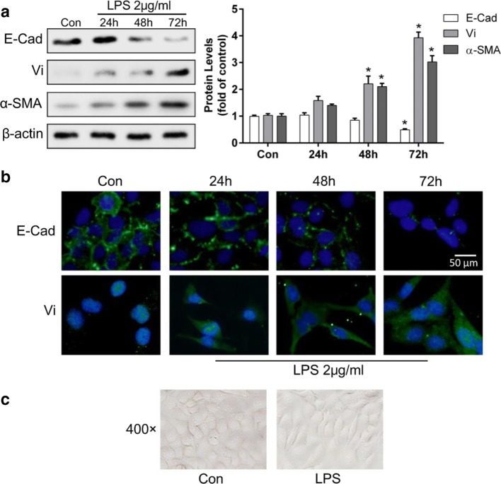
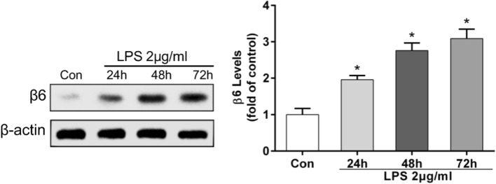
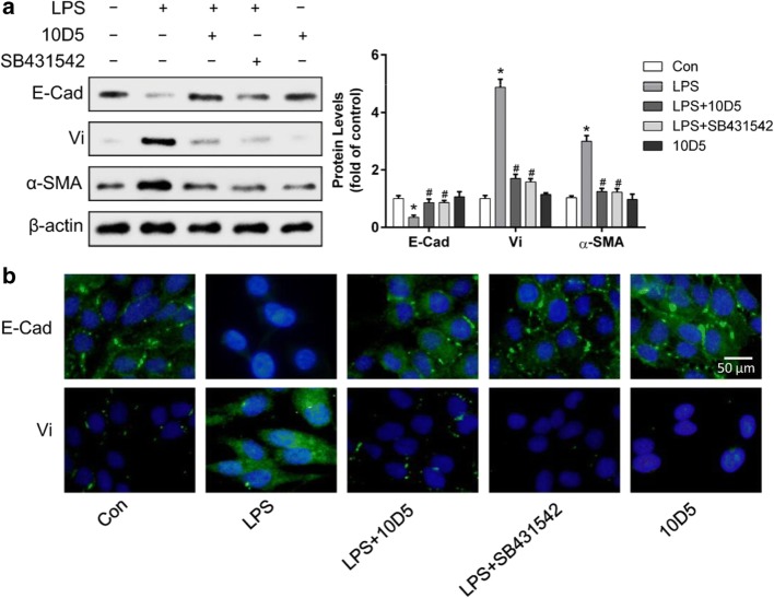
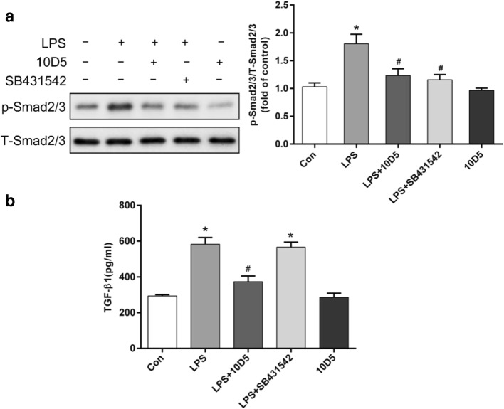
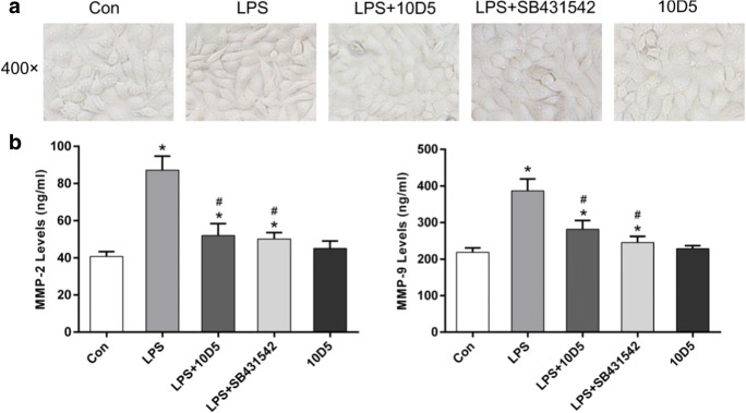
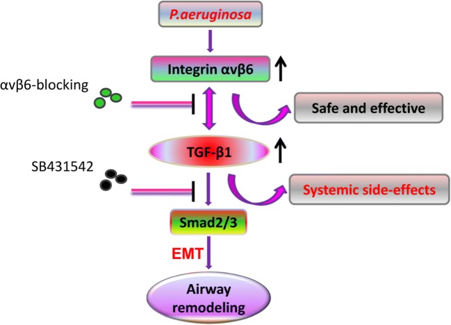
Lower respiratory tract infection due to Pseudomonas aeruginosa has become increasingly challenging, resulting in a worse morbidity and mortality. Airway remodeling is a common phenomenon in this process, to which epithelial-mesenchymal transition (EMT) may contribute as an important promoter. Previous studies showed that epithelium-specific integrin αvβ6-mediated EMT was involved in pulmonary fibrosis via transforming growth factor-β1 (TGF-β1) signaling, but whether integrin αvβ6 plays a role in the P. aeruginosa-associated airway remodeling remains unknown. BEAS-2B cells were incubated with lipopolysaccharide (LPS) from P. aeruginosa in the presence or the absence of integrin αvβ6-blocking antibodies. Morphologic changes were observed by an inverted microscopy. The EMT markers were detected using Western blotting and immunofluorescence. The activation of TGF-β1-Smad2/3 signaling pathway was assessed. Furthermore, matrix metalloproteinase (MMP)-2 and -9 in the medium were measured using ELISA. P. aeruginosa's LPS decreased the expression of the epithelial marker E-cadherin and promoted the mesenchymal markers, vimentin and α-smooth muscle actin in BEAS-2B cells. The expression of integrin αvβ6 was significantly increased during EMT process. Blocking integrin αvβ6 could attenuate P. aeruginosa's LPS-induced EMT markers' expression via TGF-β1-Smad2/3 signaling pathway. Furthermore, blocking integrin αvβ6 could prevent morphologic changes and oversecretion of MMP-2 and -9. Integrin αvβ6 mediates epithelial-mesenchymal transition in human bronchial epithelial cells induced by lipopolysaccharides of P. aeruginosa via TGF-β1-Smad2/3 signaling pathway and might be a promising therapeutic target for P. aeruginosa-associated airway remodeling.


Conflict of interest statement

The authors declare that they have no conflict of interest.

Figures

Fig. 1
Fig. 1
P. aeruginosa’s LPS-induced EMT in BEAS-2B cells. BEAS-2B cells were incubated with 2 μg/mL P. aeruginosa’s LPS for 24, 48, or 72 h. Western blotting revealed that LPS decreased the expression of E-Cad and upregulated the expression of Vi and α-SMA in a time-dependent manner (a). Immunofluorescence showed similar results of the upregulated mesenchymal marker Vi and downregulation of the epithelial marker, E-Cad, in a time-dependent manner (b). Compared with the basal condition, a fibroblast-like, spindle-shaped morphology was induced by P. aeruginosa’s LPS for 72 h in BEAS-2B cells (c). The data represent the mean ± SEM, n = 3. *p < 0.05 versus the control
Fig. 2
Fig. 2
The expression of integrin αvβ6 in BEAS-2B cells was significantly upregulated by P. aeruginosa’s LPS in a time-dependent manner. BEAS-2B cells were exposed to 2 μg/mL P. aeruginosa’s LPS for 24, 48, or 72 h. Western blotting revealed that LPS increased the expression of integrin β6 in a time-dependent manner. The data represent the mean ± SEM, n = 3. *p < 0.05 versus the control
Fig. 3
Fig. 3
Blocking integrin αvβ6 could abrogate EMT in BEAS-2B cells induced by P. aeruginosa’s LPS. BEAS-2B cells were incubated with integrin αvβ6–blocking antibody 10D5 (30 μg/mL) or specific inhibitor of TGF-β1-Smad2/3 signaling, SB431542 (10 μM), for 2 h prior to incubation with 2 μg/mL P. aeruginosa’s LPS for 72 h. Western blotting showed that the decrease of the epithelial marker, E-Cad expression, and the increase of mesenchymal markers, Vi and α-SMA, induced by P. aeruginosa’s LPS were reversed by integrin αvβ6–blocking antibody 10D5 as well as SB431542 (a). Immunofluorescence showed similar results of E-Cad and Vi expressions (b). The data represent the mean ± SEM, n = 3. *p < 0.05 versus the control (Con), #p < 0.05 versus LPS
Fig. 4
Fig. 4
Integrin αvβ6 mediated P. aeruginosa’s LPS-induced EMT in BEAS-2B cells via TGF-β1-Smad2/3 signaling pathway. BEAS-2B cells were pretreated with integrin αvβ6–blocking antibody 10D5 (30 μg/mL) or TGF-β1 inhibitor, SB431542 (10 μM), for 2 h prior to incubation with 2 μg/mL P. aeruginosa’s LPS for 72 h. Total-Smad2/3 and p-Smad2/3 were detected using Western blotting (a). The secretion of active TGF-β1 in the culture medium was measured by ELISA (b). The results showed that the phosphorylation of Smad2/3 was increased by P. aeruginosa’s LPS, which were ameliorated by integrin αvβ6–blocking antibody 10D5 as well as SB431542. In addition, elevated secretion of active TGF-β1 was induced by P. aeruginosa’s LPS, which could be inhibited by integrin αvβ6–blocking antibody 10D5. The data represent the mean ± SEM, n = 3. *p < 0.05 versus Con, #p < 0.05 versus LPS
Fig. 5
Fig. 5
Blocking integrin αvβ6 could prevent morphologic changes and the increase of MMP-2 and -9 secretion induced by P. aeruginosa’s LPS. Cell morphologic change was ameliorated by 10D5 and SB431542 (a). ELISA showed that the secretion of active MMP-2 and -9 was significantly increased by P. aeruginosa’s LPS which could be attenuated by integrin αvβ6–blocking antibody 10D5 as well as specific inhibitor of TGF-β1-Smad2/3 signaling, inhibitor SB431542 (b). The data represent the mean ± SEM, n = 3. *p < 0.05 versus Con, #p < 0.05 versus LPS
Fig. 6
Fig. 6
Summary. Integrin αvβ6 could regulate EMT in BEAS-2B cells induced by P. aeruginosa’s LPS via activation of TGF-β1-Smad2/3 signaling pathway. Compared with globally blocking TGF-β1, integrin αvβ6 blocking might be a more promising option to prevent airway remodeling induced by P. aeruginosa

References

    1. Ahmed N, Pansino F, Clyde R, Murthi P, Quinn MA, Rice GE, Agrez MV, Mok S, Baker MS. Overexpression of alpha(v)beta6 integrin in serous epithelial ovarian cancer regulates extracellular matrix degradation via the plasminogen activation cascade. Carcinogenesis. 2002;23:237–244. - PubMed
    1. Almagro P, Salvado M, Garcia-Vidal C, Rodriguez-Carballeira M, Cuchi E, Torres J, Heredia JL. Pseudomonas aeruginosa and mortality after hospital admission for chronic obstructive pulmonary disease. Respiration. 2012;84:36–43. - PubMed
    1. Ask K, Bonniaud P, Maass K, Eickelberg O, Margetts PJ, Warburton D, Groffen J, Gauldie J, Kolb M. Progressive pulmonary fibrosis is mediated by TGF-beta isoform 1 but not TGF-beta3. Int J Biochem Cell Biol. 2008;40:484–495. - PMC - PubMed
    1. Botha P, Archer L, Anderson RL, Lordan J, Dark JH, Corris PA, Gould K, Fisher AJ. Pseudomonas aeruginosa colonization of the allograft after lung transplantation and the risk of bronchiolitis obliterans syndrome. Transplantation. 2008;85:771–774. - PubMed
    1. Breuss JM, Gallo J, DeLisser HM, Klimanskaya IV, Folkesson HG, Pittet JF, Nishimura SL, Aldape K, Landers DV, Carpenter W, Et A. Expression of the beta 6 integrin subunit in development, neoplasia and tissue repair suggests a role in epithelial remodeling. J Cell Sci. 1995;108:2241–2251. - PubMed

MeSH terms

LinkOut - more resources