. 2019 Aug 21;10(8):3671-3681.
doi: 10.1021/acschemneuro.9b00248. Epub 2019 Jun 19.

Cholesterol Biosynthesis and Uptake in Developing Neurons

Affiliations

Cholesterol Biosynthesis and Uptake in Developing Neurons

Thiago C Genaro-Mattos et al. ACS Chem Neurosci. .

Abstract

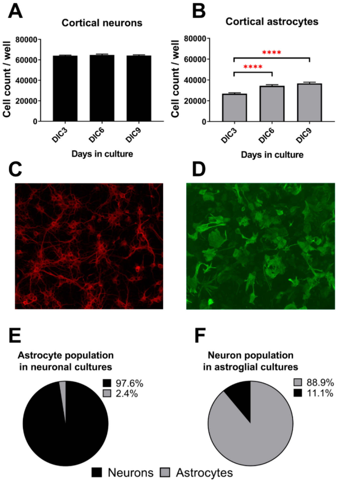
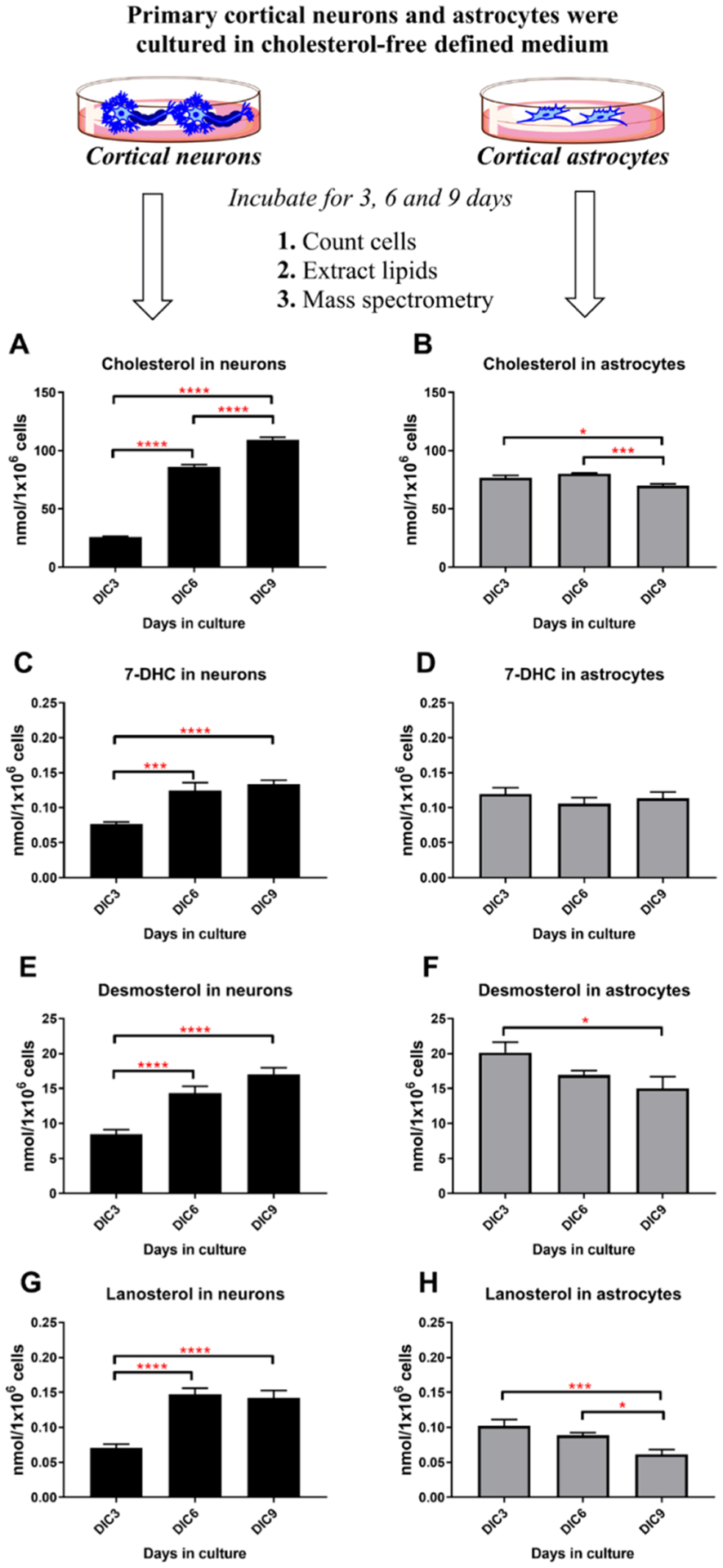
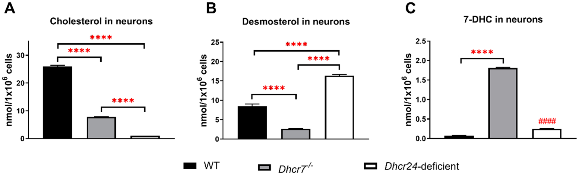
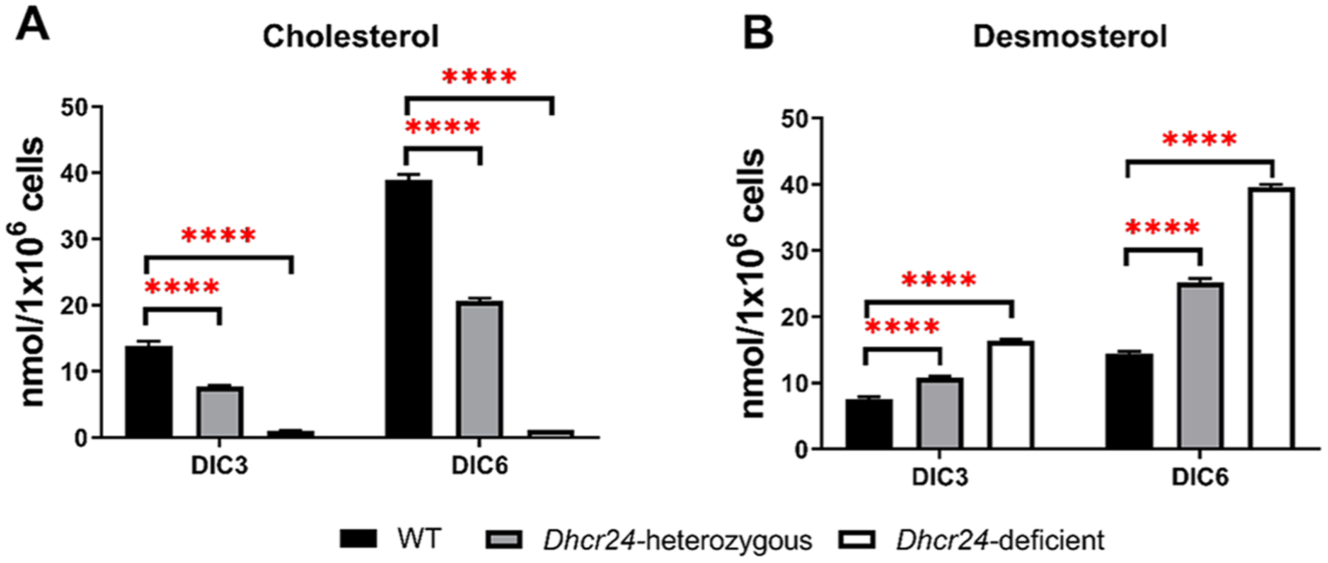
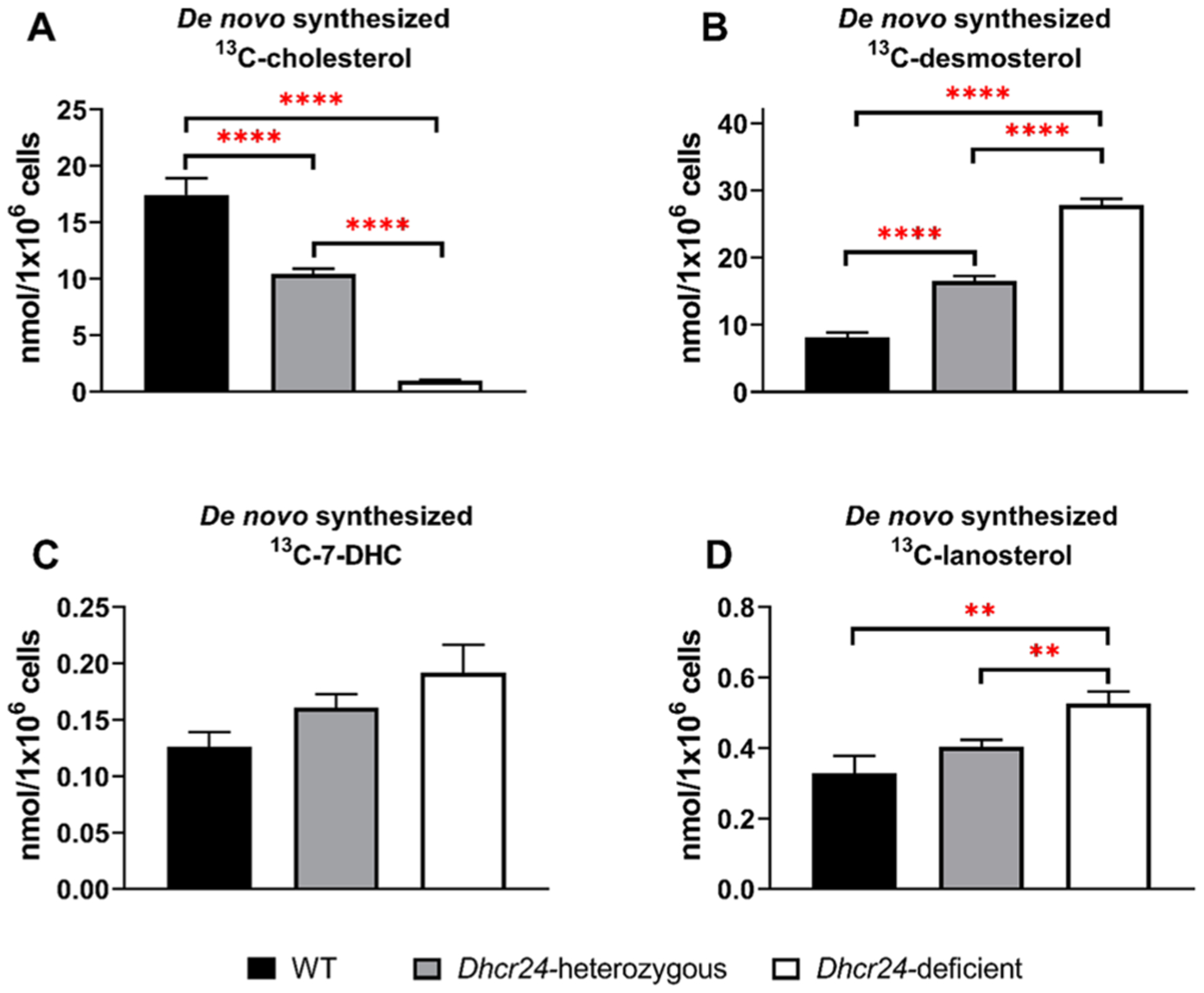
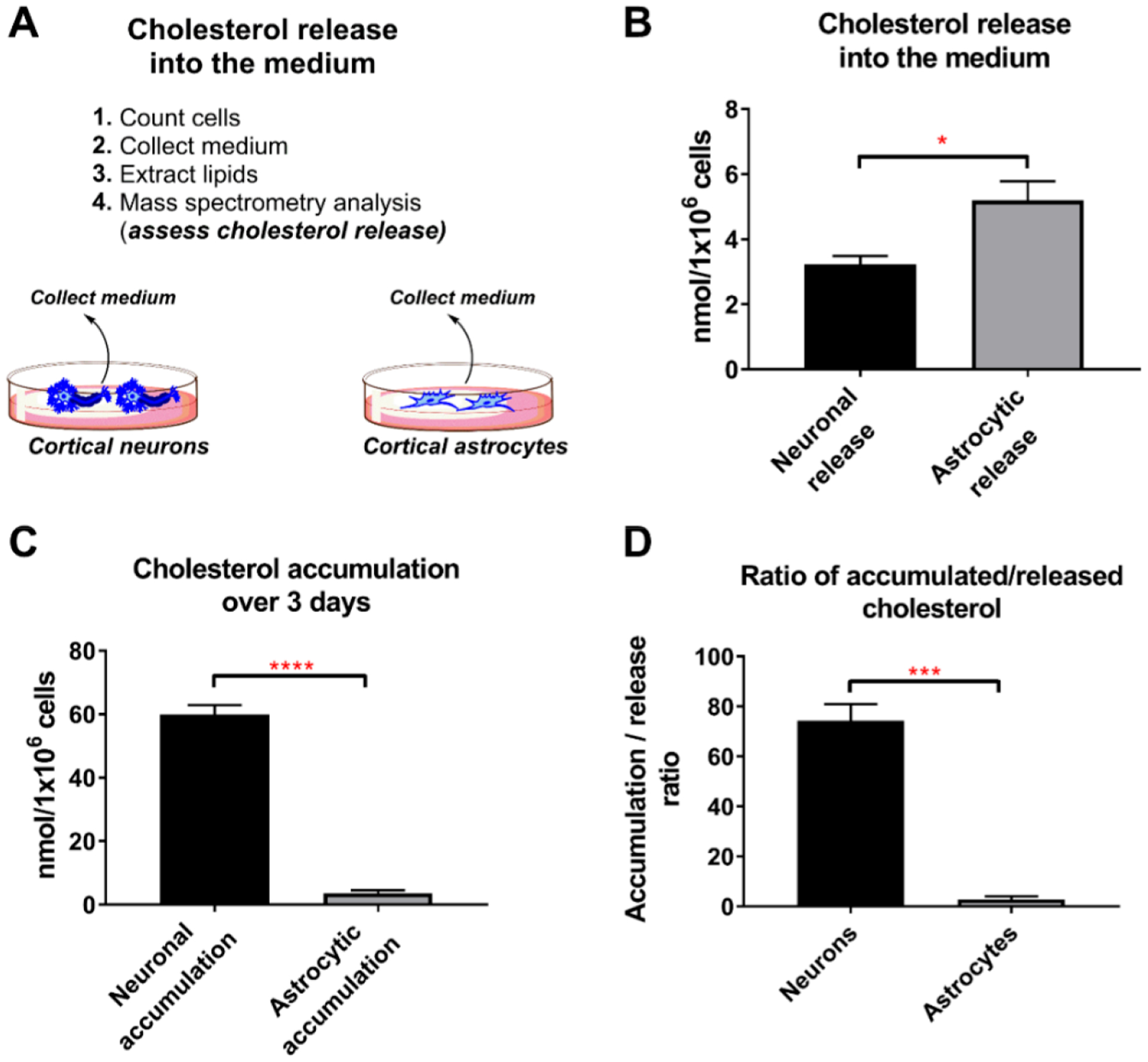
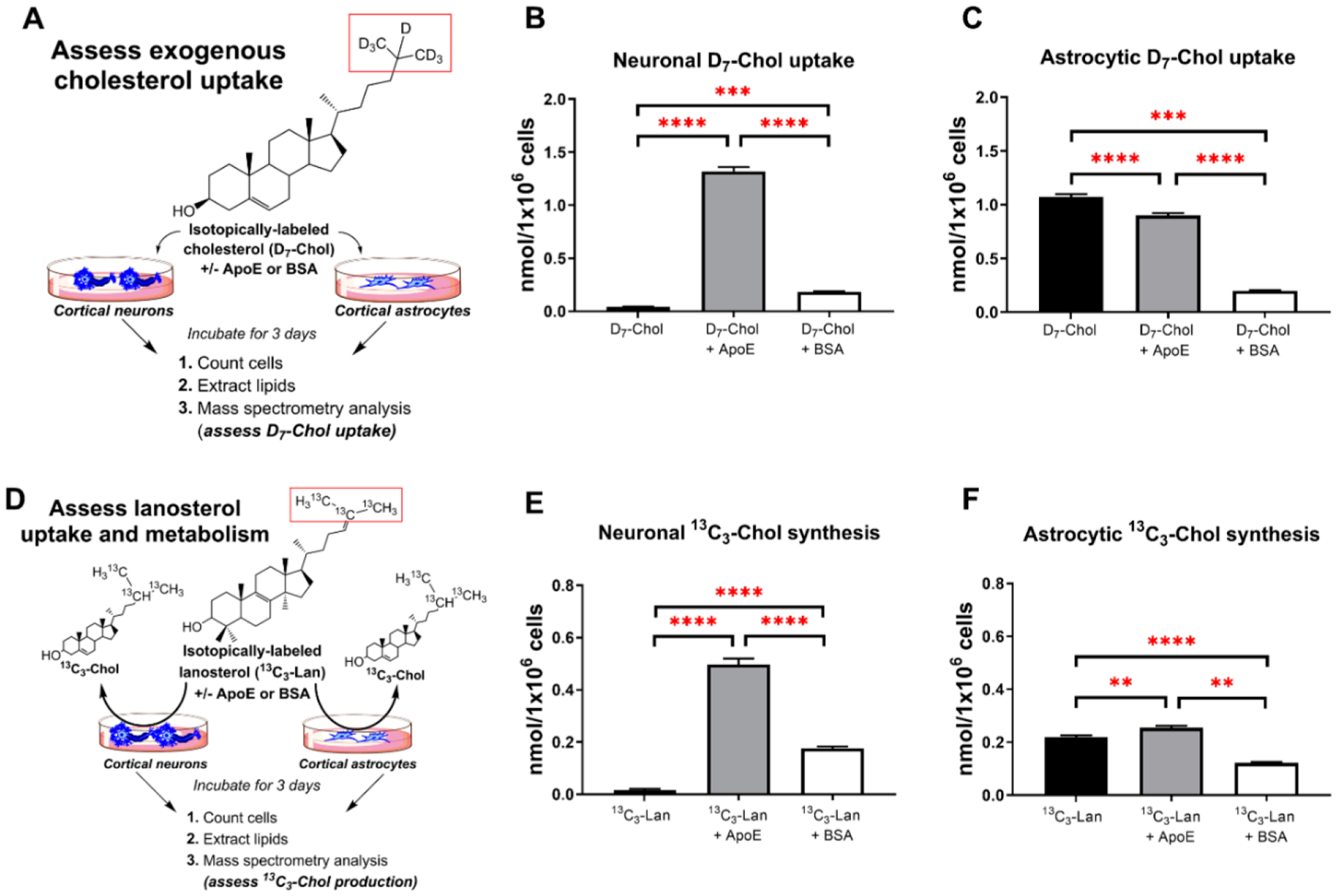
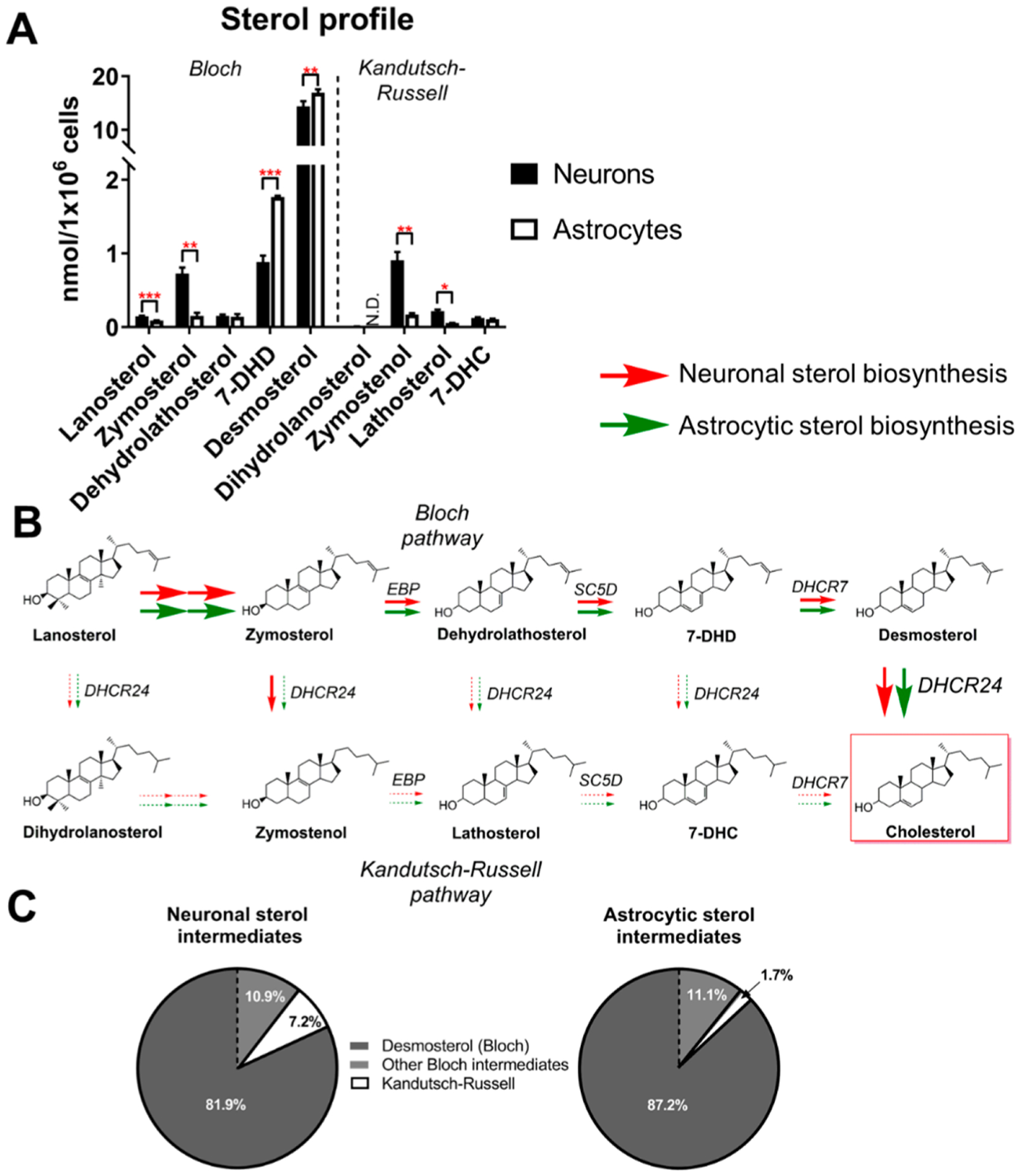
Brain cholesterol biosynthesis, a separate and distinct process from whole-body cholesterol homeostasis, starts during embryonic development. To gain a better understanding of the neuronal and glial contributions to the brain cholesterol pool, we studied this process in control, Dhcr7-/-, and Dhcr24-/- cell cultures. Our LC-MS/MS method allowed us to measure several different sterol intermediates and cholesterol during neuronal differentiation. We found that developing cortical neurons rely on endogenous cholesterol synthesis and utilize ApoE-complexed cholesterol and sterol precursors from their surroundings. Both developing neurons and astrocytes release cholesterol into their local environment. Our studies also uncovered that developing neurons produced significantly higher amounts of cholesterol per cell than the astrocytes. Finally, we established that both neurons and astroglia preferentially use the Bloch sterol biosynthesis pathway, where desmosterol is the immediate precursor to cholesterol. Overall, our studies suggest that endogenous sterol synthesis in developing neurons is a critical and complexly regulated homeostatic process during brain development.

Keywords: Dhcr24; Dhcr7; LC-MS/MS; Sterol biosynthesis; astrocytes; cholesterol; desmosterol; developing neurons.


Conflict of interest statement

The authors declare no competing financial interest.

Figures

Figure 1.
Figure 1.
Neuronal and astroglial cultures. Panels A and B show cell counts of neurons and astrocytes at different time points, respectively. Panels C and D show representative images of neurons and astrocytes in the culture wells. Neurons were labeled with anti-MAP2 antibody while astrocytes were stained with anti-GFAP antibody. Images were acquired with 20× and 40× objectives, respectively. Panels E and F denote the percent of astrocytes in neuronal cultures and the percent of neurons in astroglial cultures at 6 days in culture (DIC6), respectively. The absolute numbers of neurons and astrocytes in cultures, with statistical measures, are reported in Supplemental Table 1.
Figure 2.
Figure 2.
Cortical neurons endogenously synthesize cholesterol. Primary neurons and astrocytes were incubated for 3, 6, and 9 days in defined cholesterol-free medium. Levels of cholesterol, 7-DHC, desmosterol and lanosterol levels were measured by LC-MS/MS. Panels on the left show neuronal sterols (A, C, E, G), while panels on the right denote astrocytic sterol levels (B, D, F, H). Sterol levels were normalized to the total cell count at the end point of each experiment. Sterol values correspond to the mean ± SEM of 8–12 replicates. Statistical significance is denoted by asterisks (*p < 0.05; ***p <0.001; ****p < 0.0001). Note that all sterols accumulate in neurons in a time-dependent manner and that a similar process is not observed in astrocytes.
Figure 3.
Figure 3.
Accumulation of sterol intermediates in Dhcr7−/−and Dhcr24-deficient neurons. Comparison of cholesterol (A), desmosterol (B), and 7-DHC (C) levels between WT, Dhcr7−/−, and Dhcr24-deficient neurons (Dhcr24−/−). Neurons were cultured in defined cholesterol-free medium for 3 days and subjected to LC-MS/MS. Values correspond to mean ± SEM of 8–12 replicates. In all panels, statistical significance between individual groups is denoted by asterisks (****p < 0.0001); #### denotes the difference between WT and Dhcr24-deficient neurons (p < 0.0001).
Figure 4.
Figure 4.
Desmosterol replaces cholesterol in Dhcr24−/−neurons in a time-dependent manner. WT (Dhcr+/+), Dhcr24-heterozygous (Dhcr24±), and Dhcr24-deficient (Dhcr24−/−) neurons were cultured in defined cholesterol-free medium and cholesterol (A) and desmosterol (B) levels were assayed at DIC3 and DIC6. Values correspond to mean ± SEM of 8–12 replicates. In all panels, statistical significance between individual groups is denoted by asterisks (****p < 0.0001). Note that in Dhcr24−/− neurons, cholesterol is fully replaced by desmosterol.
Figure 5.
Figure 5.
Assessing active de novo cholesterol synthesis in neurons using13C6-glucose. Profile of selected de novo synthesized sterols in WT and Dhcr24 mutant neurons (Dhcr24± and Dhcr24−/−). Neurons were cultured in defined cholesterol-free medium where glucose was replaced by 13C6-glucose. 13C-labeled sterols were analyzed at DIC6. Values of 13C-labeled cholesterol (A), desmosterol (B), 7-DHC (C), and lanosterol (D) correspond to mean ± SEM of 8–12 replicates. In all panels, statistical significance is denoted by asterisks (**p < 0.01; ****p < 0.0001).
Figure 6.
Figure 6.
Astrocytes release more cholesterol than neurons. (A) Total cholesterol detected in the medium over 3 days in culture. Both neuronal and astroglial cells were cultured in a defined cholesterol-free medium. (B) Analyzed media were collected at DIC6 and reflect the release of cholesterol between DIC3 and DIC6. Cholesterol levels were normalized to the total cell count at DIC6. Values correspond to the mean ± SEM of 3 replicates. Note that astrocytes release 60% more cholesterol than neurons. (C) Increase in total cholesterol in neurons and astrocytes over a 3 day period. Values correspond to the mean ± SEM of 8 replicates. Note that neurons accumulated 17-fold more cholesterol than astrocytes over the 3 day period. (D) Ratio of accumulated over released cholesterol over a 3 day period. Note that neurons retain the vast majority of their newly synthesized cholesterol, while astroglial cells release more cholesterol than they accumulate. Values correspond to the mean ± SEM of 8 replicates. In all panels, statistical significance is denoted by asterisks (*p < 0.05, ***p < 0.001; ****p < 0.0001).
Figure 7.
Figure 7.
Neuronal and astrocytic uptake of exogenous sterols. Neurons and astrocytes were cultured in defined cholesterol-free medium supplemented with free isotopically labeled cholesterol (D7-Chol), D7-Chol precomplexed with ApoE, and D7-Chol precomplexed with BSA as illustrated in Panel A. The labeled moiety is highlighted in red. Final concentration of D7-Chol in all conditions was 500 nM; protein final concentration was 0.7 μg/mL (either ApoE or BSA). Panels B and C show the uptake of D7-Chol from the medium by neurons and astrocytes, respectively. D7-Chol levels were normalized to the total cell count at the end point of the experiment (DIC3). Sterol values correspond to the mean ± SEM of 8–12 replicates. Note that ApoE facilitates the uptake of D7-Chol by neurons but not by astrocytes. Panel D denotes the experimental design to test the incorporation of 13C3−Lanosterol by neurons and astrocytes. Cells were cultured in defined cholesterol-free medium and with free isotopically labeled lanosterol (13C3−Lan), 13C3−Lan precomplexed with ApoE, and 13C3−Lan precomplexed with BSA. The labeled moiety is highlighted in red. Panels E and F show 13C3−Chol levels derived from the conversion of 13C3−Lan by neurons and astrocytes, respectively. 13C3−Chol levels were normalized to the total cell count at the end point of each experiment. Sterol values correspond to the mean ± SEM of 8–12 replicates. Note that the uptake and conversion of lanosterol into cholesterol is 40-fold higher in neurons when lanosterol is precomplexed with ApoE, while in astrocytes the same treatment increases only by 15%. In all panels, statistical significance is denoted by asterisks (**p < 0.01, ***p < 0.001; ****p < 0.0001).
Figure 8.
Figure 8.
Both neurons and astrocytes synthesize cholesterol via the Bloch pathway. (A) GC-MS profile of postlanosterol cholesterol biosynthesis intermediates in cultured neurons and astrocytes. Cells were cultured in a defined cholesterol-free medium. Intermediates in the Bloch pathway are shown on the left, while those from the Kandutsch-Russell pathway are depicted on the right of the graph. Sterol levels were determined at DIC6 and values correspond to mean ± SEM of 3 replicates. Statistical significance is denoted by asterisks (*p < 0.05, **p < 0.01, ***p < 0.001, ND − nondetectable). (B) Schematic representation depicting the preferential routes for the conversion of lanosterol into cholesterol by cultured neurons and astrocytes. Red and green arrows represent the pathways utilized by neurons and astrocytes, respectively. Arrow thickness corresponds to the strength of pathway utilization. (C) Pie chart representation denoting the percent of Bloch and Kandutsch-Russell intermediates in cultured neurons and astrocytes at DIC6. Note that desmosterol is the most abundant intermediate in both neurons and astrocytes and that both cell types have higher levels of intermediates from the Bloch pathway, suggesting that the Kandutsch-Russell pathway utilization is miniscule at this developmental stage.

References

    1. Dietschy JM, and Turley SD (2001) Cholesterol metabolism in the brain. Curr. Opin. Lipidol 12, 105–12. - PubMed
    1. Dietschy JM, and Turley SD (2004) Thematic review series: brain Lipids. Cholesterol metabolism in the central nervous system during early development and in the mature animal. J. Lipid Res 45, 1375–97. - PubMed
    1. Nes WD (2011) Biosynthesis of cholesterol and other sterols. Chem. Rev 111, 6423–51. - PMC - PubMed
    1. Porter FD, and Herman GE (2011) Malformation syndromes caused by disorders of cholesterol synthesis. J. Lipid Res 52, 6–34. - PMC - PubMed
    1. Porter FD (2000) RSH/Smith-Lemli-Opitz syndrome: a multiple congenital anomaly/mental retardation syndrome due to an inborn error of cholesterol biosynthesis. Mol. Genet. Metab 71, 163–74. - PubMed

Publication types