Cerebral Dopamine Neurotrophic Factor Diffuses Around the Brainstem and Does Not Undergo Anterograde Transport After Injection to the Substantia Nigra
- PMID: 31244598
- PMCID: PMC6580362
- DOI: 10.3389/fnins.2019.00590
Cerebral Dopamine Neurotrophic Factor Diffuses Around the Brainstem and Does Not Undergo Anterograde Transport After Injection to the Substantia Nigra
Abstract
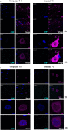
Cerebral dopamine neurotrophic factor (CDNF) has shown therapeutic potential in rodent and non-human primate models of Parkinson's disease by protecting the dopamine neurons from degeneration and even restoring their phenotype and function. Previously, neurorestorative efficacy of CDNF in the 6-hydroxydopamine (6-OHDA) model of Parkinson's disease as well as diffusion of the protein in the striatum (STR) has been demonstrated and studied. Here, experiments were performed to characterize the diffusion and transport of supra-nigral CDNF in non-lesioned rats. We injected recombinant human CDNF to the substantia nigra (SN) of naïve male Wistar rats and analyzed the brains 2, 6, and 24 h after injections. We performed immunohistochemical stainings using an antibody specific to human CDNF and radioactivity measurements after injecting iodinated CDNF. Unlike the previously reported striatonigral retrograde transport seen after striatal injection, active anterograde transport of CDNF to the STR could not be detected after nigral injection. There was, however, clear diffusion of CDNF to the brain areas surrounding the SN, and CDNF colocalized with tyrosine hydroxylase (TH)-positive neurons. Overall, our results provide insight on how CDNF injected to the SN may act in this region of the brain.
Keywords: Parkinson’s disease; cerebral dopamine neurotrophic factor; diffusion; striatum; substantia nigra; transport.
Figures


Similar articles
-
CDNF and MANF in the brain dopamine system and their potential as treatment for Parkinson's disease.Front Psychiatry. 2023 Jul 24;14:1188697. doi: 10.3389/fpsyt.2023.1188697. eCollection 2023. Front Psychiatry. 2023. PMID: 37555005 Free PMC article. Review.
-
Novel neurotrophic factor CDNF protects and rescues midbrain dopamine neurons in vivo.Nature. 2007 Jul 5;448(7149):73-7. doi: 10.1038/nature05957. Nature. 2007. PMID: 17611540
-
Chronic infusion of CDNF prevents 6-OHDA-induced deficits in a rat model of Parkinson's disease.Exp Neurol. 2011 Mar;228(1):99-108. doi: 10.1016/j.expneurol.2010.12.013. Epub 2010 Dec 24. Exp Neurol. 2011. PMID: 21185834
-
Intrastriatally Infused Exogenous CDNF Is Endocytosed and Retrogradely Transported to Substantia Nigra.eNeuro. 2017 Feb 20;4(1):ENEURO.0128-16.2017. doi: 10.1523/ENEURO.0128-16.2017. eCollection 2017 Jan-Feb. eNeuro. 2017. PMID: 28275710 Free PMC article.
-
Therapeutic potential of the endoplasmic reticulum located and secreted CDNF/MANF family of neurotrophic factors in Parkinson's disease.FEBS Lett. 2015 Dec 21;589(24 Pt A):3739-48. doi: 10.1016/j.febslet.2015.09.031. Epub 2015 Oct 9. FEBS Lett. 2015. PMID: 26450777 Review.
Cited by
-
Delivery of CDNF by AAV-mediated gene transfer protects dopamine neurons and regulates ER stress and inflammation in an acute MPTP mouse model of Parkinson's disease.Sci Rep. 2024 Jul 17;14(1):16487. doi: 10.1038/s41598-024-65735-5. Sci Rep. 2024. PMID: 39019902 Free PMC article.
-
Cerebral dopamine neurotrophic factor protects and repairs dopamine neurons by novel mechanism.Mol Psychiatry. 2022 Mar;27(3):1310-1321. doi: 10.1038/s41380-021-01394-6. Epub 2021 Dec 14. Mol Psychiatry. 2022. PMID: 34907395 Free PMC article. Review.
-
CDNF and MANF in the brain dopamine system and their potential as treatment for Parkinson's disease.Front Psychiatry. 2023 Jul 24;14:1188697. doi: 10.3389/fpsyt.2023.1188697. eCollection 2023. Front Psychiatry. 2023. PMID: 37555005 Free PMC article. Review.
-
Cerebral dopamine neurotrophic factor (CDNF) protects against quinolinic acid-induced toxicity in in vitro and in vivo models of Huntington's disease.Sci Rep. 2020 Nov 5;10(1):19045. doi: 10.1038/s41598-020-75439-1. Sci Rep. 2020. PMID: 33154393 Free PMC article.
-
Therapeutic Advances in Diabetes, Autoimmune, and Neurological Diseases.Int J Mol Sci. 2021 Mar 10;22(6):2805. doi: 10.3390/ijms22062805. Int J Mol Sci. 2021. PMID: 33802091 Free PMC article. Review.
References
LinkOut - more resources
Full Text Sources

