. 2019 Jun 6:13:230.
doi: 10.3389/fncel.2019.00230. eCollection 2019.

A Novel Transwell Blood Brain Barrier Model Using Primary Human Cells

Affiliations

A Novel Transwell Blood Brain Barrier Model Using Primary Human Cells

Nicole L Stone et al. Front Cell Neurosci. .

Abstract

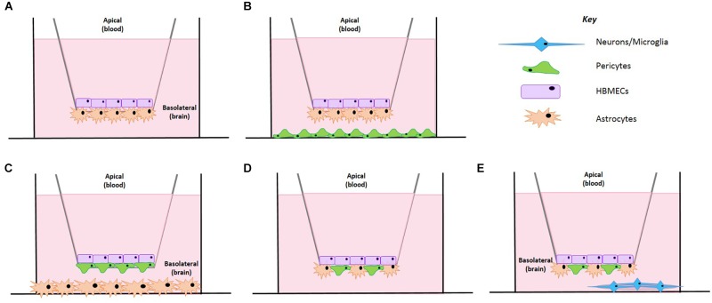
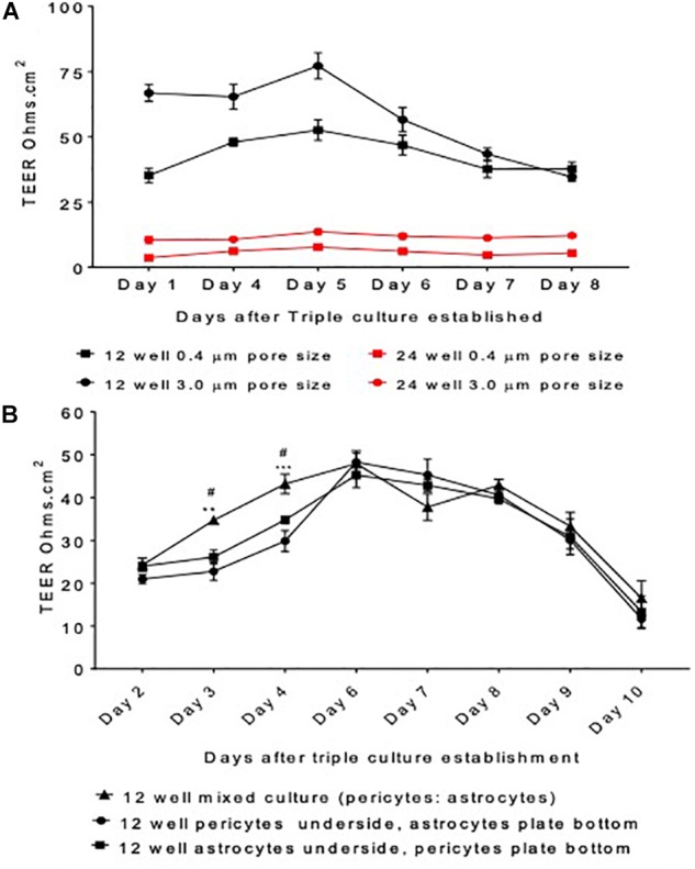
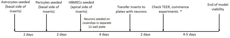
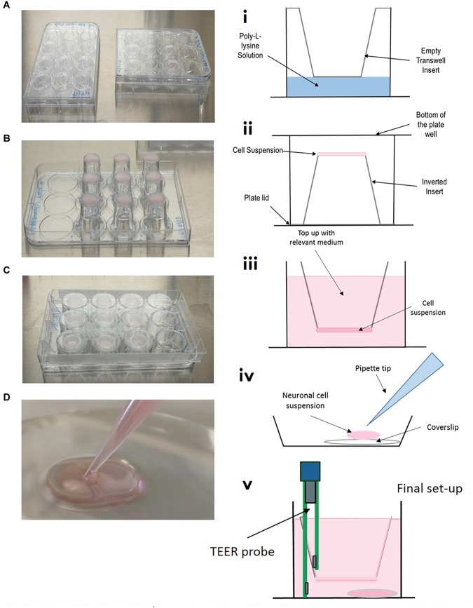
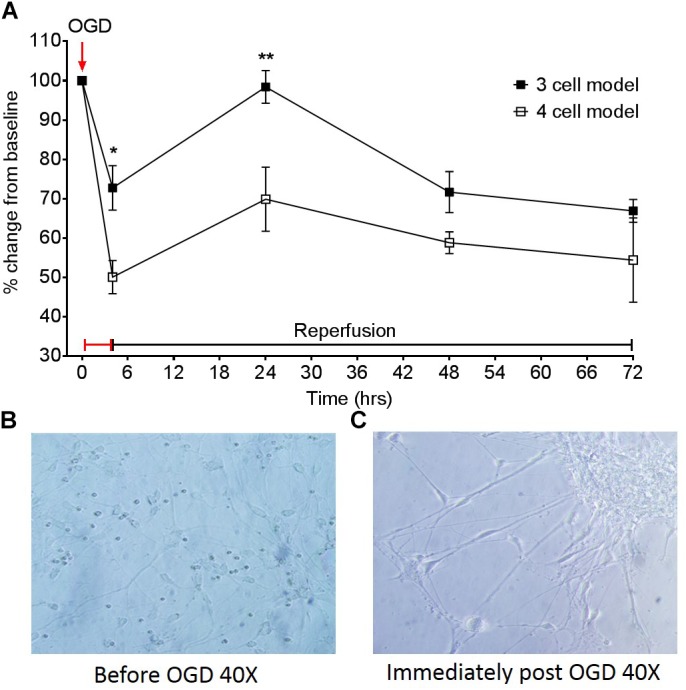
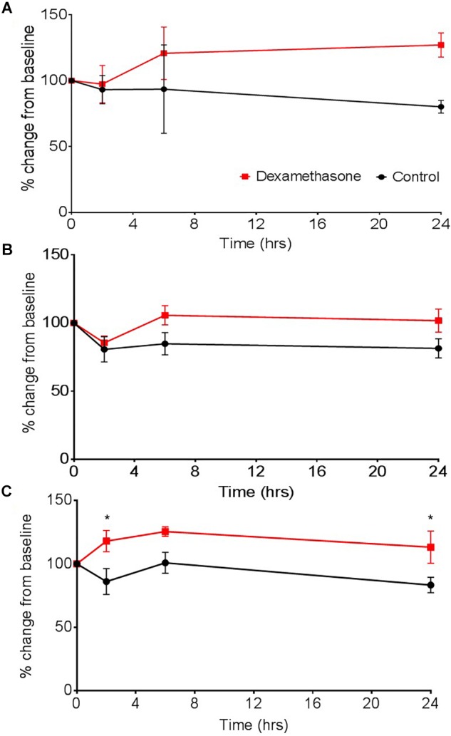
Structural alterations and breakdown of the blood brain barrier (BBB) is often a primary or secondary consequence of disease, resulting in brain oedema and the transport of unwanted substances into the brain. It is critical that effective in vitro models are developed to model the in vivo environment to aid in clinically relevant research, especially regarding drug screening and permeability studies. Our novel model uses only primary human cells and includes four of the key cells of the BBB: astrocytes, pericytes, brain microvascular endothelial cells (HBMEC) and neurons. We show that using a larger membrane pore size (3.0 μM) there is an improved connection between the endothelial cells, astrocytes and pericytes. Compared to a two and three cell model, we show that when neurons are added to HBMECs, astrocytes and pericytes, BBB integrity was more sensitive to oxygen-glucose deprivation evidenced by increased permeability and markers of cell damage. Our data also show that a four cell model responds faster to the barrier tightening effects of glucocorticoid dexamethasone, when compared to a two cell and three cell model. These data highlight the important role that neurons play in response to ischaemia, particularly how they contribute to BBB maintenance and breakdown. We consider that this model is more representative of the interactions at the neurovascular unit than other transwell models and is a useful method to study BBB physiology.

Keywords: BBB model; BBB permeability; blood-brain barrier; in vitro; primary human cells; stroke; transwell.


Figures

FIGURE 1
FIGURE 1
Schematic representation of the BBB model development. (A) A co-culture cell model containing HBMECs and astrocytes. (B) HBMECs seeded on the apical side, astrocytes seeded on the underside of the insert and pericytes seeded on the plate bottom. (C) HBMECs seeded on the apical side of the insert, pericytes seeded on the underside of the insert and astrocytes seeded on the plate bottom. (D) HBMECs seeded on the apical side of the insert with mixed culture of astrocytes and pericytes on the underside of the insert. (E) HBMECs seeded on the apical side of the insert with mixed culture of astrocytes and pericytes on the underside of the insert and neurons seeded on a poly-L-lysine coated coverslip on the plate bottom.
FIGURE 2
FIGURE 2
Model protocol development (A) measured transepithelial resistance (TEER) as a marker of barrier tightness comparing a 12 well plate transwell set up vs. a 24 well plate transwell set up and insert pore size 3.0 μm vs. 0.4 μm. HBMECs seeded on the apical side of the insert, astrocytes underneath and pericytes on the plate bottom. (B) The organization of cells was optimized by comparing the TEER generated by a mixed culture of astrocytes and pericytes, pericytes or astrocytes alone on the underside of transwell inserts and astrocytes or pericytes on the cell culture plate bottom. HBMECs were seeded on the apical side of the insert. Data given as mean ± SEM, n = 4–6 from two experimental repeats. Statistical analysis conducted using 2-way ANOVA with Turkey’s multiple comparisons test, ∗∗P < 0.01 and ∗∗∗P < 0.001 mixed culture astrocytes and pericytes vs. pericytes underside the insert and astrocytes on plate bottom. #P < 0.05 mixed culture astrocytes and pericytes vs. astrocytes underside the insert and pericytes on plate bottom.
FIGURE 3
FIGURE 3
Timeline showing stages of model establishment. On day 1, inserts were coated and astrocyte seeded on the basolateral side of transwell inserts and on day 3 pericytes were seeded on the basolateral side of inserts to form a mixed culture. On day 6 HBMECs were seeded on the apical side of inserts and neurons were seeded on coated plastic coverslips in a separate 12 well plate. On day 10/11, inserts are carefully lifted out of their current plate and placed into the second 12 well plate containing the neurons seeded on coverslips. After 2 days, TEER measurements are taken to ensure adequate barrier formation. In our lab OGD experiments were commenced at this point and were viable for 4–5 days.
FIGURE 4
FIGURE 4
Model setup (A–D) and (i–v). (A/i) Inserts are placed into 12 well plate, coated with poly-L-lysine and washed, ensuring all of the liquid is removed. (B/ii) Inserts are carefully flipped inside the plate and the plate removed. 100 μL of relevant cell suspension is carefully placed on the underside of the insert. (C/ii) The bottom of the cell culture plate acts as a “lid” and is replaced as quickly as possible, plates are then returned to the incubator for the cells to adhere for 3–4 h. (iv) In a separate 12 well plate, coverslips are placed in the bottom of the culture dish, coated with poly-L-lysine and seeded with neuronal cell suspension. (v) Once all cells have been seeded on transwells, inserts are carefully transferred to plates containing neurons on coverslips.
FIGURE 5
FIGURE 5
(A) Effect of a 4 h oxygen-glucose deprivation (OGD) protocol on transepithelial resistance (TEER) as a marker of barrier tightness in a three cell model (HBMECs, astrocytes, and pericytes) and a four cell model (HBMECs, astrocytes, pericytes, and neurons). Neuronal images taken from the four cell model (B) before OGD 40× and (C) neuronal images immediately post OGD 40×. Data given as mean ± SEM, n = 3–6 from two experimental repeats, calculated as a % change from baseline TEER readings. Statistical analysis was conducted using 2-way ANOVA with Sidak’s multiple comparisons test, P < 0.05 and ∗∗P < 0.01 was considered significant.
FIGURE 6
FIGURE 6
The effect of dexamethasone on transepithelial resistance (TEER) as a measure of barrier tightness in (A) a two cell (astrocytes and HBMECs), (B) three cell (astrocytes, HBMECs, and pericytes), and (C) four cell model (astrocytes, HBMECs, pericytes, and neurons). Dexamethasone (1 μM) was added to the luminal side and used as positive control that is known to decrease permeability, thus increase TEER. Data represented as mean ± SEM, n = 4 from two experimental repeats. Statistical analysis was conducted using 2-way ANOVA with Sidak’s multiple comparisons test, P < 0.05 was considered significant.

Similar articles

Cited by

References

    1. Abbott N. J., Patabendige A. A. K., Dolman D. E. M., Yusof S. R., Begley D. J. (2010). Structure and function of the blood-brain barrier. Neurobiol. Dis. 37 13–25. 10.1016/j.nbd.2009.07.030 - DOI - PubMed
    1. Abbott N. J., Rönnbäck L., Hansson E. (2006). Astrocyte–endothelial interactions at the blood–brain barrier. Nat. Rev. Neurosci. 7 41–53. 10.1038/nrn1824 - DOI - PubMed
    1. Allen C. L., Bayraktutan U. (2009). Antioxidants attenuate hyperglycaemia-mediated brain endothelial cell dysfunction and blood-brain barrier hyperpermeability. Diabetes Obes. Metab. 11 480–490. 10.1111/j.1463-1326.2008.00987.x - DOI - PubMed
    1. Appelt-Menzel A., Cubukova A., Günther K., Edenhofer F., Piontek J., Krause G., et al. (2017). Establishment of a human blood-brain barrier co-culture model mimicking the neurovascular unit using induced pluri- and multipotent stem cells. Stem Cell Rep. 8 894–906. 10.1016/j.stemcr.2017.02.021 - DOI - PMC - PubMed
    1. Armulik A., Genové G., Betsholtz C. (2011). Pericytes: developmental, physiological, and pathological perspectives, problems, and promises. Dev. Cell 21 193–215. 10.1016/j.devcel.2011.07.001 - DOI - PubMed

LinkOut - more resources