Review
. 2019 Jun 11:10:1290.
doi: 10.3389/fimmu.2019.01290. eCollection 2019.

Dermatitis Herpetiformis: Novel Perspectives

Affiliations
Review

Dermatitis Herpetiformis: Novel Perspectives

Emiliano Antiga et al. Front Immunol. .

Abstract

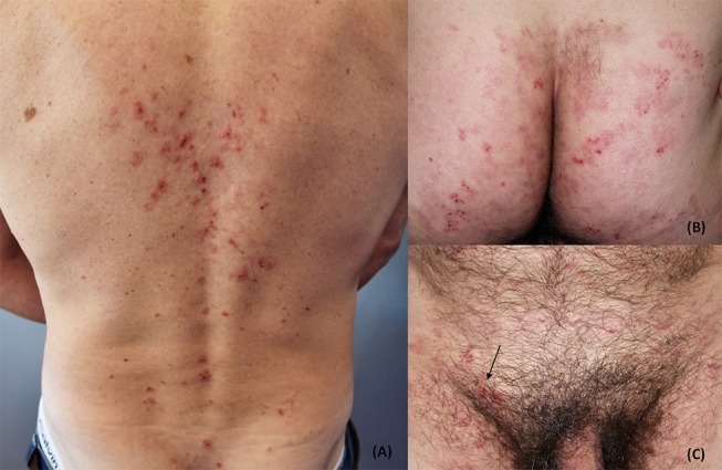
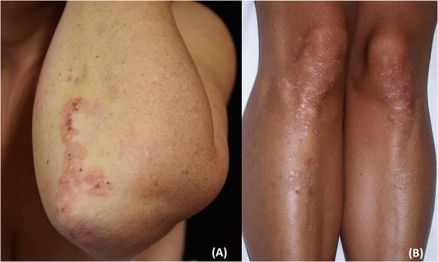
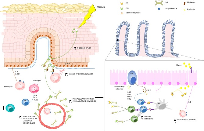
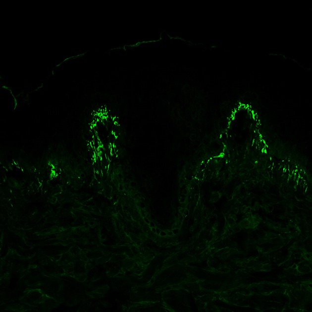
Dermatitis herpetiformis (DH) is an inflammatory disease of the skin, considered the specific cutaneous manifestation of celiac disease (CD). Both DH and CD occur in gluten-sensitive individuals, share the same Human Leukocyte Antigen (HLA) haplotypes (DQ2 and DQ8), and improve following the administration of a gluten-free diet. Moreover, almost all DH patients show typical CD alterations at the small bowel biopsy, ranging from villous atrophy to augmented presence of intraepithelial lymphocytes, as well as the generation of circulating autoantibodies against tissue transglutaminase (tTG). Clinically, DH presents with polymorphic lesions, including papules, vesicles, and small blisters, symmetrically distributed in typical anatomical sites including the extensor aspects of the limbs, the elbows, the sacral regions, and the buttocks. Intense pruritus is almost the rule. However, many atypical presentations of DH have also been reported. Moreover, recent evidence suggested that DH is changing. Firstly, some studies reported a reduced incidence of DH, probably due to early recognition of CD, so that there is not enough time for DH to develop. Moreover, data from Japanese literature highlighted the absence of intestinal involvement as well as of the typical serological markers of CD (i.e., anti-tTG antibodies) in Japanese patients with DH. Similar cases may also occur in Caucasian patients, complicating DH diagnosis. The latter relies on the combination of clinical, histopathologic, and immunopathologic findings. Detecting granular IgA deposits at the dermal-epidermal junction by direct immunofluorescence (DIF) from perilesional skin represents the most specific diagnostic tool. Further, assessing serum titers of autoantibodies against epidermal transglutaminase (eTG), the supposed autoantigen of DH, may also serve as a clue for the diagnosis. However, a study from our group has recently demonstrated that granular IgA deposits may also occur in celiac patients with non-DH inflammatory skin diseases, raising questions about the effective role of eTG IgA autoantibodies in DH and suggesting the need of revising diagnostic criteria, conceivably emphasizing clinical aspects of the disease along with DIF. DH usually responds to the gluten-free diet. Topical clobetasol ointment or dapsone may be also applied to favor rapid disease control. Our review will focus on novel pathogenic insights, controversies, and management aspects of DH.

Keywords: coeliac disease; dermatitis herpetiformis; direct immunofluorescence; epidermal transglutaminase; non-coeliac gluten sensitivity.


Figures

Figure 1
Figure 1
Clinical presentation of dermatitis herpetiformis (DH): erythematous grouped papules and vesicles associated with excoriations and crusts at the back (A), sacral region and buttocks (B). Rarely, DH may also affect the groin and pubis (arrow) (C). The patient gave written informed consent for the publication of these pictures.
Figure 2
Figure 2
Clinical presentation of dermatitis herpetiformis: grouped papules and vesicles associated with excoriations and crusts at the elbows (A) and lower limbs (B). Post-inflammatory pigmentary changes such as hypo-pigmentation could be also appreciated (B). The patient gave written informed consent for the publication of these pictures.
Figure 3
Figure 3
Pathogenesis of dermatitis herpetiformis (DH): Production of IgA autoantibodies against eTG occurs in the gut, probably as a result of an epitope spreading phenomenon (1), due to the high sequence homology between tissue TG, which is a major autoantigen in coeliac disease, and eTG. Activation of innate immunity in the gut leads to increased release of IL-8, which is thought to be responsible for the initial priming of neutrophils (2). One theory suggests that, in region of trauma, keratinocyte damage leads to shedding of eTG to the dermal-epidermal junction (3), where it binds to anti-eTG IgA. An alternative hypothesis suggests that eTG/IgA complexes exists as circulating immune complexes, which can deposit both at the dermal-epidermal junction and around dermal vessels (4). A complex interplay between inflammatory cytokines and the activation of fibrinogen stimulate neutrophil adherence to the activated endothelium (5) and migration to the dermal papillae. Herein neutrophils, which probably bind to IgA aggregates through the Fc IgA receptor, release proteases, which finally induce the cleavage of the dermal-epidermal junction (6). In parallel, hyper-activation of Th2 cells activate eosinophils, which also co-operate with neutrophils to the cleavage of the dermal-epidermal junction. Down-regulation of Treg cells do also occur, favoring the maintenance of a pro-inflammatory microenvironment in DH skin. Abbreviations in the figure: eTG, epidermal transglutaminase; tTG, tissue transglutaminase; IL, interleukin; Th, T helper cell; Treg, regulatory T cell; APC, antigen presenting cell.
Figure 4
Figure 4
Direct immunofluorescence of perilesional skin specimens from patients with dermatitis herpetiformis (DH). Granular IgA deposits at the dermal papillary tips are considered a pathognomonic finding of DH (magnification 400×).

References

    1. Duhring LA. Landmark article, Aug 30, 1884: Dermatitis herpetiformis. JAMA. (1983) 250:212–16. 10.1001/jama.250.2.212 - DOI - PubMed
    1. Esteves J, Brandao FN. Effect of sulfonamides and sulfones on Duhring's disease. Clin Lat. (1952) 2:34–9. - PubMed
    1. van der Meer JB. Granular deposits of immunoglobulins in the skin of patients with dermatitis herpetiformis. An immunofluorescent study. Br J Dermatol. (1969) 81:493–503. 10.1111/j.1365-2133.1969.tb16024.x - DOI - PubMed
    1. Marks J, Shuster S, Watson AJ. Small-bowel changes in dermatitis herpetiformis. Lancet. (1966) 2:1280–2. 10.1016/S0140-6736(66)91692-8 - DOI - PubMed
    1. Katz SI, Falchuk ZM, Dahl MV, Rogentine GN, Strober W. HL-A8: a genetic link between dermatitis herpetiformis and gluten-sensitive enteropathy. J Clin Invest. (1972) 51:2977–80. 10.1172/JCI107123 - DOI - PMC - PubMed

Publication types

MeSH terms