. 2019 Jun 18:13:54.
doi: 10.1186/s13036-019-0183-2. eCollection 2019.

Engineered mosaic protein polymers; a simple route to multifunctional biomaterials

Affiliations

Engineered mosaic protein polymers; a simple route to multifunctional biomaterials

Daniel T Peters et al. J Biol Eng. .

Abstract

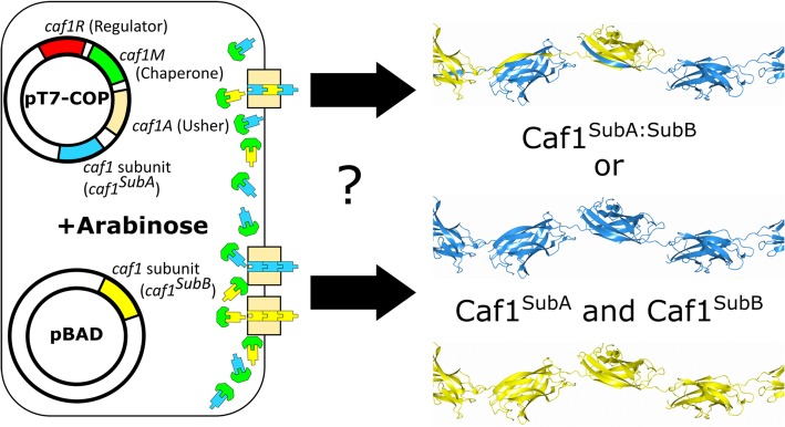
Background: Engineered living materials (ELMs) are an exciting new frontier, where living organisms create highly functional materials. In particular, protein ELMs have the advantage that their properties can be manipulated via simple molecular biology. Caf1 is a protein ELM that is especially attractive as a biomaterial on account of its unique combination of properties: bacterial cells export it as a massive, modular, non-covalent polymer which is resistant to thermal and chemical degradation and free from animal material. Moreover, it is biologically inert, allowing the bioactivity of each 15 kDa monomeric Caf1 subunit to be specifically engineered by mutagenesis and co-expressed in the same Escherichia coli cell to produce a mixture of bioactive Caf1 subunits.

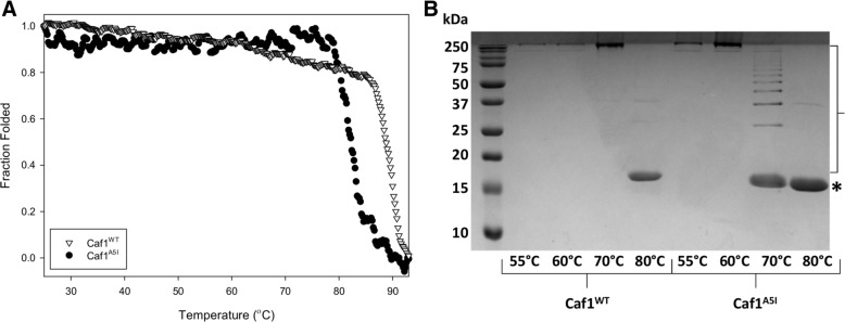
Results: Here, we show by gel electrophoresis and transmission electron microscopy that the bacterial cells combine these subunits into true mosaic heteropolymers. By combining two separate bioactive motifs in a single mosaic polymer we demonstrate its utility by stimulating the early stages of bone formation by primary human bone marrow stromal cells. Finally, using a synthetic biology approach, we engineer a mosaic of three components, demonstrating that Caf1 complexity depends solely upon the variety of monomers available.

Conclusions: These results demonstrate the utility of engineered Caf1 mosaic polymers as a simple route towards the production of multifunctional biomaterials that will be useful in biomedical applications such as 3D tissue culture and wound healing. Additionally, in situ Caf1 producing cells could create complex bacterial communities for biotechnology.

Keywords: Biomaterials; Bone; Electron microscopy; Protein engineering; Synthetic biology; Tissue scaffolds.


Conflict of interest statement

Competing interestsThe authors declare no competing interests.

Figures

Fig. 1
Fig. 1
Production of mosaic Caf1 polymers by E. coli. In this diagram, E. coli have been transformed with the pT7-COP plasmid, which contains the caf1 operon with a caf1 subunit “A” gene (caf1SubA, blue), and a pBad plasmid containing a caf1 subunit “B” gene (caf1SubB, yellow) under the control of an arabinose inducible promoter. When arabinose is added to cells growing in culture at 35 °C, both the caf1 operon and the additional subunit genes are expressed. Subunits are exported to the periplasm where they are bound by the chaperone, Caf1M (green). Caf1M delivers the subunits to the outer membrane usher, Caf1A (tan), which assembles the subunits into a polymer. Both subunits have been detected in the extracellular fraction of cell cultures expressing both genes, but it was not known whether they form a mosaic homopolymer containing a mixture of the two subunits (Caf1SubA:SubB, top), or two separate homopolymers (Caf1SubA and Caf1SubB, bottom). Caf1 polymer models were prepared from the Caf1:Caf1:Caf1M crystal structure (PDB: 1P5U), and visualised using the CCP4MG software package [50]
Fig. 2
Fig. 2
Characterisation of the Caf1A5I low stability mutant. (a) Near-UV circular dichroism thermal melt of the Caf1WT (adapted from [13]) and Caf1A5I proteins. Circular dichroism at 290 nm was followed as 1 mg/mL protein was heated between 25 and 95 °C, and the signal converted into fraction of protein folded. Data represent the average of three independent replicates. (b) SDS-PAGE analysis of Caf1WT and Caf1A5I proteins heated for 5 min in SDS containing sample buffer at the indicated temperatures. The position of the monomeric species is denoted by a star, with oligomeric breakdown products seen as a ladder within the region bounded by the bracket
Fig. 3
Fig. 3
Characterisation of Caf1 mosaic polymers by SDS-PAGE. (a) Three mutant Caf1 subunits are depicted as cartoons: Caf1WT in yellow, Caf1His in blue and Caf1A5I in orange, with a yellow star showing the location of the A5I mutation in the N-terminal β-strand that forms the subunit-subunit interface. Caf1WT: His, Caf1A5I and Caf1A5I: His proteins, as well as a mixture of Caf1A5I and Caf1WT: His, are shown in the top panel, with their expected state when heated to 70 °C shown in the bottom panel. Species which span the box represent full length Caf1 polymers. At 70 °C, the Caf1A5I subunit-subunit interactions break, leading to a pattern of oligomers (Monomer – M, Dimer – D, Trimer – T). The Caf1WT and Caf1His subunits have wild-type subunit-subunit interfaces, and do not break down at 70 °C (Polymer – P). If the Caf1A5I subunit forms mosaic heteropolymers when co-expressed with the Caf1His subunit, extra bands corresponding to oligomers containing the higher molecular weight Caf1His subunit will be present. (b) Expected gel result if co-expression of Caf1A5I: His subunits leads to mosaic heteropolymers. The expected monomer (M), dimer (D) and trimer (T) bands are shown as lines for samples 1–4 from (a) when they are heated at 70 °C. If the co-expressed subunits form separate homopolymers, samples 2, 3 and 4 would appear identical. (c) SDS-PAGE analysis of Caf1 mosaic polymers. Four samples consisting of a Caf1WT:His mosaic polymer, a Caf1A5I mutant Caf1 polymer, an Caf1A5I:His mosaic polymer and an equimolar mixture of the Caf1WT:His and Caf1A5I polymers, were heated in SDS sample buffer for 5 min at either 70 °C or 100 °C. The Caf1A5I and Caf1WT subunits have similar molecular weights (15.6 kDa), whereas the Caf1His subunit has a molecular weight which is ~ 2 kDa higher (17.2 kDa). The positions of these monomeric subunits are highlighted, and oligomeric breakdown products can be observed within the region bounded by the bracket. Oligomeric breakdown products containing the higher molecular weight Caf1His subunits in the Caf1A5I:His mosaic polymer sample are highlighted using small white triangles, with the dimer region from sample 3 expanded and shown to the left of the gel
Fig. 4
Fig. 4
Characterisation of Caf1 mosaic polymers by transmission electron microscopy (a) Schematic of the electron microscopy experiment. Mosaic Caf1 polymers containing a His tag (blue, Caf1His) and a biotinylated cysteine (yellow, Caf1Cys(Biotin)) are mixed with Nickel-NTA-10 nm and Streptavidin-20 nm gold conjugates. These recognise and bind to the His tags (blue triangles) and biotinylated cysteines (yellow semicircles) respectively. On the electron micrograph (grey box), these should appear as light grey beads (Caf1 polymer) surrounded by small and large black dots, representing the two different size gold conjugates. (b) Negative stain electron micrographs of the His-tagged, biotinylated cysteine containing mosaic polymer (Caf1His:Cys(Biotin)). His-tagged subunits were labelled with Nickel-NTA-10 nm gold particles and biotinylated cysteine containing subunits labelled with Streptavidin-20 nm gold particles. Images were taken at 92000x magnification. Dotted lines show the position of the Caf1 polymer. Additional data for WT unlabelled polymers are shown in Supplementary Information
Fig. 5
Fig. 5
Caf1 mosaic polymers direct the early stages of bone formation. Phase microscopy images of primary human bone marrow stromal cells grown on plastic surfaces that were either uncoated (a) or coated with Caf1WT (b) or Caf1OPN:BMP2 (c) mosaic polymers (scale bar = 200 μm). Areas of mineralisation triggered by the differentiation of the cells are highlighted with black arrows. (d) qRT-PCR analysis of Runx2 (blue) and BMP2 (orange) expression from each of the cultures at 14 days. Error bars represent the standard deviation from three biological replicates
Fig. 6
Fig. 6
Design and production of a 3-subunit mosaic Caf1 polymer. (a) Diagram depicting the pBad-Caf1OPN and pBad2x-Caf1OPN:BMP2 plasmids used in this study. The pBad plasmid was used as a template for the insertion of a caf1 mutant gene containing the osteopontin sequence (Caf1OPN, yellow), a second ribosome binding site (RBS, green) and a caf1 mutant gene containing the BMP2 peptide sequence (Caf1BMP2, blue) in order to construct the pBad2x-Caf1OPN:BMP2 plasmid, which was thus designed to express two caf1 genes at once under the control of the arabinose inducible promoter. The pBad-Caf1OPN plasmid, designed to express only the one caf1 gene, is shown alongside as a comparison. (b) SDS-PAGE analysis showing the expression of a 3-subunit mosaic Caf1 polymer. Cultures of E. coli BL21(DE3) cells transformed with pCOP and either pBad-Caf1OPN, pBad-Caf1BMP2 or pBad2x OPN:BMP2 were grown for 22 h in the presence and absence of 1% w/v arabinose. Samples of the extracellular fraction (flocculent layer and supernatant) were then heated to 100 °C for 5 min in SDS containing buffer and applied to the gel. The “U” lane represents the pCOP/pBad2x-Caf1OPN:BMP2 1% arabinose sample that was not heated to 100 °C before application, and shows most Caf1 subunits are present in high molecular weight polymers. The monomeric subunits corresponding to each mutant are shown by numbers next to the relevant band: 1 is Caf1WT, 2 is Caf1OPN and 3 is Caf1BMP2
Fig. 7
Fig. 7
Model of mosaic Caf1 polymer production by bacterial cells. A diagram of a bacterium is shown with the cytoplasm, periplasm and extracellular medium sections labelled. The cytoplasm contains the pCOP and pBad2x-Caf1OPN:BMP2 plasmids needed for Caf1 mosaic polymer production where genes are colour coded as follows: caf1R regulator (red), caf1M chaperone (green), caf1A usher (tan), caf1WT subunit (orange), caf1OPN mutant subunit (yellow) and caf1BMP2 mutant (cyan). Caf1M and Caf1 subunits are targeted to the periplasm and form chaperone:subunit complexes. These complexes are assembled into a Caf1 polymer by the Caf1A usher, which resides in the outer membrane. The usher does not discriminate between the different types of Caf1 subunit, and so the polymer formed contains a random mixture of the expressed subunits, and is thus a three-subunit mosaic heteropolymer

Similar articles

Cited by

References

    1. Gilbert C, Ellis T. Biological engineered living materials: growing functional materials with genetically programmable properties. ACS Synth Biol. 2019;8(1):1–15. doi: 10.1021/acssynbio.8b00423. - DOI - PubMed
    1. Nguyen PQ, Courchesne N-MD, Duraj-Thatte A, Praveschotinunt P, Joshi NS. Engineered living materials: prospects and challenges for using biological systems to direct the assembly of smart materials. Adv Mater. 2018;30(19):1704847. doi: 10.1002/adma.201704847. - DOI - PMC - PubMed
    1. Muiznieks LD, Keeley FW. Biomechanical Design of Elastic Protein Biomaterials: a balance of protein structure and conformational disorder. ACS Biomaterials Science & Engineering. 2017;3(5):661–679. doi: 10.1021/acsbiomaterials.6b00469. - DOI - PubMed
    1. Bracalello A, Santopietro V, Vassalli M, Marletta G, Del Gaudio R, Bochicchio B, et al. Design and production of a chimeric Resilin-, elastin-, and collagen-like engineered polypeptide. Biomacromolecules. 2011;12(8):2957–2965. doi: 10.1021/bm2005388. - DOI - PubMed
    1. Parenteau-Bareil R, Gauvin R, Berthod F. Collagen-based biomaterials for tissue engineering applications. Materials. 2010;3(3):1863–1887. doi: 10.3390/ma3031863. - DOI

LinkOut - more resources