

CD137 promotes bone metastasis of breast cancer by enhancing the migration and osteoclast differentiation of monocytes/macrophages
- PMID: 31244935
- PMCID: PMC6568184
- DOI: 10.7150/thno.29617
CD137 promotes bone metastasis of breast cancer by enhancing the migration and osteoclast differentiation of monocytes/macrophages
Abstract
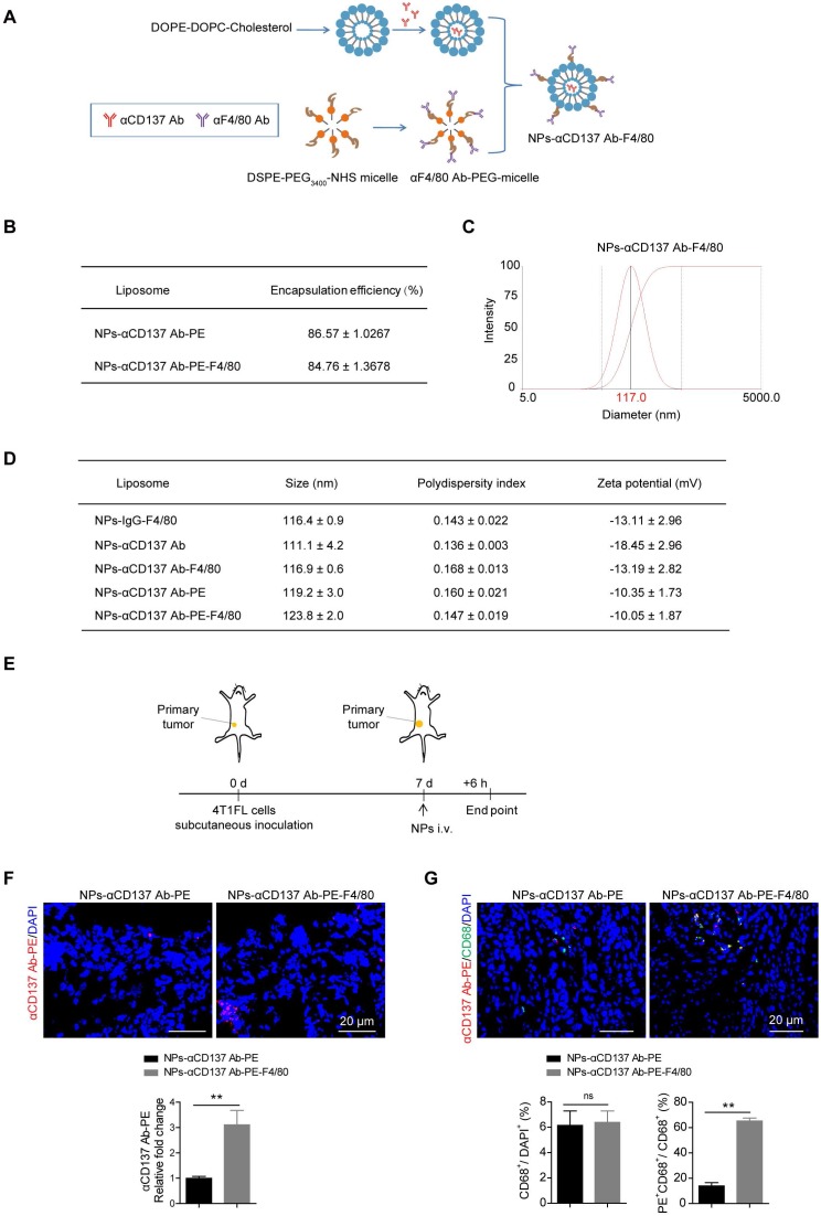
Rationale: Bone is one of the most common metastatic sites of breast cancer. CD137 (4-1BB), a member of the tumor necrosis factor (TNF) receptor superfamily, is mainly expressed in activated leukocytes. Previous study demonstrates the effect of CD137-CD137L bidirectional signaling pathway on RANKL-mediated osteoclastogenesis. However, the role of CD137 in bone metastasis of breast cancer needs further study. Methods: Stable monocyte/macrophage cell lines with Cd137 overexpression and silencing were established. Western blot, real-time PCR, transwell and tartrate-resistant acid phosphatase staining were used to detect the regulatory effect of CD137 on migration and osteoclastogenesis of monocytes/macrophages in vitro. Spontaneous bone metastasis mouse model was established, bioluminescent images, immunohistochemistry and histology assay were performed to detect the function of CD137 in bone metastasis in vivo. Results: We found that CD137 promotes the migration of monocytes/macrophages to tumor microenvironment by upregulating the expression of Fra1. It also promoted the differentiation of monocytes/macrophages into osteoclasts at the same time, thus providing a favorable microenvironment for the colonization and growth of breast cancer cells in bone. Based on these findings, a novel F4/80-targeted liposomal nanoparticle encapsulating the anti-CD137 blocking antibody (NP-αCD137 Ab-F4/80) was synthesized. This nanoparticle could inhibit both bone and lung metastases of 4T1 breast cancer cells with high efficacy in vivo. In addition, it increased the therapeutic efficacy of Fra1 inhibitor on tumor metastasis. Conclusions: Taken together, these findings reveal the promotion effect of macrophage/monocyte CD137 on bone metastases and provide a promising therapeutic strategy for metastasis of breast cancer.
Keywords: Fra1; anti-CD137 antibody; bone metastasis; breast cancer; liposomal nanoparticles; metastatic niche..
Conflict of interest statement
Competing Interests: The authors have declared that no competing interest exists.
Figures


Similar articles
-
Plumbagin inhibits breast tumor bone metastasis and osteolysis by modulating the tumor-bone microenvironment.Curr Mol Med. 2012 Sep;12(8):967-81. doi: 10.2174/156652412802480871. Curr Mol Med. 2012. PMID: 22574935
-
CD137-CD137L Signaling Affects Angiogenesis by Mediating Phenotypic Conversion of Macrophages.J Cardiovasc Pharmacol. 2020 Feb;75(2):148-154. doi: 10.1097/FJC.0000000000000772. J Cardiovasc Pharmacol. 2020. PMID: 31651672
-
Interleukin-4 inhibits RANKL-induced expression of NFATc1 and c-Fos: a possible mechanism for downregulation of osteoclastogenesis.Biochem Biophys Res Commun. 2005 Apr 15;329(3):839-45. doi: 10.1016/j.bbrc.2005.02.049. Biochem Biophys Res Commun. 2005. PMID: 15752732
-
CD137 / CD137 ligand signalling regulates the immune balance: A potential target for novel immunotherapy of autoimmune diseases.J Autoimmun. 2020 Aug;112:102499. doi: 10.1016/j.jaut.2020.102499. Epub 2020 Jun 4. J Autoimmun. 2020. PMID: 32505443 Review.
-
Parathyroid hormone-related protein and bone metastases.Cancer. 1997 Oct 15;80(8 Suppl):1572-80. doi: 10.1002/(sici)1097-0142(19971015)80:8+<1572::aid-cncr7>3.3.co;2-d. Cancer. 1997. PMID: 9362424 Review.
Cited by
-
Anterior Gradient 2 is a Significant Prognostic Biomarker in Bone Metastasis of Breast Cancer.Pathol Oncol Res. 2022 Nov 3;28:1610538. doi: 10.3389/pore.2022.1610538. eCollection 2022. Pathol Oncol Res. 2022. PMID: 36405393 Free PMC article.
-
The Comprehensive Analysis Illustrates the Role of CDCA5 in Breast Cancer: An Effective Diagnosis and Prognosis Biomarker.Int J Genomics. 2023 May 30;2023:7150141. doi: 10.1155/2023/7150141. eCollection 2023. Int J Genomics. 2023. PMID: 37287817 Free PMC article.
-
Transcriptome Reprogramming of CD11b+ Bone Marrow Cells by Pancreatic Cancer Extracellular Vesicles.Front Cell Dev Biol. 2020 Nov 27;8:592518. doi: 10.3389/fcell.2020.592518. eCollection 2020. Front Cell Dev Biol. 2020. PMID: 33330473 Free PMC article.
-
Immune Microenvironment in Breast Cancer Metastasis.Adv Exp Med Biol. 2025;1464:413-432. doi: 10.1007/978-3-031-70875-6_20. Adv Exp Med Biol. 2025. PMID: 39821036 Review.
-
TNFSF9 promotes metastasis of pancreatic cancer through Wnt/Snail signaling and M2 polarization of macrophages.Aging (Albany NY). 2021 Sep 13;13(17):21571-21586. doi: 10.18632/aging.203497. Epub 2021 Sep 13. Aging (Albany NY). 2021. PMID: 34517345 Free PMC article.
References
-
- Siegel RL, Miller KD, Jemal A. Cancer statistics, 2018. CA Cancer J Clin. 2018;68:7–30. - PubMed
-
- Sterling JA, Edwards JR, Martin TJ, Mundy GR. Advances in the biology of bone metastasis: how the skeleton affects tumor behavior. Bone. 2011;48:6–15. - PubMed
-
- Paget S. The distribution of secondary growths in cancer of the breast. 1889. Cancer metastasis reviews. 1989;8:98–101. - PubMed
-
- Fokas E, Engenhart-Cabillic R, Daniilidis K, Rose F, An HX. Metastasis: the seed and soil theory gains identity. Cancer metastasis reviews. 2007;26:705–15. - PubMed
-
- Sica G, Chen L. Biochemical and immunological characteristics of 4-1BB (CD137) receptor and ligand and potential applications in cancer therapy. Archivum immunologiae et therapiae experimentalis. 1999;47:275–9. - PubMed
Publication types
MeSH terms
Substances
LinkOut - more resources
Full Text Sources
Medical
Research Materials
Miscellaneous
