

Acute thrombotic vascular events complicating influenza-associated pneumonia
- PMID: 31245274
- PMCID: PMC6582236
- DOI: 10.1016/j.rmcr.2019.100884
Acute thrombotic vascular events complicating influenza-associated pneumonia
Abstract
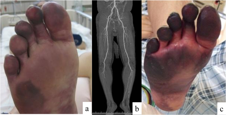
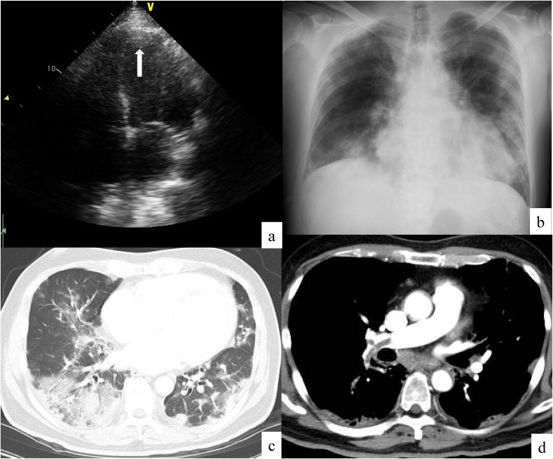
A 58-year-old man with previous myocardial infarction presented to our hospital with fever, cough, and dyspnea. PCR testing with nasopharyngeal swabs confirmed influenza virus infection, and enhanced computed tomography and transthoracic echocardiography revealed bilateral ground-glass opacities and consolidation, deep venous thrombosis, acute pulmonary artery embolism, and acute arterial embolism that appeared to originate from thrombus in the left ventricle. Combination of a neuraminidase inhibitor, antibiotics, an anticoagulant, and anti-platelet agent improved these complications; however, amputation of the patient's right foot was required. Because influenza can cause vascular events, physicians should pay attention to this complication in patients with influenza-associated pneumonia.
Keywords: Acute arterial embolism; Deep venous thrombosis; Gangrene; Influenza; Pneumonia.
Figures
References
-
- Bunce P.E., High S.M., Nadjafi M., Stanley K., Liles W.C., Christian M.D. Pandemic H1N1 influenza infection and vascular thrombosis. Clin. Infect. Dis. 2011;52:e14–e17. - PubMed
-
- Kwong J.C., Schwartz K.L., Campitelli M.A., Chung H., Crowcroft N.S., Karnauchow T., Katz K., Ko D.T., McGeer A.J., McNally D., Richardson D.C., Rosella L.C., Simor A., Smieja M., Zahariadis G., Gubbay J.B. Acute myocardial infarction after laboratory-confirmed influenza infection. N. Engl. J. Med. 2018;378(4):345–353. - PubMed
Publication types
LinkOut - more resources
Full Text Sources
