

Hoffa's Fat Pad-associated Solitary Neurofibroma as the Cause of Anterior Knee Pain: A Case Report
- PMID: 31245314
- PMCID: PMC6588136
- DOI: 10.13107/jocr.2250-0685.1294
Hoffa's Fat Pad-associated Solitary Neurofibroma as the Cause of Anterior Knee Pain: A Case Report
Abstract
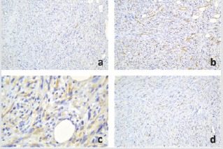
Introduction: Infrapatellar peripheral neural tumors, particularly neurofibromas, are rather rare entities reported in the literature. They are slow-growing lesions that usually do not exhibit clinical manifestations other than interspecific swelling or pain; hence, their diagnosis can be quite challenging. Therefore, scrutiny should include not only traditional clinical assessment and imaging but also more specific molecular biology techniques, such as immunohistochemistry.
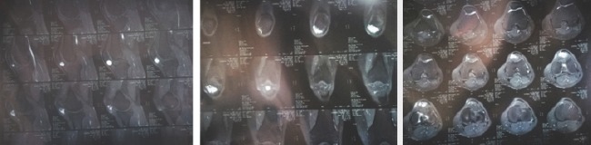
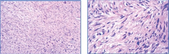
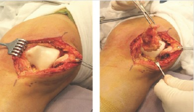
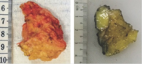
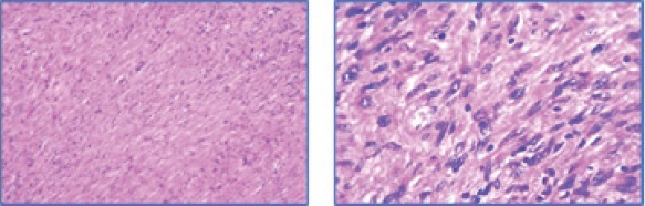
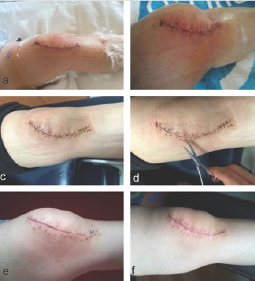
Case report: We present the clinical, imaging, histological, and immunohistochemical features of a unique case of solitary neurofibromain a 33-year-old female presenting chronic anterior knee pain. The tumor was completely removed through a surgical approach.
Conclusions: Although cases of a solitary neurofibroma originating within Hoffa's fat pad are extremely rare; the entity should be considered in the differential diagnosis when symptomatology is not alleviated with appropriate treatments.
Keywords: Anterior knee pain; Hoffa’s fat pad; immunohistochemistry; solitary neurofibroma.
Conflict of interest statement
Conflict of Interest: Nil
Figures
Similar articles
-
[Hoffa's disease of the adipose pad: magnetic resonance versus surgical findings].Radiol Med. 1998 Apr;95(4):278-85. Radiol Med. 1998. PMID: 9676203 Italian.
-
Solitary synovial chondromatosis as a cause of Hoffa's fat pad impingement.Med Pregl. 2015 Jan-Feb;68(1-2):49-52. doi: 10.2298/mpns1502049m. Med Pregl. 2015. PMID: 26012244
-
Presence of a t(12;18)(q14;q21) Chromosome Translocation and Fusion of the Genes for High-mobility Group AT-Hook 2 (HMGA2) and WNT Inhibitory Factor 1 (WIF1) in Infrapatellar Fat Pad Cells from a Patient With Hoffa's Disease.Cancer Genomics Proteomics. 2022 Sep-Oct;19(5):584-590. doi: 10.21873/cgp.20343. Cancer Genomics Proteomics. 2022. PMID: 35985683 Free PMC article.
-
Magnetic resonance imaging of Hoffa's fat pad and relevance for osteoarthritis research: a narrative review.Osteoarthritis Cartilage. 2016 Mar;24(3):383-97. doi: 10.1016/j.joca.2015.09.018. Epub 2015 Oct 9. Osteoarthritis Cartilage. 2016. PMID: 26455999 Review.
-
Posterior Hoffa's fat pad impingement secondary to a thickened infrapatellar plica: a case report and review of the literature.J Radiol Case Rep. 2015 Mar 31;9(3):20-6. doi: 10.3941/jrcr.v9i3.2037. eCollection 2015 Mar. J Radiol Case Rep. 2015. PMID: 25926930 Free PMC article. Review.
Cited by
-
Ewing sarcoma of Hoffa fat pad in the knee: a case report and review of the literature on primary intraarticular sarcomas and Hoffa fat pad masses.Skeletal Radiol. 2023 Jul;52(7):1415-1420. doi: 10.1007/s00256-022-04239-7. Epub 2022 Dec 9. Skeletal Radiol. 2023. PMID: 36484842 Review.
-
Intra-articular Knee Neurofibroma in a Patient with Neurofibromatosis: A Case Report and Review of Literature.J Orthop Case Rep. 2024 Oct;14(10):140-145. doi: 10.13107/jocr.2024.v14.i10.4840. J Orthop Case Rep. 2024. PMID: 39381300 Free PMC article.
References
-
- Pradilla-Gordillo MP. Solitary neurofibroma in cervical nerve root C7. Rev Iberoam Cir Mano. 2016;44:35–8.
-
- Argenyi ZB. Neural and neuroendocrine neoplasm (other than neurofibromatosis) In: Bolognia J, Jorizzo J, Rapini R, editors. Dermatology. 2nd ed. Madrid: Mosby; 2008. pp. 1795–811.
-
- Reyes-Martínez G, Plascencia-Gómez JA, de Anda-Juárez MC, Gómez-Villa R, Sáenz-Corral C, Carreón AA, et al. Solitary neurofibroma:Its frequency in a general hospital in Mexico City. Dermatol CQM. 2012;10:8–12.
-
- McDonald JS. Tumors of the oral soft tissues and cysts and tumors of the bones. In: Dean JA, editor. McDonald and Avery's Dentistry for the Child and Adolescent. 10th ed. USA: Elsevier; 2016. pp. 603–26.
-
- Zheng H, Chang L, Patel N, Yang J, Lowe L, Burns DK, et al. Induction of abnormal proliferation by nonmyelinating Schwann cells triggers neurofibroma formation. Cancer Cell. 2008;13:117–28. - PubMed
Publication types
LinkOut - more resources
Full Text Sources