. 2015 Feb 19:1:18-29.
doi: 10.1016/j.reth.2014.12.004. eCollection 2015 Jun.

Xenogeneic-free defined conditions for derivation and expansion of human embryonic stem cells with mesenchymal stem cells

Affiliations

Xenogeneic-free defined conditions for derivation and expansion of human embryonic stem cells with mesenchymal stem cells

Hidenori Akutsu et al. Regen Ther. .

Abstract

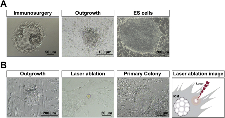
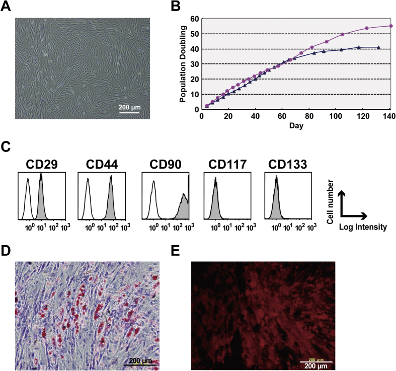
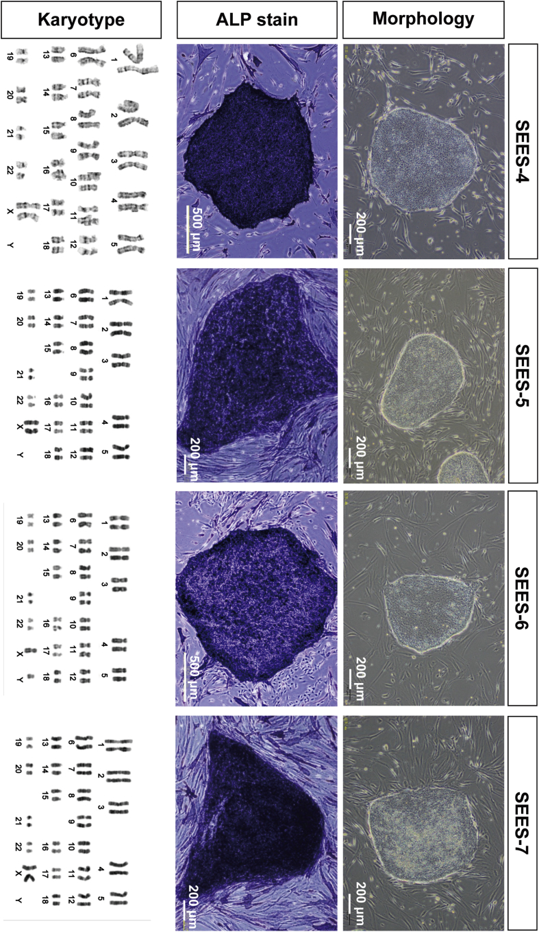
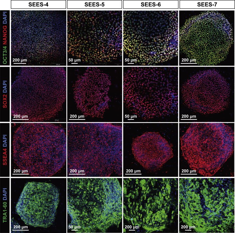
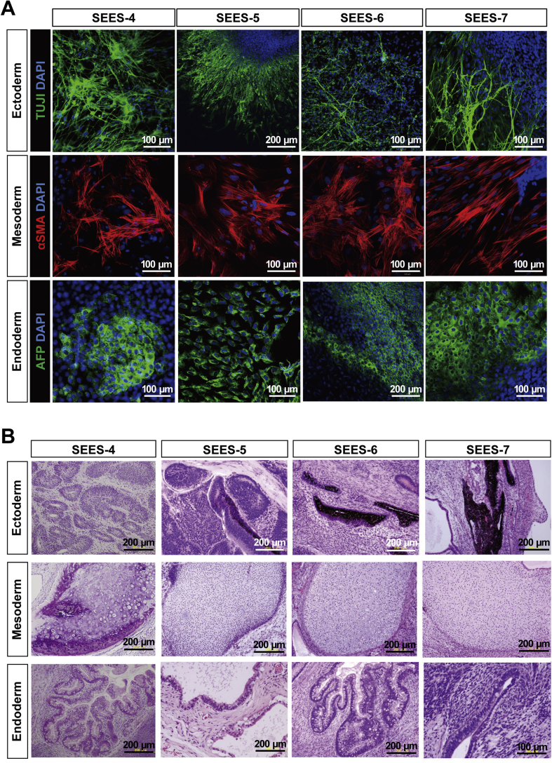
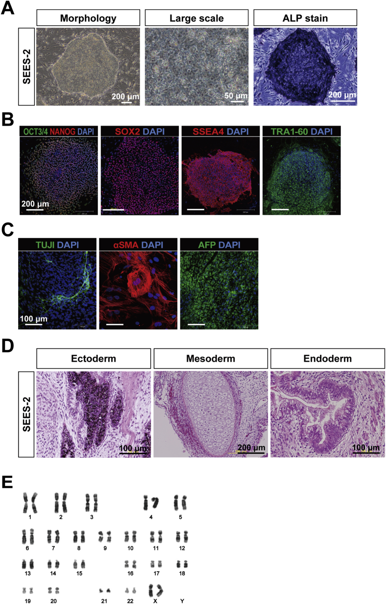
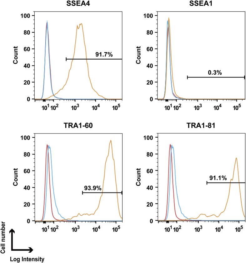
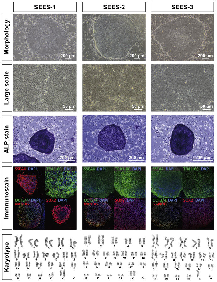
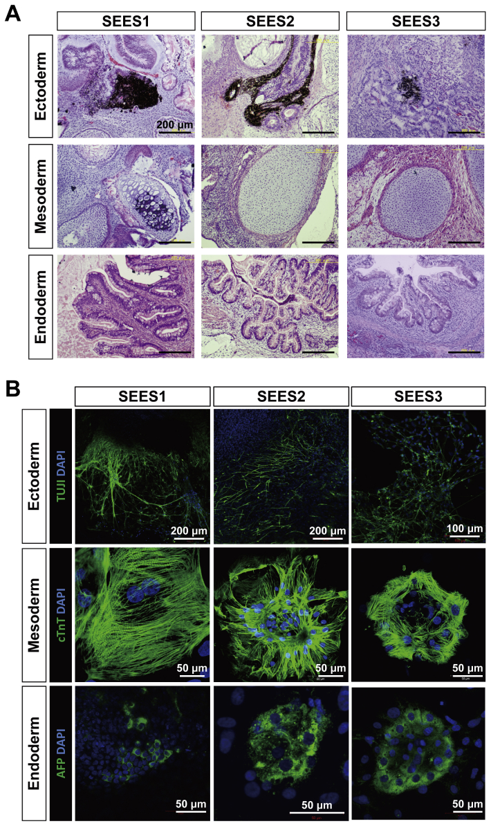
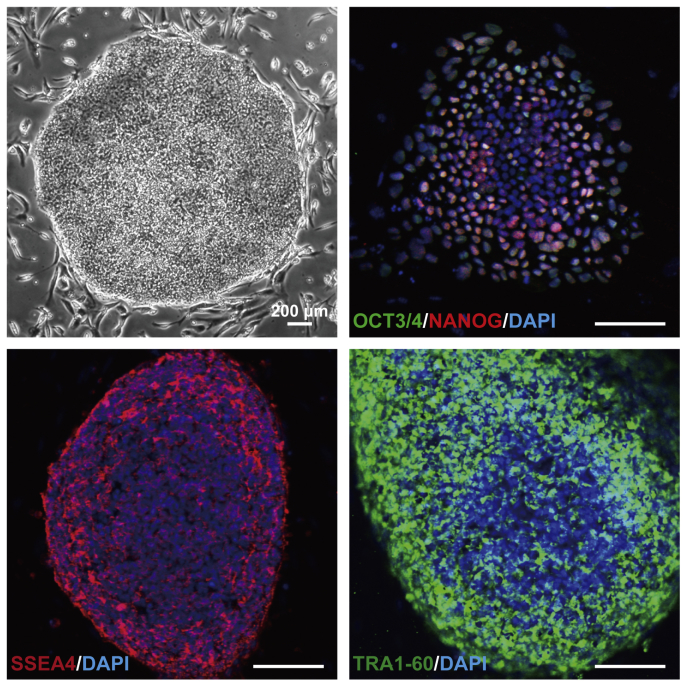
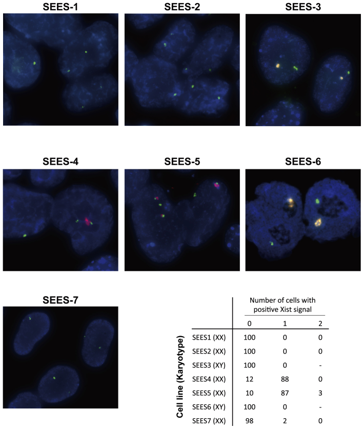
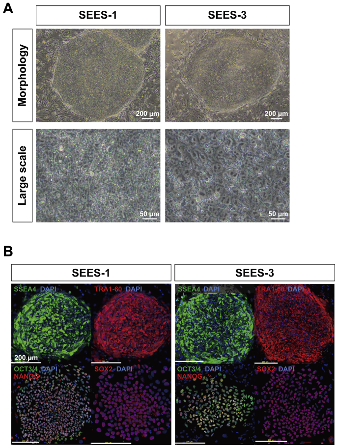
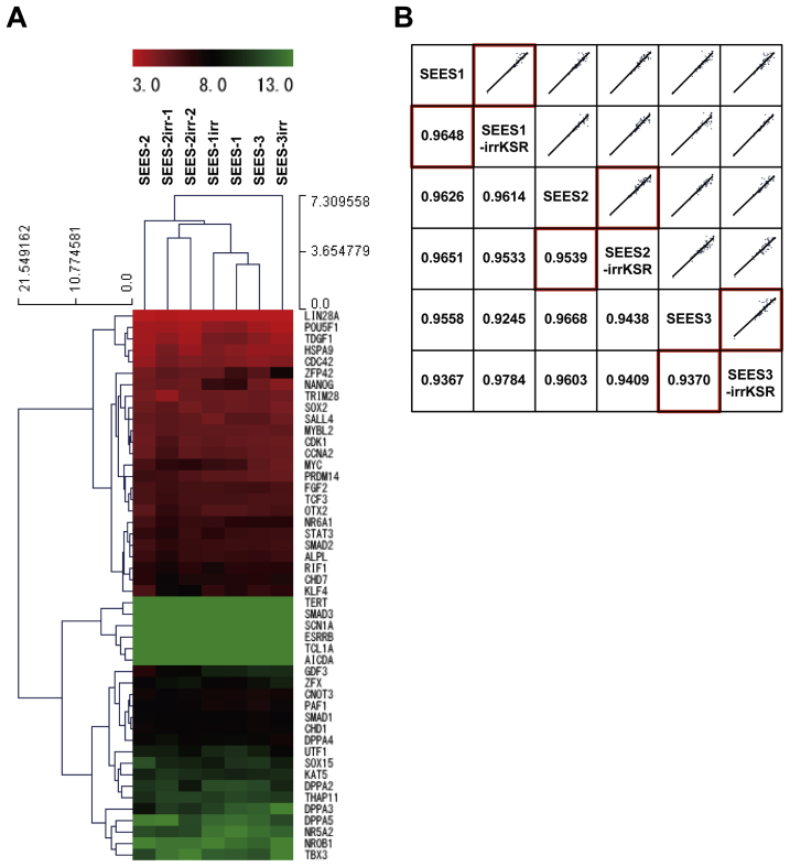
The potential applications of human embryonic stem cells (hESCs) in regenerative medicine and developmental research have made stem cell biology one of the most fascinating and rapidly expanding fields of biomedicine. The first clinical trial of hESCs in humans has begun, and the field of stem cell therapy has just entered a new era. Here, we report seven hESC lines (SEES-1, -2, -3, -4, -5, -6, and -7). Four of them were derived and maintained on irradiated human mesenchymal stem cells (hMSCs) grown in xenogeneic-free defined media and substrate. Xenogeneic-free hMSCs isolated from the subcutaneous tissue of extra fingers from individuals with polydactyly showed appropriate potentials as feeder layers in the pluripotency and growth of hESCs. In this report, we describe a comprehensive characterization of these newly derived SEES cell lines. In addition, we developed a scalable culture system for hESCs having high biological safety by using gamma-irradiated serum replacement and pharmaceutical-grade recombinant basic fibroblast growth factor (bFGF, also known as trafermin). This is first report describing the maintenance of hESC pluripotency using pharmaceutical-grade human recombinant bFGF (trafermin) and gamma-irradiated serum replacement. Our defined medium system provides a path to scalability in Good Manufacturing Practice (GMP) settings for the generation of clinically relevant cell types from pluripotent cells for therapeutic applications.

Keywords: Gamma irradiation; Human embryonic stem cells; Human feeder layer; Mesenchymal stem cells; Stem cell expansion; Xenogeneic-free medium.


Figures

Fig. 1
Fig. 1
Derivation of human ES cells by immunosurgery and laser ablation A) The intact inner cell mass (ICM) was isolated from blastocysts by immunosurgery. Cells of the trophectoderm are destroyed by brief exposure to antibodies directed against human cells in tandem with complement activity. Attachment and outgrowth of the ICM grew into an ESC (SEES-1) colony. B) The intact ICM was isolated by laser ablation without any animal products. Outgrowth of the blastocyst grown on human MSC feeder layers. Trophectoderm cells were targeted with multiple pulses of laser ablation. Typical morphology of ESC colonies was readily visible at high magnification. The image shows a schematic of ICM isolation by laser ablation, which is commonly used in artificial reproductive technology (ART).
Fig. 2
Fig. 2
Isolation and characterization of the xenogeneic-free hMSC feeder layer A) Primary human mesenchymal cells isolated from subcutaneous tissue of the polydactyly were expanded in xenogeneic-free media. B) Cells grew well over PD 30 under xenogeneic-free conditions. Two differentially isolated cell lines (red circle and blue triangle) showed similar cellular proliferation characteristics. C) Mesenchymal markers such as CD29, CD44, and CD90 were observed. D,E) The cells differentiated into adipogenic and osteogenic cells.
Fig. 3
Fig. 3
Derivation of xenogeneic-free hESCs on the hMSC feeder layer Under completely xenogeneic-free conditions, four hESC lines were derived from the inactivated hMSC feeder layer using the laser ablation system. Typical ESC morphology was readily visible. Alkaline phosphatase (ALP) activity was detected in each SEES cell line. Chromosome analysis of SEES-4, SEES-5, SEES-6, and SEES-7 cells showed normal karyotypes: 46,XX, 46,XX, 46,XY, and 46,XX, respectively.
Fig. 4
Fig. 4
Pluripotent marker expression of xenogeneic-free SEES cell lines In the undifferentiated state, xenogeneic-free SEES cell lines expressed markers characteristic of pluripotent hESCs, including OCT4, NANOG, SOX2, SSEA4, and TRA1-60. SEES-4: scale bars are 200 μm; SEES-5: scale bars are 50 μm in OCT3/4 and TRA1-60 and 200 μm in SOX2 and SSEA4; SEES-6: scale bars are 50 μm in OCT3/4 and SOX2 and 200 μm in SSEA4; SEES-7: scale bars are 200 μm.
Fig. 5
Fig. 5
Differentiation of three germ layers of xenogeneic-free SEES cell lines A) SEES cells differentiated in vitro via EBs expressed markers of the primary germ layers. Immunohistochemical analyses of markers of the ectoderm (TUJ1), mesoderm (αSMA), and endoderm (AFP) layers are shown. SEES-4: scale bars are 100 μm; SEES-5: scale bars are 200 μm for TUJ1 and 100 μm for αSMA and AFP; SEES-6: scale bars are 100 μm for TUJ1 and αSMA and 200 μm for AFP; SEES-7: scale bars are 100 μm. B) SEES cells differentiated in vivo via teratoma formation. Hematoxylin and eosin staining revealed germ layer derivatives, such as neural tissues, pigmented epithelium (ectoderm), cartilage (mesoderm), and gut epithelial tissues (endoderm). Scale bars are 200 μm.
Fig. 6
Fig. 6
Characterization of the pluripotency of SEES-2 maintained using a modified conventional hESC culture medium SEES-2 cells were stably maintained over 20 passages on the qualified MEF feeder layer in the modified medium, which contained pharmaceutical-grade recombinant human bFGF (trafermin) and high-dose (35-K) gamma-irradiated KO-SR without antibiotics. A) Typical hESC colony morphology was readily visible. ALP activity was detected. B) SEES-2 cells expressed undifferentiated hESC markers, including OCT4, NANOG, SOX2, SSEA4, and TRA1-60. SEES-2 cells could differentiate into three embryonic germ layers in vitro and in vivo. Scale bars are 200 μm. C) SEES cells that had been differentiated in vitro via EBs expressed markers of the primary germ layers, ectoderm (TUJ1), mesoderm (αSMA), and endoderm (AFP). Scale bars are 100 μm. D) Histological analysis of teratomas containing multidifferentiated tissues derived from SEES-2 cells. Pigmented epithelium (ectoderm), cartilage (mesoderm), and gut epithelial tissues (endoderm). E) Chromosomal analysis of SEES-2 cells cultivated through 16 passages using a modified conventional hESC culture medium showed a normal 46,XX karyotype.
Fig. 7
Fig. 7
Expression of pluripotency markers in SEES-2 cells was maintained using a modified conventional hESC culture medium Flow cytometric analysis of hESC-specific marker expression in SEES-2 cells. The isotype control is indicated by the blue line, and the unlabeled sample, which was used as a control, is indicated by the red line. Surface staining is shown by the yellow line for SSEA4, SSEA1, TRA-1-60, and TRA-1-81.
Supplemental Fig. 1
Supplemental Fig. 1
Characterization of SEES-1, SEES-2, and SEES-3 cells Typical ESC morphology is shown for SEES-1, SEES-2, and SEES-3 cells. Alkaline phosphatase (ALP) activity was detected in each SEES cell line. Three SEES cell lines, grown in conventional hESC culture medium, expressed markers characteristic of pluripotent hESCs, including OCT4, NANOG, SOX2, SSEA4, and TRA1-60. Chromosomal analysis of SEES-1, SEES-2, and SEES-3 cells showed normal karyotype, 46,XX, 46,XX, and 46,XY, respectively.
Supplemental Fig. 2
Supplemental Fig. 2
Differentiation potential of SEES-1, SEES-2, and SEES-3 cells A) Histological analysis of teratomas containing multidifferentiated tissues derived from SEES-1, SEES-2, and SEES-3 cells. Pigmented epithelium (ectoderm), cartilage (mesoderm), and gut epithelial tissues (endoderm). Scale bars are 200 μm. B) SEES-1, SEES-2, and SEES-3 cells that were differentiated in vivo via EB formation expressed markers of the primary germ layers, ectoderm (TUJ1), mesoderm (cTnT∗), and endoderm (AFP). ∗cTnT: cardiac troponin T, a cardiac marker. SEES-1 and SEES-2 cells: scale bars are 200 μm for TUJ1 and 50 μm for cTnT and AFP. SEES-3 cells: scale bars are 100 μm for TUJ1 and 50 μm for cTnT and AFP.
Supplemental Fig. 3
Supplemental Fig. 3
Establishment of human iPSCs under xenogeneic-free conditions Human iPSCs were generated from XF MSCs (Yub-1896 cells) by transduction of three reprogramming factors (OCT3/4, SOX2 and KLF4) under hXF culture conditions using XF hESC medium and XF hMSC feeder layers. Derived human XF iPSCs express pluripotent markers including OCT4, NANOG, SSEA4, and TRA1-60. Scale bars are 100 μm
Supplemental Fig. 4
Supplemental Fig. 4
Detection of Neu5Gc and Neu5Ac on SEES cells N-glycolylneuraminic acid (Neu5Gc) was not detected in xenogeneic-free SEES cells, but was observed in SEES-1, SEES-2, and SEES-3 cells. N-acetylneuraminic acid (Neu5Ac) was found in all examined SEES cell lines. The detection limit of sialic acids is 0.01 nmol/mg protein.
Supplemental Fig. 5
Supplemental Fig. 5
Blood group ABO antigen typing of SEES cell lines and hMSCs ABO blood typing was performed by sequencing of ABO gene polymorphisms as described previously [S1] with some modifications and confirmed by exome sequencing. ABO genotyping was also performed by the PCR restriction-fragment length polymorphism method [S2] and allele-specific primers method [S3]. SEES-1, SEES-3, SEES-4, and SEES-6 cells were AO type; SEES-2 and SEES-5 cells were O type; and SEES-7 cells were AA type; hMSCs feeder layer (Yub-1896 cells) was BO type.
Supplemental Fig. 6
Supplemental Fig. 6
Xist expression in SEES cells Fluorescent in situ hybridization (FISH) of Xist in SEES cells was used to characterize the epigenetic status of X-chromosome inactivation. FISH was performed at the Chromosome Science Laboratory, Hokkaido, Japan. A total 100 cells were analyzed for Xist expression in each cell line. FISH analysis showed that SEES-1, SEES-2 and SEES-7 cells would have activated X chromosomes. RNA and DNA FISH staining with probes detecting XIST RNA (red), the X (green) and Y (yellow) chromosome, and DAPI counterstain.
Supplemental Fig. 7
Supplemental Fig. 7
Expression of pluripotency markers in SEES-1 and SEES-3 cells was maintained using a modified conventional hESC culture medium SEES-1 and SEES-3 cells were stably maintained in the modified medium containing pharmaceutical-grade recombinant human bFGF, trafermin, and 35-K gamma-irradiated KO-SR without antibiotics. A) Typical hESC colony morphology was readily visible. B) SEES-1 and SEES-3 cells expressed undifferentiated hESC markers, including SEES-4, TRA1-60, OCT4, NANOG, and SOX2. Scale bars are 200 μm.
Supplemental Fig. 8
Supplemental Fig. 8
Hierarchical clustering analysis of expression data from the PCR array across the 48 pluripotency-related genes We used a Human Embryonic Stem Cell PCR Array and RT2 qPCR Mastermix (Qiagen, Germany) for quantitative reverse transcription-polymerase chain reaction (qRT-PCR). Transcript levels were determined using the QuantStudio 12K Flex real-time PCR system (Life Technologies). Relative quantification was carried out using ACTB, GAPDH, and RPL13A as endogenous control genes. Hierarchical clustering analyses across the 48 pluripotent marker genes was performed using delta Ct values for gene expression data with MEV v4.8 statistical analysis software. A) Hierarchical clustering analysis of SEES cell lines was performed using a modified conventional hESC culture medium. Gene expression levels in each sample, relative to the median level of expression of that gene across all the samples, is represented using a red-black-green color scale, as shown in the key (green: below median; black: equal to median; red: above median). B) Correlation analysis for gene expression. Scatter plots (upper diagonal) of the gene expression of 48 pluripotency markers across all pair-wise comparisons. Correlation coefficients are shown in corresponding squares below the diagonal.

Similar articles

Cited by

References

    1. Thomson J.A., Itskovitz-Eldor J., Shapiro S.S., Waknitz M.A., Swiergiel J.J., Marshall V.S. Embryonic stem cell lines derived from human blastocysts. Science. 1998;282:1145–1147. - PubMed
    1. Schuldt B.M., Guhr A., Lenz M., Kobold S., MacArthur B.D., Schuppert A. Power-laws and the use of pluripotent stem cell lines. PLoS One. 2013;8:e52068. - PMC - PubMed
    1. Amit M., Carpenter M.K., Inokuma M.S., Chiu C.P., Harris C.P., Waknitz M.A. Clonally derived human embryonic stem cell lines maintain pluripotency and proliferative potential for prolonged periods of culture. Dev Biol. 2000;227:271–278. - PubMed
    1. Cowan C.A., Klimanskaya I., McMahon J., Atienza J., Witmyer J., Zucker J.P. Derivation of embryonic stem-cell lines from human blastocysts. N Engl J Med. 2004;350:1353–1356. - PubMed
    1. Chen A.E., Egli D., Niakan K., Deng J., Akutsu H., Yamaki M. Optimal timing of inner cell mass isolation increases the efficiency of human embryonic stem cell derivation and allows generation of sibling cell lines. Cell Stem Cell. 2009;4:103–106. - PMC - PubMed

References in supplemental figures

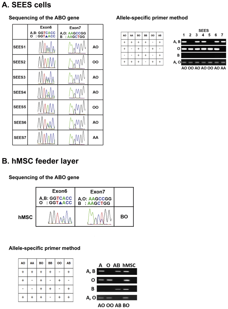
    1. Hosoi E. Biological and clinical aspects of ABO blood group system. J Med Invest. 2008;55:174–182. (For Supplemental Figure S1) - PubMed
    1. Ota M., Fukushima H., Kulski J.K., Inoko H. Single nucleotide polymorphism detection by polymerase chain reaction-restriction fragment length polymorphism. Nat Protoc. 2007;2:2857–2864. (For Supplemental Figure S2) - PubMed
    1. Muro T., Fujihara J., Imamura S., Nakamura H., Kimura-Kataoka K., Toga T. Determination of ABO genotypes by real-time PCR using allele-specific primers. Leg Med Tokyo. 2012;14:47–50. (For Supplemental Figure S3) - PubMed