

A hybrid of cells and pancreatic islets toward a new bioartificial pancreas
- PMID: 31245475
- PMCID: PMC6581840
- DOI: 10.1016/j.reth.2016.03.004
A hybrid of cells and pancreatic islets toward a new bioartificial pancreas
Abstract
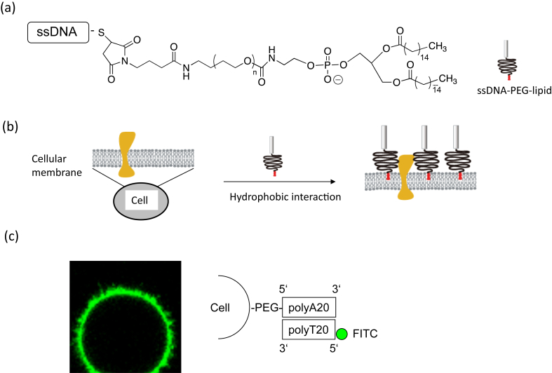
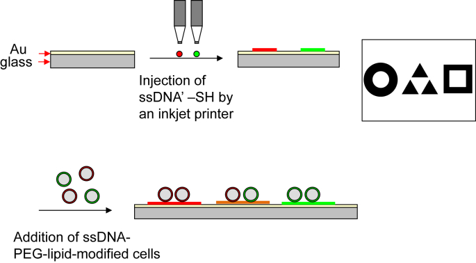
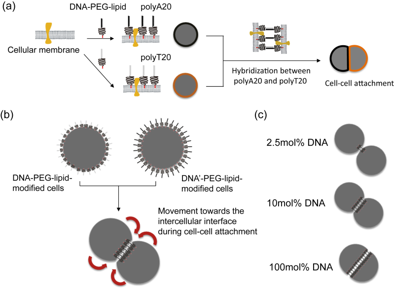
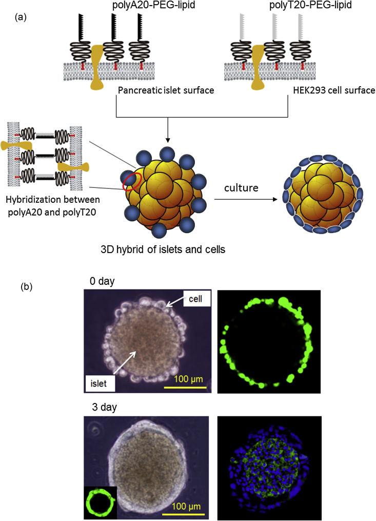
Cell surface engineering using single-stranded DNA-poly(ethylene glycol)-conjugated phospholipid (ssDNA-PEG-lipid) is useful for inducing cell-cell attachment two and three dimensionally. In this review, we summarize our recent techniques for cell surface engineering and their applications to islet transplantation. Because any DNA sequence can be immobilized onto the cell surface by hydrophobic interactions between ssDNA-PEG-lipid and the cellular membrane without impairing cell function, a cell-cell hybrid can be formed through the DNA hybridization. With this technique, it would be possible to create three-dimensional hybrid structures of pancreatic islets coated with various accessory cells, such as patients' own cells, mesenchymal and adipose-derived stem cells, endothelial progenitor cells, neural crest stem cells or regulatory T cells, which might significantly improve the outcome of islet transplantation in diabetic patients.
Keywords: Cell surface modification; Diabetes; IBMIR, instant blood-mediated inflammatory reaction; Instant blood-mediated inflammatory reaction (IBMIR); PEG-conjugated phospholipid (PEG-lipid); PEG-lipid, poly(ethylene glycol)-conjugated phospholipid; PMPC, poly(2-methacryloyloxyethyl phosphorylcholine); Pancreatic islet; islets, islets of Langerhans.
Figures
Similar articles
-
Improvement of graft survival by surface modification with poly(ethylene glycol)-lipid and urokinase in intraportal islet transplantation.Transplantation. 2011 Feb 15;91(3):271-8. doi: 10.1097/tp.0b013e3182034fa4. Transplantation. 2011. PMID: 21344734
-
Encapsulation of pancreatic islets within nano-thin functional polyethylene glycol coatings for enhanced insulin secretion.Tissue Eng Part A. 2010 Jul;16(7):2217-28. doi: 10.1089/ten.TEA.2009.0640. Tissue Eng Part A. 2010. PMID: 20163204
-
Islet-encapsulation in ultra-thin layer-by-layer membranes of poly(vinyl alcohol) anchored to poly(ethylene glycol)-lipids in the cell membrane.Biomaterials. 2007 Nov;28(32):4818-25. doi: 10.1016/j.biomaterials.2007.07.050. Epub 2007 Aug 14. Biomaterials. 2007. PMID: 17698188
-
Isolated human islets trigger an instant blood mediated inflammatory reaction: implications for intraportal islet transplantation as a treatment for patients with type 1 diabetes.Ups J Med Sci. 2000;105(2):125-33. doi: 10.1517/03009734000000059. Ups J Med Sci. 2000. PMID: 11095109 Review.
-
Control of instant blood-mediated inflammatory reaction to improve islets of Langerhans engraftment.Curr Opin Organ Transplant. 2011 Dec;16(6):620-6. doi: 10.1097/MOT.0b013e32834c2393. Curr Opin Organ Transplant. 2011. PMID: 21971510 Review.
Cited by
-
Nanotechnology in cell replacement therapies for type 1 diabetes.Adv Drug Deliv Rev. 2019 Jan 15;139:116-138. doi: 10.1016/j.addr.2019.01.013. Epub 2019 Feb 2. Adv Drug Deliv Rev. 2019. PMID: 30716349 Free PMC article. Review.
-
Elastomer-Hydrogel Systems: From Bio-Inspired Interfaces to Medical Applications.Polymers (Basel). 2022 Apr 29;14(9):1822. doi: 10.3390/polym14091822. Polymers (Basel). 2022. PMID: 35566990 Free PMC article. Review.
-
Engineering immunomodulatory biomaterials for type 1 diabetes.Nat Rev Mater. 2019 Jun;4(6):429-450. doi: 10.1038/s41578-019-0112-5. Epub 2019 May 17. Nat Rev Mater. 2019. PMID: 32617176 Free PMC article.
-
Bioencapsulation technologies in tissue engineering.J Appl Biomater Funct Mater. 2016 Nov 2;14(4):e395-e403. doi: 10.5301/jabfm.5000299. J Appl Biomater Funct Mater. 2016. PMID: 27716872 Free PMC article. Review.
-
Scaffold-free endocrine tissue engineering: role of islet organization and implications in type 1 diabetes.BMC Endocr Disord. 2025 Apr 21;25(1):107. doi: 10.1186/s12902-025-01919-y. BMC Endocr Disord. 2025. PMID: 40259265 Free PMC article. Review.
References
-
- Ballinger W.F., Lacy P.E. Transplantation of intact pancreatic-islets in rats. Surgery. 1972;72:175–186. - PubMed
-
- Ryan E.A., Lakey J.R., Rajotte R.V., Korbutt G.S., Kin T., Imes S. Clinical outcomes and insulin secretion after islet transplantation with the Edmonton protocol. Diabetes. 2001;50:710–719. - PubMed
-
- Shapiro A.M., Lakey J.R., Ryan E.A., Korbutt G.S., Toth E., Warnock G.L. Islet transplantation in seven patients with type 1 diabetes mellitus using a glucocorticoid-free immunosuppressive regimen. N Engl J Med. 2000;343:230–238. - PubMed
-
- Carlsson P.O., Palm F., Mattsson G. Low revascularization of experimentally transplanted human pancreatic islets. J Clin Endocrinol Metab. 2002;87:5418–5423. - PubMed
-
- Korsgren O., Jansson L., Andersson A., Sundler F. Reinnervation of transplanted pancreatic islets. A comparison among islets implanted into the kidney, spleen, and liver. Transplantation. 1993;56:138–143. - PubMed
Publication types
LinkOut - more resources
Full Text Sources
