Review
. 2019 Feb;1(2):177-188.
doi: 10.1038/s42255-019-0032-0. Epub 2019 Feb 11.

Metabolic signatures of cancer cells and stem cells

Affiliations
Review

Metabolic signatures of cancer cells and stem cells

Andrew M Intlekofer et al. Nat Metab. 2019 Feb.

Abstract

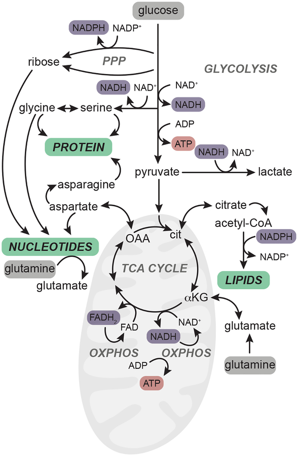
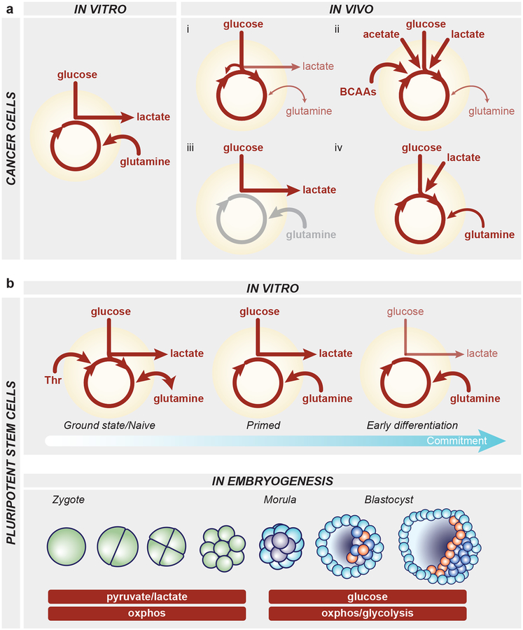
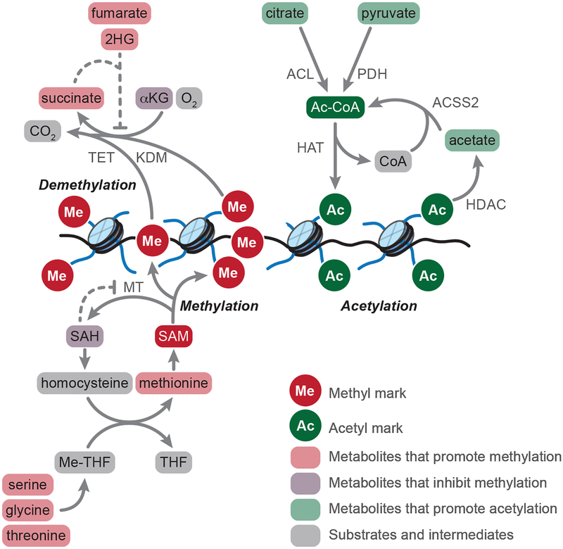
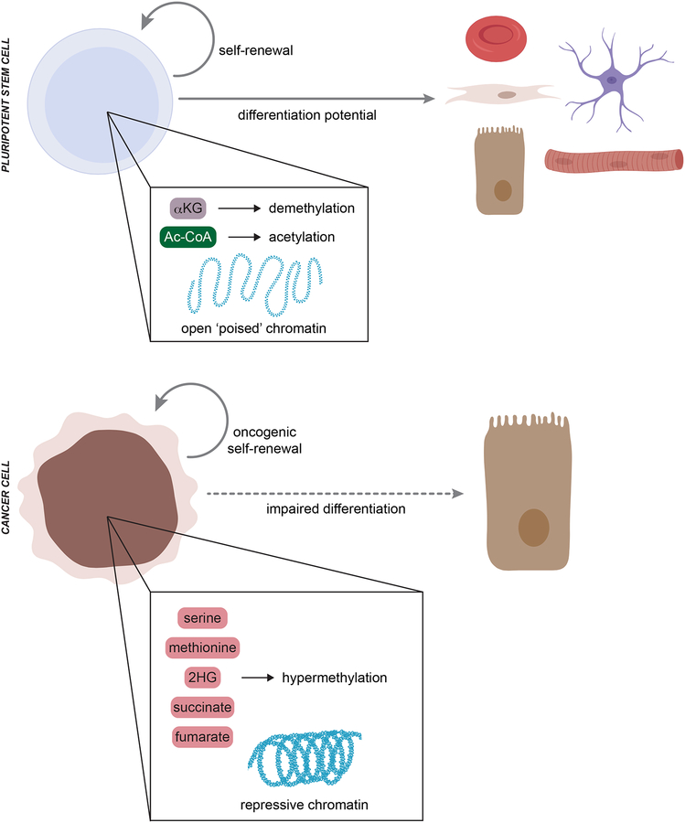
In contrast to terminally differentiated cells, cancer cells and stem cells retain the ability to re-enter the cell cycle and proliferate. In order to proliferate, cells must increase the uptake and catabolism of nutrients to support anabolic cell growth. Intermediates of central metabolic pathways have emerged as key players that can influence cell differentiation 'decisions', processes relevant for both oncogenesis and normal development. Consequently, how cells rewire metabolic pathways to support proliferation may have profound consequences for cellular identity. Here, we discuss the metabolic programs that support proliferation and explore how metabolic states are intimately entwined with the cell fate decisions that characterize stem cells and cancer cells. By comparing the metabolism of pluripotent stem cells and cancer cells, we hope to illuminate common metabolic strategies as well as distinct metabolic features that may represent specialized adaptations to unique cellular demands.


Conflict of interest statement

Competing Interests. A.I. has previously consulted for Foundation Medicine, Inc.

Figures

Figure 1.
Figure 1.
Glucose and glutamine are critical inputs for major anabolic pathways. In proliferating cells, glucose and glutamine (highlighted in grey) are taken up from the extracellular environment and catabolized through major metabolic pathways including glycolysis, the pentose phosphate pathway (PPP) and the tricarboxylic acid (TCA) cycle to provide the reducing equivalents (purple) and high-energy carriers (ATP, red) required to synthesize major macromolecules (green). A subset of the non-essential amino acids that are synthesized from glucose and glutamine are shown. Reducing equivalents (NADH, FADH2) in the mitochondria fuel the electron transport chain and enable synthesis of ATP through oxidative phosphorylation (oxphos). TCA cycle intermediates such as citrate and oxaloacetate (OAA, converted to aspartate) likewise contribute to lipid and nucleotide biosynthesis, respectively.
Figure 2.
Figure 2.
Metabolic strategies utilized by cancer cells and pluripotent stem cells. a, Left, cancer cells in vitro take up high levels of glucose and glutamine. Much of the glucose is converted to lactate via aerobic glycolysis and glutamine-derived carbons provide tricarboxylic acid (TCA) cycle anaplerosis to fuel oxidative phosphorylation. Right, a sampling of the diverse metabolic strategies utilized by cancer cells in vivo. i) Many tumors including lung and brain tumors exhibit high glucose uptake and catabolism in the TCA cycle, often in addition to aerobic glycolysis. Glucose-derived carbons can fuel the TCA cycle through forward, pyruvate dehydrogenase flux or via pyruvate carboxylase-mediated anaplerotic entry as oxaloacetate. In this scenario, glutamine is a relatively minor contributor to TCA cycle metabolites and often tumors can net produce glutamine de novo from glucose-derived carbons. ii) Increasingly, substrates beyond glucose and glutamine are recognized to server as major substrates for anabolic pathways and the TCA cycle, including acetate, lactate and branched chain amino acids in lung, brain and liver tumors. iii) In human clear cell renal carcinoma, tumors exhibit high glycolysis and lactate production, consistent with the canonical Warburg effect. Glutamine metabolism has not been assessed and is therefore shaded in grey. iv) Studies in mouse pancreatic cancer reveal that glucose, lactate and glutamine all contribute to TCA cycle intermediates. b, Top, PSCs exhibit a variety of metabolic strategies depending on the culture condition or stage of differentiation. Naïve mouse PSCs in the ground state of pluripotency can synthesize glutamine de novo from glucose-derived carbons; naïve mouse PSCs also can catabolize threonine (Thr). Both naïve and primed PSCs in culture exhibit high consumption of glucose and glutamine as well as extensive production of lactate. Upon differentiation, the proliferative hallmark of aerobic glycolysis decreases along with proliferation rate. Bottom, while little is known about pluripotent cell metabolism in vivo, studies of embryos developing ex vivo have identified two major metabolic stages: from zygote to morula, embryos are dependent upon the moncarboxylates pyruvate and lactate and are highly oxidative; around the morula stage, embryos begin to use glycolysis to fuel metabolic pathways and the trophectoderm exhibits features consistent with oxidative phosphorylation while the inner cell mass may rely more on aerobic glycolysis. Green, blastomeres; light blue, trophectoderm; purple, inner cell mass; orange, primitive endoderm; dark blue, epiblast.
Figure 3.
Figure 3.
Metabolic regulation of chromatin marks. Methyltransferase (MT) enzymes transfer methyl (Me) groups from S-adenosylmethionine (SAM) to histones and DNA. Methylation reactions generate S-adenosylhomocysteine (SAH) as a product; SAH inhibits methylation reactions, making MT activity responsive to the relative ratio of SAM:SAH. Methionine serves as the direct precursor for SAM, and methionine pools are regulated both by cellular uptake (not shown) and the methionine cycle, which regenerates methionine from homocysteine. The methionine cycle is intimately connected to the folate cycles, in which serine, glycine, and (in mouse PSCs) threonine provide one-carbon units to tetrahydrofolate (THF) for transfer to homocysteine. Lysine demethylases (KDM) and ten-eleven translocation (TET) enzymes catalyze demethylation of histones and DNA, respectively, using alpha-ketoglutarate (αKG), oxygen, and ferrous iron (not shown) as substrates. Succinate, fumarate, and 2-hydroxyglutarate (2HG) function as competitive inhibitors of KDM and TET enzymes, thereby promoting accumulation of methyl marks. Histone acetyltransferases (HAT) transfer acetyl (Ac) groups from acetyl-coenzyme A (Ac-CoA) to histones. Ac-CoA derives from cytosolic and/or nuclear citrate, pyruvate, and acetate, which serve as substrates for the Ac-CoA generating enzymes ATP citrate lyase (ACL), pyruvate dehydrogenase (PDH), or acyl-coenzyme A synthetase short-chain family member 2 (ACSS2). Moreover, class I, II, and IV histone deacetylase (HDAC) reactions generate acetate as a product, which can be captured by nuclear ACSS2 to regenerate Ac-CoA.
Figure 4.
Figure 4.
Metabolic control of differentiation in stem cells and cancer cells. Naïve pluripotent stem cell (PSC) self-renewal and differentiation potential depend on a characteristic open (‘poised’) chromatin structure. Metabolic inputs from alpha-ketoglutarate (αKG) and acetyl-CoA (Ac-CoA) drive active demethylation and histone acetylation to maintain naïve PSC identity. In contrast, cancer cells generally exhibit a hypermethylated chromatin landscape that represses expression of both tumor suppressors and gene expression programs required for normal lineage differentiation. The hypermethylated state in cancer cells can be driven, at least in part, by metabolic rewiring that results in accumulation of serine, methionine, 2-hydroxyglutarate (2HG), succinate, and/or fumarate.

References

    1. Vander Heiden MG, Cantley LC & Thompson CB Understanding the Warburg effect: the metabolic requirements of cell proliferation. Science 324, 1029–1033, doi:10.1126/science.1160809 (2009). - DOI - PMC - PubMed
    1. Beaudin AE & Stover PJ Insights into metabolic mechanisms underlying folate-responsive neural tube defects: a minireview. Birth Defects Res A Clin Mol Teratol 85, 274–284, doi:10.1002/bdra.20553 (2009). - DOI - PMC - PubMed
    1. Luengo A, Gui DY & Vander Heiden MG Targeting Metabolism for Cancer Therapy. Cell Chem Biol 24, 1161–1180, doi:10.1016/j.chembiol.2017.08.028 (2017). - DOI - PMC - PubMed
    1. Kong H & Chandel NS Regulation of redox balance in cancer and T cells. The Journal of biological chemistry 293, 7499–7507, doi:10.1074/jbc.TM117.000257 (2018). - DOI - PMC - PubMed
    1. Chantranupong L, Wolfson RL & Sabatini DM Nutrient-sensing mechanisms across evolution. Cell 161, 67–83, doi:10.1016/j.cell.2015.02.041 (2015). - DOI - PMC - PubMed

Publication types