

Bilateral brachial synovial cysts in systemic juvenile idiopathic arthritis: Case report and literature review
- PMID: 31245900
- PMCID: PMC6771648
- DOI: 10.1111/1756-185X.13618
Bilateral brachial synovial cysts in systemic juvenile idiopathic arthritis: Case report and literature review
Abstract
Aim: To review the clinical features of brachial synovial cyst.
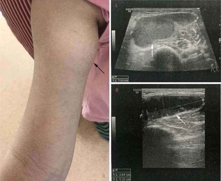
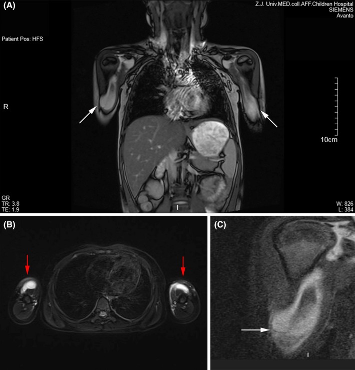
Method: A case of bilateral brachial synovial cysts is described in a child suffering from systemic juvenile idiopathic arthritis during a relapse. Magnetic resonance imaging and ultrasonography were conducted to further evaluate the nature of the cysts. The case is compared with known cases in a literature review.
Results: Review of the literature showed that brachial synovial cysts occur most commonly in systemic juvenile idiopathic arthritis. It is considered that uncontrolled systemic inflammation and recurrent disease activity might be the cause of synovial cysts.
Conclusion: Brachial synovial cyst is a rare manifestation of juvenile idiopathic arthritis. Uncontrolled systemic inflammation inducing chronic damage to joint structure may be the primary cause of synovial cyst formation.
Keywords: brachial synovial cyst; juvenile idiopathic arthritis; subclinical synovitis.
© 2019 The Authors. International Journal of Rheumatic Diseases published by Asia Pacific League of Associations for Rheumatology and John Wiley & Sons Australia, Ltd.
Conflict of interest statement
None.
Figures
Similar articles
-
Synovial cyst in juvenile idiopathic arthritis.Clin Rheumatol. 2008 Dec;27 Suppl 2:S43-5. doi: 10.1007/s10067-008-0860-x. Epub 2008 Mar 11. Clin Rheumatol. 2008. PMID: 18330610
-
Bicipital synovial cyst as the sole musculoskeletal presentation of systemic juvenile idiopathic arthritis-case report.Mod Rheumatol Case Rep. 2021 Jul;5(2):246-249. doi: 10.1080/24725625.2020.1869509. Epub 2021 Jan 25. Mod Rheumatol Case Rep. 2021. PMID: 33430713
-
Bicipital synovial cyst in systemic-onset juvenile idiopathic arthritis.J Pediatr. 2010 Jul;157(1):168. doi: 10.1016/j.jpeds.2010.02.014. Epub 2010 Mar 24. J Pediatr. 2010. PMID: 20338579 No abstract available.
-
Presence of riziform bodies in a patient with juvenile idiopathic arthritis: case report and literature review.Rev Bras Reumatol Engl Ed. 2017 Nov-Dec;57(6):610-612. doi: 10.1016/j.rbre.2014.09.004. Epub 2014 Nov 28. Rev Bras Reumatol Engl Ed. 2017. PMID: 29173696 Review. English, Portuguese. No abstract available.
-
Ultrasound imaging of synovial inflammation in juvenile idiopathic arthritis.Pediatr Radiol. 2017 Aug;47(9):1160-1170. doi: 10.1007/s00247-017-3934-6. Epub 2017 Aug 4. Pediatr Radiol. 2017. PMID: 28779188 Review.
References
-
- Fang CJ, McCarthy CL, McNally EG. Intramuscular dissection of Baker's cysts: report on three cases. Skeletal Radiol. 2004;33:367‐371. - PubMed
-
- Mizuta M, Shimizu M, Nakagishi Y, et al. Bicipital synovial cyst associated with systemic juvenile idiopathic arthritis: new insights obtained from unique pathological findings. Int J Rheum Dis. 2017;20(12):2242‐2244. - PubMed
-
- Shimizu M, Yokoyama T, Wada T, et al. Bicipital synovial cyst in systemic‐onset juvenile idiopathic arthritis. J Pediatr. 2010;157(1):168. - PubMed
-
- Dell'Era L, Vercellesi P, Forzenigo LV, et al. Synovial cyst in juvenile idiopathic arthritis. Clin Rheumatol. 2008;27(Suppl 2):S43‐S45. - PubMed
-
- Roth J, Scheer I, Kraft S, et al. Uncommon synovial cysts in children. Eur J Pediatr. 2006;165(3):178‐181. - PubMed
Publication types
MeSH terms
Grants and funding
LinkOut - more resources
Full Text Sources
Medical
