

A Rare Complication of Oropharyngeal Tularemia: Dacryocystitis
- PMID: 31245979
- PMCID: PMC6624460
- DOI: 10.4274/tjo.galenos.2018.96337
A Rare Complication of Oropharyngeal Tularemia: Dacryocystitis
Abstract
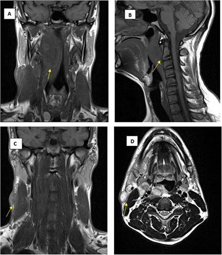
Tularemia is a zoonotic disease caused by Francisella tularensis, a highly virulent gram-negative coccobacillus. Oropharyngeal tularemia, one of the clinical subtypes, is the most common clinical form of the disease in Eastern Europe, including Turkey. This clinical form affects mostly the head and neck region and the most common complaints of patients are mass in the neck, sore throat, and fever. This form of tularemia may be confused with tonsillitis, pharyngitis, or cervical lymphadenitis caused by other microbial agents due to the nonspecific clinical and laboratory features. In this study, we present a patient with nasolacrimal duct obstruction and dacryocystitis caused by oropharyngeal tularemia.
Keywords: Tularemia; nasolacrimal duct obstruction; dacryocystitis.
Conflict of interest statement
Figures

Similar articles
-
[Evaluation of the oropharyngeal tularemia cases admitted to our hospital from the provinces of Central Anatolia].Mikrobiyol Bul. 2011 Jan;45(1):58-66. Mikrobiyol Bul. 2011. PMID: 21341160 Turkish.
-
Leprosy as a cause of nasolacrimal duct obstruction.Orbit. 2025 Jun;44(3):353-356. doi: 10.1080/01676830.2024.2393793. Epub 2024 Aug 28. Orbit. 2025. PMID: 39193776
-
[Oropharyngeal tularemia: a case report].Kulak Burun Bogaz Ihtis Derg. 2012 Nov-Dec;22(6):337-41. doi: 10.5606/kbbihtisas.2012.064. Kulak Burun Bogaz Ihtis Derg. 2012. PMID: 23176698 Turkish.
-
Tularemia presenting as tonsillopharyngitis and cervical lymphadenitis: a case report and review of the literature.Eur Arch Otorhinolaryngol. 2003 Jul;260(6):298-300. doi: 10.1007/s00405-002-0565-8. Epub 2003 Jan 10. Eur Arch Otorhinolaryngol. 2003. PMID: 12883950 Review.
-
Primary tubercular dacryocystitis - a case report and review of 18 cases from the literature.Orbit. 2019 Aug;38(4):331-334. doi: 10.1080/01676830.2018.1513044. Epub 2018 Aug 24. Orbit. 2019. PMID: 30142013 Review.
Cited by
-
Tularemia treatment: experimental and clinical data.Front Microbiol. 2024 Jan 17;14:1348323. doi: 10.3389/fmicb.2023.1348323. eCollection 2023. Front Microbiol. 2024. PMID: 38298538 Free PMC article. Review.
References
-
- Mörner T. The ecology of tularemia. Rev Sci Tech. 1992;11:1123–1130. - PubMed
-
- Boyce JM. Recent trends in the epidemiology of tularemia in the United States. J Infect Dis. 1975;131:197–199. - PubMed
-
- Berdal BP, Mehl R, Haaheim H, Loksa M, Grunow R, Burans J, Morgan C, Meyer H. Field detection of Francisella tularensis. Scand J Infect Dis. 2000;32:287–291. - PubMed
Publication types
MeSH terms
LinkOut - more resources
Full Text Sources
Medical