. 2019 Jul 23;58(29):3169-3184.
doi: 10.1021/acs.biochem.9b00197. Epub 2019 Jul 12.

Analysis of RNA Methylation by Phylogenetically Diverse Cfr Radical S-Adenosylmethionine Enzymes Reveals an Iron-Binding Accessory Domain in a Clostridial Enzyme

Affiliations

Analysis of RNA Methylation by Phylogenetically Diverse Cfr Radical S-Adenosylmethionine Enzymes Reveals an Iron-Binding Accessory Domain in a Clostridial Enzyme

James D Gumkowski et al. Biochemistry. .

Abstract

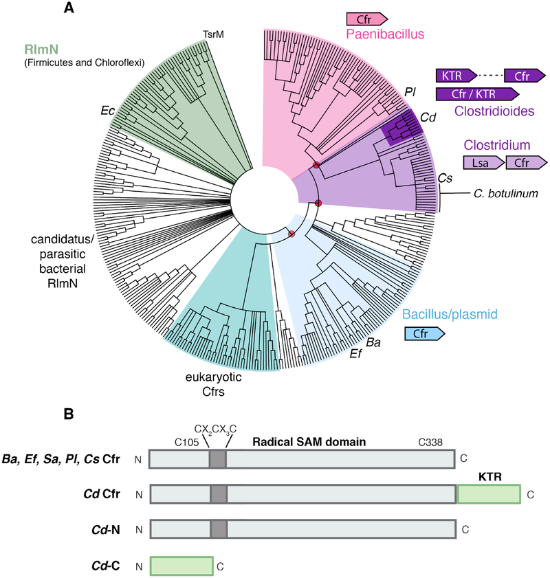
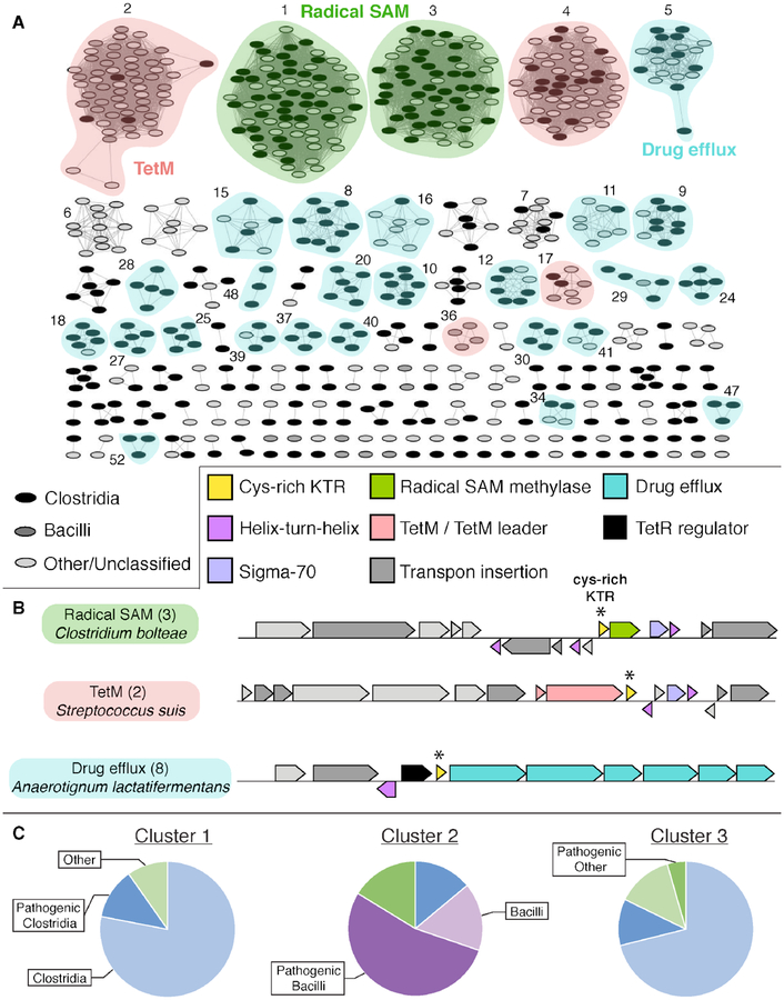
Cfr is a radical S-adenosylmethionine (SAM) RNA methylase linked to multidrug antibiotic resistance in bacterial pathogens. It catalyzes a chemically challenging C-C bond-forming reaction to methylate C8 of A2503 (Escherichia coli numbering) of 23S rRNA during ribosome assembly. The cfr gene has been identified as a mobile genetic element in diverse bacteria and in the genome of select Bacillales and Clostridiales species. Despite the importance of Cfr, few representatives have been purified and characterized in vitro. Here we show that Cfr homologues from Bacillus amyloliquefaciens, Enterococcus faecalis, Paenibacillus lautus, and Clostridioides difficile act as C8 adenine RNA methylases in biochemical assays. C. difficile Cfr contains an additional Cys-rich C-terminal domain that binds a mononuclear Fe2+ ion in a rubredoxin-type Cys4 motif. The C-terminal domain can be truncated with minimal impact on C. difficile Cfr activity, but the rate of turnover is decreased upon disruption of the Fe2+-binding site by Zn2+ substitution or ligand mutation. These findings indicate an important purpose for the observed C-terminal iron in the native fusion protein. Bioinformatic analysis of the C. difficile Cfr Cys-rich domain shows that it is widespread (∼1400 homologues) as a stand-alone gene in pathogenic or commensal Bacilli and Clostridia, with >10% encoded adjacent to a predicted radical SAM RNA methylase. Although the domain is not essential for in vitro C. difficile Cfr activity, the genomic co-occurrence and high abundance in the human microbiome suggest a possible functional role for a specialized rubredoxin in certain radical SAM RNA methylases that are relevant to human health.


Figures

Figure 1.
Figure 1.. Proposed mechanism of C8 adenine methylation by Cfr., ,
Schematic diagram of the catalytic Cys residues located in the Cfr RNA binding pocket (gray box) and the A2503 rRNA target nucleobase.
Figure 2.
Figure 2.. Selection of phylogenetically diverse Cfr enzymes for biochemical characterization.
(A) Minimum evolution phylogenetic tree containing 361 representative sequences obtained by NCBI BLAST. The sequence of Sa Cfr was the initial query. Bootstrap values (1000 replicates) were calculated for branch support and red circles denote values > 85% in selected branches. Dominant genera and the genomic context in each cluster are indicated in colored text/schematics. (B) Cartoon diagram of the domain architecture in Cfrs selected for study. Locations of conserved Cys residues (Sa numbering) in the radical SAM domain are shown.
Figure 3.
Figure 3.. Methylation of a 155mer rRNA substrate by Cfr enzymes under limiting substrate conditions initiated with a protein-based reductant.
Reactions used the Ec Flv/Flx/NADPH reducing system in mixtures containing 100 mM EPPS, pH 8, 10 mM MgCl2, 12.5 % glycerol, 250 mM KCl, 200 μM Flv, 20 μM Flx, 2 mM NADPH, 1 mM SAM, 50 μM 155mer RNA, and 48 μM protein. (A) LC-MS analysis of methylated RNA products via single-ion monitoring at m/z = 282.1 (bottom trace, m8A) and m/z = 296.1 (top trace, m2,8A). Time course experiment monitoring formation and decay of singly-methylated m8A (solid bars) and doubly methylated m2,8a (patterned bars) with (B) Ba Cfr, (C) Ef Cfr, (D) Pl Cfr, and (E) Cd Cfr. Error bars correspond to standard deviation of three averaged trials.
Figure 4.
Figure 4.. Methylation of a 155mer rRNA by Cfr enzymes with excess substrate in reactions initiated with a protein-based reductant.
Reactions used the Ec Flv/Flx/NADPH reducing system in mixtures containing 100 mM EPPS, pH 8, 10 mM MgCl2, 12.5 % glycerol, 250mM KCl, 200 μM Flv, 20 μM Flx, 2 mM NADPH, 1 mM SAM, 100 μM 155mer RNA, and 3 μM protein. Time course experiment monitoring formation and decay of singly-methylated m8A with (A) Ba Cfr, (B) Ef Cfr, (C) Pl Cfr, and (D) Cd Cfr. Error bars correspond to standard deviation of three averaged trials. The inset in panel D shows that Cd Cfr can promote multiple turnovers (red line).
Figure 5.
Figure 5.. Mössbauer spectra of Cfr enzymes.
Left panel: comparison of reconstituted samples of Cfr from Bacillus amyloliquefaciens (Ba), Enterococcus faecalis (Ef), Paenibacillus lautus (Pl), and Clostridioides difficile (Cd). Spectra of N-terminal and C-terminal truncations of Cd Cfr are also shown; features of the [4Fe4S]2+ cluster and ferrous rubredoxin-like site are denoted by red and blue dashed lines, respectively. Right panel: Mössbauer spectra of the C-terminal domain of Cd Cfr in varying applied magnetic fields. Experimental data are displayed as black vertical bars, with simulations overlaid (blue solid lines).
Figure 6.
Figure 6.. Methylation of a 155mer rRNA substrate by variant Cd Cfr enzymes under limiting substrate and excess substrate conditions and initiated by a protein-based reductant.
Reactions were performed with the Ec Flv/Flx/NADPH reducing system in mixtures containing 100 mM EPPS, pH 8, 10 mM MgCl2, 12.5 % glycerol, 250 mM KCl, 200 μM Flv, 20 μM Flx, and 2mM NADPH. (A) Limiting substrate LC-MS analysis of methylated RNA products via single-ion monitoring at m/z = 282.1 (bottom trace, m8A) and m/z = 296.1 (top trace, m2,8A). Cd-N = radical SAM domain only, Cd-C = C-terminal KTR domain with Fe2+ or Zn2+, Cd-M = C363A variant (full-length), rescue (res) experiments contain Cd-C added to Cd-N in trans. Endpoint analysis (165 min) of the formation of singly-methylated m8A under (B) limiting substrate and (C) excess substrate conditions. Error bars correspond to standard deviation of three averaged trials.
Figure 7.
Figure 7.. Electrochemical and UV-visible characterization of Cd C-terminal domain.
(A) UV-visible difference spectrum of the oxidation of the C-terminal domain of Cd Cfr. Absorbance maxima are visible at 370 nm, 485 nm, and 582 nm. (B) Square-wave voltammogram of KTR domain of Cd Cfr. The midpoint potential is +106 mV. (C) Size exclusion chromatogram illustrating the interaction of Cd-N and Cd-C through co-elution. (D) Comparison of the C-terminal domain of Cd Cfr (Cd-C), the KTR from Cb, and the rubredoxin from Cp. Metal coordinating cysteine pairs are highlighted in yellow, the structurally important residues directly following are highlighted in green, the positively charged residues are colored blue, and the negatively charged residues are colored red. The number of positively and negatively charged residues (bottom, numbers in parentheses) differentiates cys-rich KTRs from typical rubredoxins.
Figure 8.
Figure 8.. Sequence similarity and genome neighborhood network analyses of stand-alone Cys-rich KTR proteins.
(A) SSN of IPR025957 constructed with an alignment score of 31. Nodes are arranged in an organic layout and each represents a single sequence. Nodes are shaded by host organism and colored by co-occurring gene type. Clusters without numbering represent groups that do not fall into one of the three major gene type categories. Additional clusters numbering two or less are not shown. (B) Representative gene clusters from each major category as determined by GNN analysis (±10 ORFs, ≥5% co-occurrence). Host organism and cluster number are displayed (left). KTR proteins are marked by a “*” symbol. (C) Breakdown by organism type in each of the first three SSN clusters. Clostridia = blue, Bacilli = purple, and other = green. Pathogenic organisms are highlighted with dark shading and non-pathogens are shown in light shading.
Figure 9.
Figure 9.. Metagenome abundance of KTR proteins in healthy human microbiome samples.
(A) Heatmap for clusters 1–10 in the KTR SSN (see Fig. 8) showing marker abundance in 380 HMP datasets collected from six different areas of the body. Cluster 4 is omitted due to low abundance in the metagenomic sample set. (B-D) Box plots of the per-site abundance for the top three clusters. This analysis shows that the KTR proteins encoded adjacent to radical SAM enzymes are abundant in the gut microbiome.

References

    1. Finn RD, Attwood TK, Babbitt PC, Bateman A, Bork P, Bridge AJ, Chang HY, Dosztanyi Z, El-Gebali S, Fraser M, Gough J, Haft D, Holliday GL, Huang H, Huang X, Letunic I, Lopez R, Lu S, Marchler-Bauer A, Mi H, Mistry J, Natale DA, Necci M, Nuka G, Orengo CA, Park Y, Pesseat S, Piovesan D, Potter SC, Rawlings ND, Redaschi N, Richardson L, Rivoire C, Sangrador-Vegas A, Sigrist C, Sillitoe I, Smithers B, Squizzato S, Sutton G, Thanki N, Thomas PD, Tosatto SC, Wu CH, Xenarios I, Yeh LS, Young SY, and Mitchell AL (2017) InterPro in 2017-beyond protein family and domain annotations, Nucleic Acids Res 45, D190–D199. - PMC - PubMed
    1. Frey PA, Hegeman AD, and Ruzicka FJ (2008) The radical SAM superfamily, Crit Rev Biochem Mol Biol 43, 63–88. - PubMed
    1. Broderick JB, Duffus BR, Duschene KS, and Shepard EM (2014) Radical S-adenosylmethionine enzymes, Chem Rev 114, 4229–4317. - PMC - PubMed
    1. Landgraf BJ, McCarthy EL, and Booker SJ (2016) Radical S-adenosylmethionine enzymes in human health and disease, Annu Rev Biochem 85, 485–514. - PubMed
    1. Kimura S, and Suzuki T (2015) Iron-sulfur proteins responsible for RNA modifications, Biochim Biophys Acta 1853, 1272–1283. - PubMed

Publication types