. 2019 Jun 27;14(6):e0218949.
doi: 10.1371/journal.pone.0218949. eCollection 2019.

A novel direct co-culture assay analyzed by multicolor flow cytometry reveals context- and cell type-specific immunomodulatory effects of equine mesenchymal stromal cells

Affiliations

A novel direct co-culture assay analyzed by multicolor flow cytometry reveals context- and cell type-specific immunomodulatory effects of equine mesenchymal stromal cells

Aline Hillmann et al. PLoS One. .

Abstract

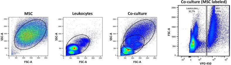
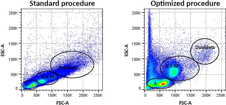
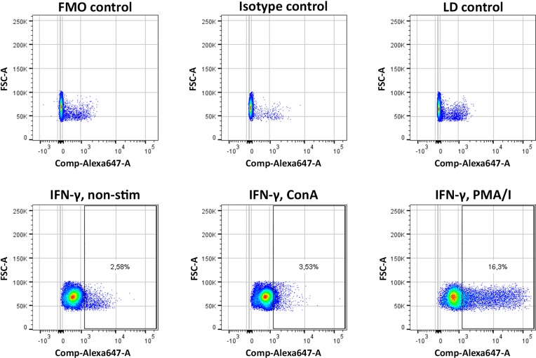
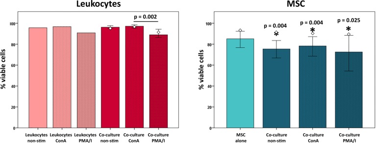
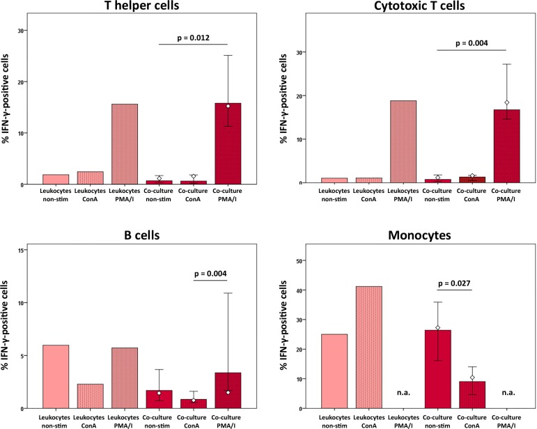
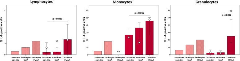
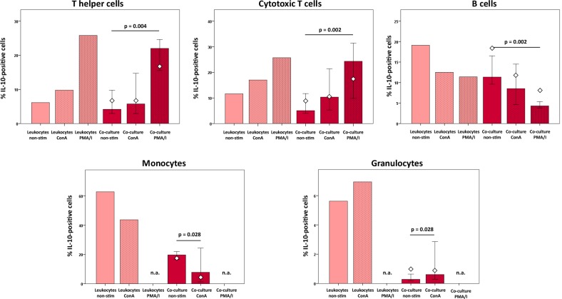
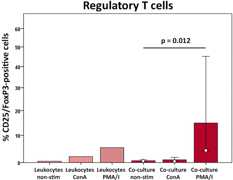
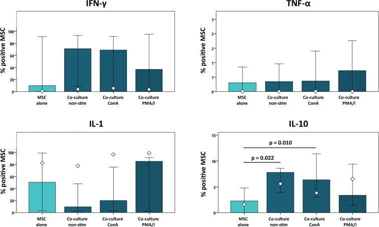
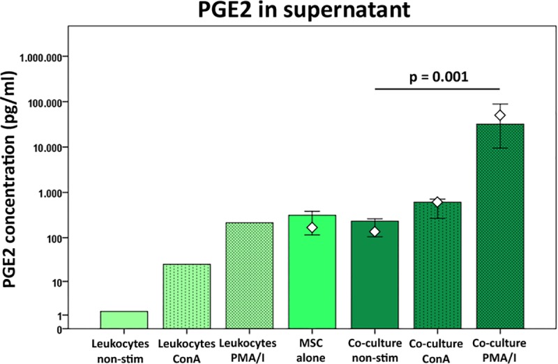
The immunomodulatory potential of multipotent mesenchymal stromal cells (MSC) provides a basis for current and future regenerative therapies. In this study, we established an approach that allows to address the effects of pro-inflammatory stimulation and co-culture with MSC on different specific leukocyte subpopulations. Equine peripheral blood leukocyte recovery was optimized to preserve all leukocyte subpopulations and leukocyte activation regimes were evaluated. Allogeneic labeled equine adipose-derived MSC were then subjected to direct co-culture with either non-stimulated, concanavalin A (ConA)-activated or phosphate 12-myristate 13-acetate and ionomycin (PMA/I)-activated leukocytes. Subsequently, production of the cytokines interferon-γ (IFN- γ), interleukin-1 (IL-1) and tumor necrosis factor-α (TNF-α) and presence of FoxP3 were determined in specific cell populations using multicolor flow cytometry. Prostaglandin E2 (PGE2) was measured in the supernatants. ConA-stimulation induced mild activation of leukocytes, whereas PMA/I-stimulation led to strong activation. In T cells, PMA/I promoted production of all cytokines, with no distinct suppressive effects of MSC. However, increased numbers of CD25/FoxP3-positive cells indicated that MSC supported regulatory T cell differentiation in PMA/I-activated leukocyte cultures. MSC also reduced numbers of cytokine-producing B cells and granulocytes, mostly irrespective of preceding leukocyte activation, and reversed the stimulatory effect of ConA on IFN-γ production in monocytes. Illustrating the possible suppressive mechanisms, higher numbers of MSC produced IL-10 when co-cultured with non-stimulated or ConA-activated leukocytes. This was not observed in co-culture with PMA/I-activated leukocytes. However, PGE2 concentration in the supernatant was highest in the co-culture with PMA/I-activated leukocytes, suggesting that PGE2 could still mediate modulatory effects in strongly inflammatory environment. These context- and cell type-specific modulatory effects observed give insight into the interactions between MSC and different types of immune cells and highlight the roles of IL-10 and PGE2 in MSC-mediated immunomodulation. The approach presented could provide a basis for further functional MSC characterization and the development of potency assays.


Conflict of interest statement

This work has no affiliation to the company Vaxxinova GmbH diagnostics. There are no competing interests. This does not alter our adherence to PLOS ONE policies on sharing data and materials.

Figures

Fig 1
Fig 1. MSC discrimination based on VDP 450 labeling.
The figure shows representative scatter plots of MSC and leukocytes cultured alone as well as in co-culture, analyzed by flow cytometry. In the left three plots, side scatter area (SSC-A) is plotted against forward scatter area (FSC-A), which did not enable a clear discrimination between MSC and leukocytes. Therefore, MSC were labeled with Violet Proliferation Dye 450 (VPD 450; BD) prior to co-culture. The right plot, with FSC-A being plotted against VPD 450, shows that a precise cut-off could then be placed between labeled MSC and the non-labeled leukocytes.
Fig 2
Fig 2. Leukocyte recovery.
The scatter plots illustrate leukocyte fractions obtained following standard density gradient centrifugation (left) and the optimized procedure using Leuko Spin Medium (pluri select) (right). Cells were analyzed by flow cytometry; side scatter area (SSC-A) is plotted against forward scatter area (FSC-A). The peripheral blood leukocyte subpopulations were better preserved and less doublets were found using the optimized procedure. L: lymphocytes; G: granulocytes; M: monocytes.
Fig 3
Fig 3. Lymphocyte activation.
Representative scatter plots of lymphocytes stained for IFN-γ using a monoclonal antibody conjugated with Alexa Fluor 647 (Alexa647) after leukocytes had been left unstimulated (non-stim) or activated with ConA or PMA/I (lower row). Cells were analyzed by flow cytometry and gated based on viability staining as well as forward and side scatter. Forward scatter area (FSC-A) is plotted against Alexa647. The upper row shows all corresponding controls (FMO, isotype and viability (LD) Fixable E-Fluor 780 single stained). Data on monocyte and granulocyte populations is not shown.
Fig 4
Fig 4. Cell viability at different culture conditions.
Diagrams display the percentage of viable cells in the whole leukocyte and MSC populations, as determined by Fixable E-Fluor 780 (Thermo Fisher Scientific) staining and flow cytometry. Bars represent the median values, error bars the 95% confidence intervals. The white rhombs indicate the results obtained with the pooled MSC sample. P values in the left diagram are based on Friedman- and Wilcoxon post-hoc tests (n = 6). Stars within the right diagram indicate differences between MSC and leukocyte viability in the corresponding groups, with p values based on Mann-Whitney U-tests (n = 6).
Fig 5
Fig 5. IFN-γ production in leukocyte subpopulations at different culture conditions.
Data were obtained by multicolor flow cytometry following intracellular cytokine staining. Bars represent the median values, error bars the 95% confidence intervals. The white rhombs indicate the results obtained with the pooled MSC sample. P values are based on Friedman- and Wilcoxon post-hoc tests (n = 6). Groups designated as not assessed (n.a.) could not be analyzed as degranulation of granulocytes following PMA/I-stimulation had hampered monocyte-specific gating.
Fig 6
Fig 6. IL-1 production in leukocyte subpopulations at different culture conditions.
Data were obtained by multicolor flow cytometry following intracellular cytokine staining. Bars represent the median values, error bars the 95% confidence intervals. The white rhombs indicate the results obtained with the pooled MSC sample. P values are based on Friedman- and Wilcoxon post-hoc tests (n = 6). Groups designated as not assessed (n.a.) could not be analyzed due to difficulties with gating of the monocytes due to possible CD14 surface antigen loss.
Fig 7
Fig 7. IL-10 production in leukocyte subpopulations at different culture conditions.
Data were obtained by multicolor flow cytometry following intracellular cytokine staining. Bars represent the median values, error bars the 95% confidence intervals. The white rhombs indicate the results obtained with the pooled MSC sample. P values are based on Friedman- and Wilcoxon post-hoc tests (n = 6). Groups designated as not assessed (n.a.) could not be analyzed as degranulation of granulocytes following PMA/I-stimulation had hampered monocyte- and granulocyte-specific gating.
Fig 8
Fig 8. CD25/FoxP3-positive regulatory T cells at different culture conditions.
Data were obtained by multicolor flow cytometry following surface antigen and intranuclear FoxP3 staining. CD25/FoxP3-positive cells are given as percentage of the CD4-positive T cell population. Bars represent the median values, error bars the 95% confidence intervals. The white rhombs indicate the results obtained with the pooled MSC sample. P values are based on Friedman- and Wilcoxon post-hoc tests (n = 6).
Fig 9
Fig 9. Cytokine production in MSC at different culture conditions.
Data were obtained by multicolor flow cytometry following intracellular cytokine staining. Bars represent the median values, error bars the 95% confidence intervals. The white rhombs indicate the results obtained with the pooled MSC sample. P values are based on Friedman- and Wilcoxon post-hoc tests (n = 6).
Fig 10
Fig 10. PGE2 concentrations in cell culture supernatants at different culture conditions.
PGE2 was measured in the supernatants by ELISA. Bars represent the median values, error bars the 95% confidence intervals. The white rhombs indicate the results obtained with the pooled MSC sample. P values are based on Friedman- and Wilcoxon post-hoc tests (n = 6).

References

    1. Hillmann A, Ahrberg AB, Brehm W, Heller S, Josten C, Paebst F, Burk J. Comparative Characterization of Human and Equine Mesenchymal Stromal Cells: A Basis for Translational Studies in the Equine Model. Cell Transplant. 2015;25:109–24. 10.3727/096368915X687822 - DOI - PubMed
    1. Jones E, Schäfer R. Where is the common ground between bone marrow mesenchymal stem/stromal cells from different donors and species? Stem Cell Res Ther 2015; 6:143 10.1186/s13287-015-0144-8 - DOI - PMC - PubMed
    1. Dominici M, Le Blanc K, Mueller I, Slaper-Cortenbach I, Marini F, Krause D, et al. Minimal criteria for defining multipotent mesenchymal stromal cells. The International Society for Cellular Therapy position statement. Cytotherapy. 2006;8:315–7. 10.1080/14653240600855905 - DOI - PubMed
    1. Samsonraj RM, Raghunath M, Nurcombe V, Hui JH, van Wijnen AJ, Cool SM. Concise Review: Multifaceted Characterization of Human Mesenchymal Stem Cells for Use in Regenerative Medicine. Stem Cells Transl Med. 2017;6:2173–85. 10.1002/sctm.17-0129 - DOI - PMC - PubMed
    1. Galipeau J, Krampera M, Barrett J, Dazzi F, Deans RJ, DeBruijn J, et al. International Society for Cellular Therapy perspective on immune functional assays for mesenchymal stromal cells as potency release criterion for advanced phase clinical trials. Cytotherapy. 2016;18:151–9. 10.1016/j.jcyt.2015.11.008 - DOI - PMC - PubMed

Publication types