. 2019 Jun 3;60(7):2753-2763.
doi: 10.1167/iovs.18-25750.

Visual Cortical Plasticity in Retinitis Pigmentosa

Affiliations

Visual Cortical Plasticity in Retinitis Pigmentosa

Claudia Lunghi et al. Invest Ophthalmol Vis Sci. .

Abstract

Purpose: Retinitis pigmentosa is a family of genetic diseases inducing progressive photoreceptor degeneration. There is no cure for retinitis pigmentosa, but prospective therapeutic strategies are aimed at restoring or substituting retinal input. Yet, it is unclear whether the visual cortex of retinitis pigmentosa patients retains plasticity to react to the restored visual input.

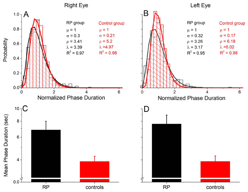
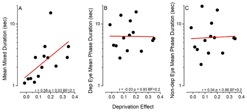
Methods: To investigate short-term visual cortical plasticity in retinitis pigmentosa, we tested the effect of short-term (2 hours) monocular deprivation on sensory ocular dominance (measured with binocular rivalry) in a group of 14 patients diagnosed with retinitis pigmentosa with a central visual field sparing greater than 20° in diameter.

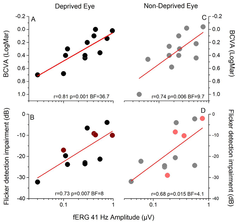
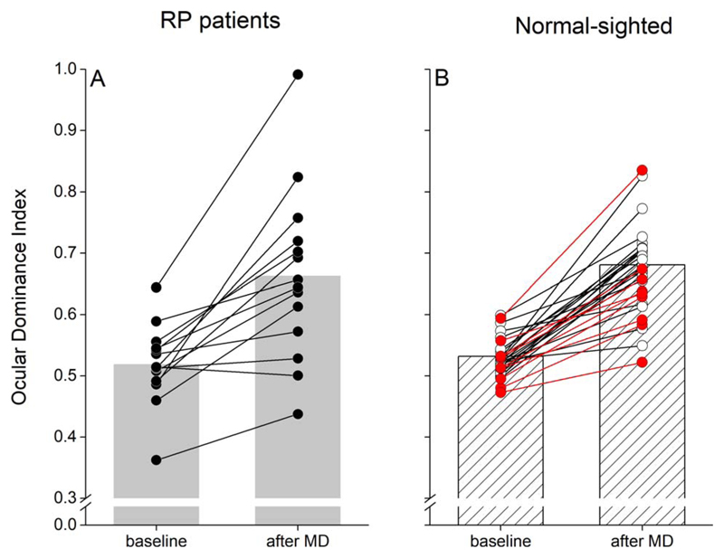
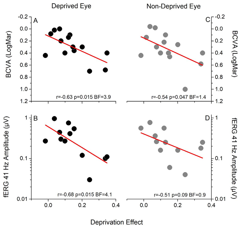
Results: After deprivation most patients showed a perceptual shift in ocular dominance in favor of the deprived eye (P < 0.001), as did control subjects, indicating a level of visual cortical plasticity in the normal range. The deprivation effect correlated negatively with visual acuity (r = -0.63, P = 0.015), and with the amplitude of the central 18° focal electroretinogram (r = -0.68, P = 0.015) of the deprived eye, revealing that in retinitis pigmentosa stronger visual impairment is associated with higher plasticity.

Conclusions: Our results provide a new tool to assess the ability of retinitis pigmentosa patients to adapt to altered visual inputs, and suggest that in retinitis pigmentosa the adult brain has sufficient short-term plasticity to benefit from prospective therapies.


Figures

Figure 1
Figure 1
Binocular rivalry phase durations in RP patients. Phase duration distributions of the right (A) and left (B) eye are well approximated by a gamma distribution (Equation 1), both for RP patients (black) and for an age-matched sample of normally sighted participants (red), indicating normal binocular rivalry dynamics in RP patients. (C, D) RP patients show longer mean phase durations in binocular rivalry than control subjects both for the right (C) and left (D).
Figure 2
Figure 2
Correlation between the amplitude of the fERG response and measures of visual functions in RP patients. The amplitude of the fERG correlates across subjects with visual acuity (A–C) and with the impairment in flicker detection (B–D) both for the deprived (A, B) and nondeprived (C, D) eye. Dark and light red symbols in (B) and (D) represent the four patients tested with a different setup probing motion direction discrimination.
Figure 3
Figure 3
Effect of monocular deprivation on binocular rivalry. (A) Following 2 hours of monocular deprivation, ocular dominance measured by means of binocular rivalry (ocular dominance index; Equation 2) shifts in favor of the deprived eye (***P < 0.001), indicating normal short-term visual plasticity in RP patients. (B) Effect of monocular deprivation in normally sighted subjects (white symbols: data adapted from Lunghi and Sale; red symbols: data from nine age-matched control subjects).
Figure 4
Figure 4
Correlations between the effect of monocular deprivation, visual acuity, and the amplitude of the fERG. Across patients, the effect of monocular deprivation (difference between the ocular dominance index measured before and after deprivation) strongly correlates both with visual acuity (BCVA) and with the amplitude of the fERG of the deprived eye (A, B). A trend for correlation between BCVA and the amplitude of the fERG of the nondeprived eye is also observed (C, D).
Figure 5
Figure 5
Correlation between the effect of monocular deprivation and binocular rivalry parameters. The effect of monocular deprivation correlates across subjects with the average duration of mixed percepts (A), but not with the mean phase duration of either the deprived (B) or nondeprived (C) eye measured before deprivation.

References

    1. Hamel C. Retinitis pigmentosa. Orphanet J Rare Dis. 2006;1:40. - PMC - PubMed
    1. Verbakel SK, van Huet RAC, Boon CJF, et al. Non-syndromic retinitis pigmentosa. Prog Retin Eye Res. 2018;66:157–186. - PubMed
    1. Fahim AT, Daiger SP, Weleber RG. Nonsyndromic Retinitis Pigmentosa Overview. Seattle, WA: University of Washington, Seattle; 1993.
    1. Berson EL. Long-term visual prognoses in patients with retinitis pigmentosa: the Ludwig von Sallmann lecture. Exp Eye Res. 2007;85:7–14. - PMC - PubMed
    1. Rachitskaya AV, Yuan A. Argus II retinal prosthesis system: an update. Ophthalmic Genet. 2016;37:260–266. - PubMed

Publication types