Review
. 2019 Aug;34(8):1393-1406.
doi: 10.1002/jbmr.3822. Epub 2019 Jul 26.

Nerves in Bone: Evolving Concepts in Pain and Anabolism

Affiliations
Review

Nerves in Bone: Evolving Concepts in Pain and Anabolism

Jennifer M Brazill et al. J Bone Miner Res. 2019 Aug.

Abstract

The innervation of bone has been described for centuries, and our understanding of its function has rapidly evolved over the past several decades to encompass roles of subtype-specific neurons in skeletal homeostasis. Current research has been largely focused on the distribution and function of specific neuronal populations within bone, as well as their cellular and molecular relationships with target cells in the bone microenvironment. This review provides a historical perspective of the field of skeletal neurobiology that highlights the diverse yet interconnected nature of nerves and skeletal health, particularly in the context of bone anabolism and pain. We explore what is known regarding the neuronal subtypes found in the skeleton, their distribution within bone compartments, and their central projection pathways. This neuroskeletal map then serves as a foundation for a comprehensive discussion of the neural control of skeletal development, homeostasis, repair, and bone pain. Active synthesis of this research recently led to the first biotherapeutic success story in the field. Specifically, the ongoing clinical trials of anti-nerve growth factor therapeutics have been optimized to titrated doses that effectively alleviate pain while maintaining bone and joint health. Continued collaborations between neuroscientists and bone biologists are needed to build on this progress, leading to a more complete understanding of neural regulation of the skeleton and development of novel therapeutics. © 2019 The Authors. Journal of Bone and Mineral Research published by Wiley Periodicals, Inc.

Keywords: ANALYSIS/QUANTITATION OF BONE, OTHER; BONE-BRAIN-NERVOUS SYSTEM INTERACTIONS; SYSTEMS BIOLOGY - BONE INTERACTORS, OTHER; THERAPEUTICS; THERAPEUTICS, ANABOLICS.


Figures

Figure 1
Figure 1
Multifactorial neural regulation of bone and joint. (A) Anteroposterior X‐ray of the knee joint of a 62‐year‐old patient with unilateral Charcot neuroarthropathy with characteristic joint collapse, bone loss, and fragmentation. Patient presented with chronic polyneuropathy and total sensorineural loss following a failed spinal stenosis surgery. Reprinted with permission from Cıvan and colleagues.182 (B, C) X‐ray of the knee joints of Eloesser's cat showing a frontal projection of the control side (B) and lateral view of the affected side (C); the affected knee was subjected to an acute thermocautery trauma following sensory posterior root resection as depicted in D (scissors) and subsequently developed deformity and grating of joint surfaces within 3 weeks. Reprinted with permission from Eloesser.3 (D) Conceptual representation of neurotrophic, neurotraumatic, and neurovascular theories of neural regulation of bone and joint, where sympathetic nerves regulate vascular tone (red), and sensory nerves provide trophic signals and mediate protective pain perception, as well as vasoregulation (blue). Collectively, the peripheral nerve carries different types of neurons that contribute to bone and joint homeostasis; destruction of these, eg, upon dorsal column degeneration (gray hatched region of spinal cord) with tabes dorsalis or dorsal root resection (scissors) and degeneration of the central primary sensory axon, contributes to joint collapse and bone loss.
Figure 2
Figure 2
Peripheral neurons’ route to the skeleton. A cross‐section of the lumbar spinal cord, where peripheral neurons that innervate the lower limbs either originate or terminate. Primary sensory neurons (blue) are pseudo‐unipolar: cell bodies reside in dorsal root ganglia from which a single axonal process bifurcates into a centrally projecting axon that targets the dorsal horn of the spinal cord and a long peripheral axon that projects out to the target tissue. The sympathetic system communicates to peripheral targets via a two‐neuron relay. Preganglionic sympathetic axons (orange) originate in the intermediolateral cell column of the spinal cord and project to sympathetic ganglia, mostly the sympathetic chain ganglia for innervation of the lower limb, to synapse with postganglionic sympathetic neurons. Postganglionic sympathetic neurons (red) project long axons towards their targets. The sensory and sympathetic axons are carried through major mixed nerves, as depicted for the right lower limb in relation to the skeleton, posterior view. Proximal to the bone, fine nerve branches containing the small sensory and autonomic axons described leave to supply the periosteum, bone, and bone marrow.
Figure 3
Figure 3
Classification of skeletal axons. The axons in peripheral nerves supplying the bone can be classified on the basis of their size and myelination status, both of which contribute to conduction velocity.14 The majority of skeletal fibers are unmyelinated, small diameter axons (yellow, 0.2 to 1.5 µm) that conduct slowly (0.5 to 2 m/s). These consist of sympathetic fibers, which can have either an adrenergic or cholinergic phenotype, and sensory C fibers. The second most prevalent axon type found in the bone are Aδ fibers: lightly myelinated, medium diameter axons (green, 1‐µm to 5‐µm diameter) that conduct at medium speeds (5 to 30 m/s). Last, Aβ fibers are large‐diameter, thickly myelinated axons (6 to 12 µm) that conduct much faster (35 to 75 m/s) but are relatively scarce or absent in bone. Each fiber subclass has a unique set of markers that are often used to define it immunohistochemically. Small‐sized and medium‐sized neurons of both sensory and/or autonomic origin also have the capacity to release specific neurotransmitters to the local environment. TrkA = tyrosine kinase receptor type 1; TH = tyrosine hydroxylase; VAChT = vesicular acetylcholine transporter; VIP = vasoactive intestinal peptide; TRPV1 = transient receptor potential cation channel subfamily V member 1; CGRP = calcitonin gene‐related peptide; SP = substance P; IB4 = isolectin‐B4; NF200 = neurofilament 200; PGP9.5 = protein gene product 9.5; GAP43 = growth associated protein 43; NE = norepinephrine; NPY = neuropeptide Y; ACh = acetylcholine.
Figure 4
Figure 4
Distribution and patterning of nerves in bone. (A) Schematic of the peptidergic sensory axons and adrenergic sympathetic axons, highlighting their relationship to the vasculature, as well their relative distributions and typical patterning in the periosteum, as they enter the cortical bone, and within the marrow space. (BD) Representative confocal micrographs of the distal mouse femur with immunostaining for TH + sympathetic fibers (yellow), CGRP + peptidergic sensory fibers (green), CD31 + endothelial cells of blood vessels (red), showing cell nuclei (DAPI, blue) and overlaid on an image captured by DIC microscopy to provide orientation in the periosteum (B) and marrow (C, D). Z‐stack thickness and scale bars as indicated. BD reprinted and adapted with permission from Chartier and colleagues.15 DIC = differential interference contrast.

References

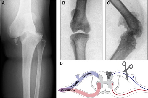
    1. Sanders LJ. The Charcot foot: historical perspective 1827–2003. Diabetes Metab Res Rev. 2004;20 Suppl 1:S4–8. - PubMed
    1. Corbin KB, Hinsey JC. Influence of the nervous system on bone and joints. Anat Rec. 1939;75(3):307–17.
    1. Eloesser L. On the nature of neuropathic affections of the joints. Ann Surg. 1917;66(2):201–7. - PMC - PubMed
    1. Edmonds ME, Clarke MB, Newton S, Barrett J, Watkins PJ. Increased uptake of bone radiopharmaceutical in diabetic neuropathy. Q J Med. 1985;57(224):843–55. - PubMed
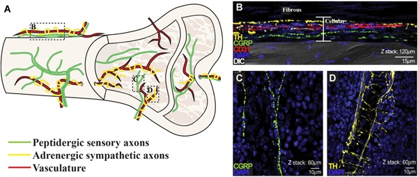
    1. Bjurholm A, Kreicbergs A, Brodin E, Schultzberg M. Substance P‐ and CGRP‐immunoreactive nerves in bone. Peptides. 1988;9(1):165–71. - PubMed

Publication types