Review
. 2019 Aug:57:204-214.
doi: 10.1016/j.sbi.2019.05.015. Epub 2019 Jun 24.

Structures of vertebrate Patched and Smoothened reveal intimate links between cholesterol and Hedgehog signalling

Affiliations
Review

Structures of vertebrate Patched and Smoothened reveal intimate links between cholesterol and Hedgehog signalling

Christiane Kowatsch et al. Curr Opin Struct Biol. 2019 Aug.

Abstract

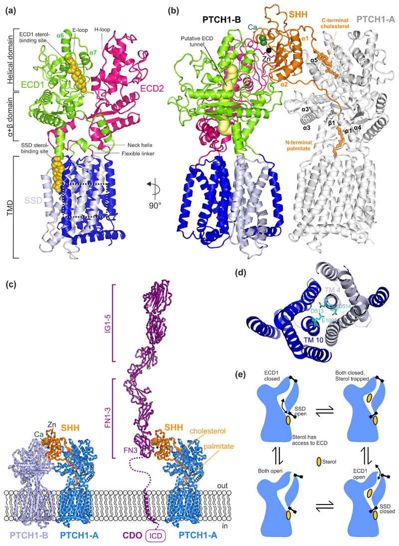
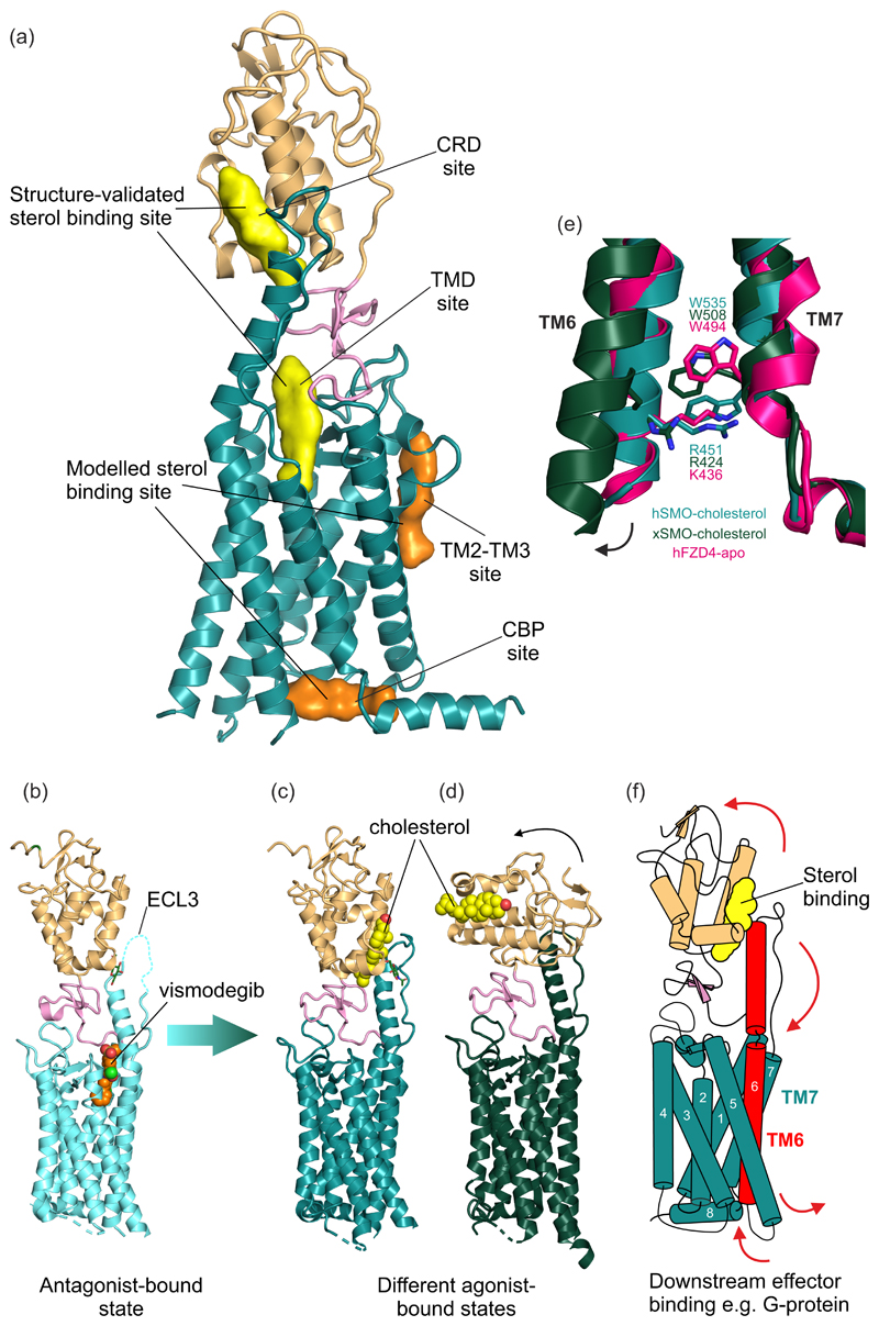
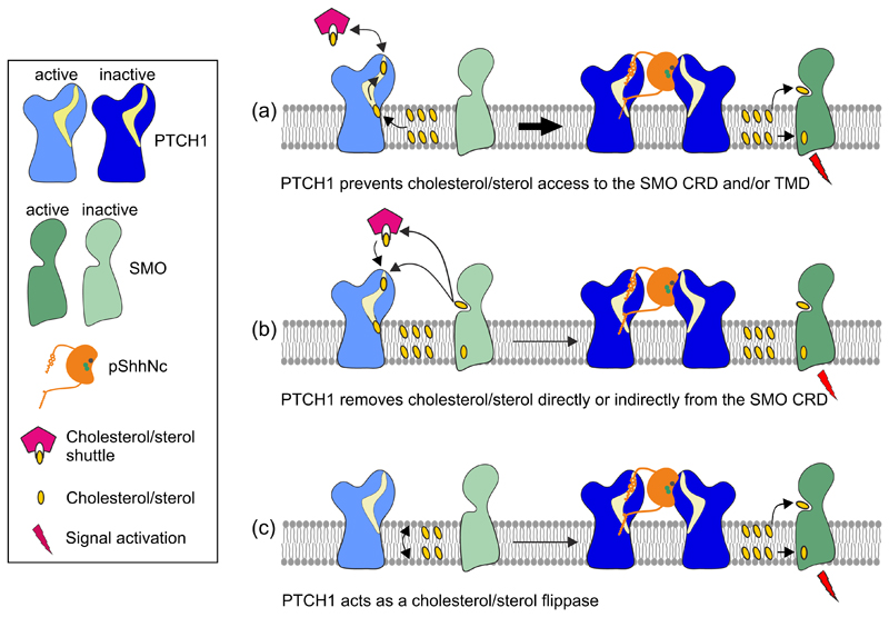
The Hedgehog (HH) signalling pathway is a cell-cell communication system that controls the patterning of multiple tissues during embryogenesis in metazoans. In adults, HH signals regulate tissue stem cells and regenerative responses. Abnormal signalling can cause birth defects and cancer. The HH signal is received on target cells by Patched (PTCH1), the receptor for HH ligands, and then transmitted across the plasma membrane by Smoothened (SMO). Recent structural and biochemical studies have pointed to a sterol lipid, likely cholesterol itself, as the elusive second messenger that communicates the HH signal between PTCH1 and SMO, thus linking ligand reception to transmembrane signalling.


Figures

Figure 1
Figure 1. Transmission of the Hedgehog (HH) signal across the plasma membrane.
(a) The receptor for HH ligands, PTCH1 (a 12-pass TM protein) inhibits SMO (a 7-pass TM protein) and thereby prevents it from initiating signalling in the cytoplasm of target cells. The domain architecture of both TM proteins is highlighted. PTCH1 is composed of two tandem RND (resistance-nodulation-division) domains: ECD1, extracellular domain 1; ECD2, extracellular domain 2; NTD, N-terminal domain; CTD, C-terminal domain; ICL3, intracellular loop 3; SSD, sterol-sensing domain (light blue). SMO is composed of an extracellular region which is made up of a cysteine rich domain (CRD) and linker domain (LD) followed by a 7-pass transmembrane bundle characteristic of GPCRs and then a unique intracellular domain (ICD). (b) HH ligands (such as Sonic Hedgehog (SHH)) bind and inhibit PTCH1, allowing SMO activation, likely by sterol ligands. Activated SMO overcomes the negative influence of PKA on the GLI transcription factors, which control gene expression.
Figure 2
Figure 2. Structures of PTCH1 and their functional implications.
(a) The overall structural arrangement of monomeric PTCH1 (PDB 6DMB [19]). Each ECD is connected to the TMD through a flexible linker at the N-terminus and a neck helix at the C-terminus. Each ECD contains an α+β domain, located immediately above the TM, followed by a helical domain consisting of α-helices and lengthy loops. The E loop on ECD1 and the H loop on ECD2 contain residues important for SHH binding. Two sterol-like molecules have been identified in the structures, one located in ECD1 and another one in the SSD at the level of the outer leaflet of the membrane. (b) Cryo-EM structure of the 2:1 PTCH1:SHH complex (PDB 6E1H [21]) containing two PTCH1 molecules labelled PTCH1-A and PTCH1-B. A putative tunnel (yellow) through PTCH1 is proposed to be the conduit for sterol flow from the ECD1 to the SSD. The N-terminal palmitoyl appendage of SHH occludes this tunnel and the C-terminal cholesterol appendage occludes the sterol binding site in ECD1, presumably blocking transport. These lipidic appendages, together with α1 and α2 of SHH, form the major SHH-PTCH1-A binding interface. SHH interacts with PTCH1-B through an interface organized by the Ca2+-binding site in SHH. (c) Structural model of the CDO:SHH:PTCH1 co-receptor complex. CDO occupies a similar position as PTCH-B. The model of CDO:SHH was generated using the HHPRED [63] webserver as well as the crystal strcture structure of the CDO-FN3:SHH complex (PDB ID. 3D1M [25]). Ig: immunoglobulin-like domain, FN: fibronectin type III-like domain, ICD: intracellular domain. (d) Close-up of a charged triad, formed by PTCH1 residues D513, D514 (TM4) and E1095 (TM10) that may facilitate cation flow down a concentration gradient to power sterol transport. Mutations in this region have been shown to abolish the function of PTCH1. (e) Ordered conformational cycling of PTCH1, driven by cation flow, may enforce the directionality of sterol movement through PTCH1. An “alternating gate” mechanism is proposed where conformational changes (stylized as gates) either lead to the opening of the gate in the membrane, near the SSD site, or the mouth of the ECD1. Sterol flow could be in either direction, from the SSD site to the ECD1 (as shown) or vice versa.
Figure 3
Figure 3. Structure of the HH signal transducer Smoothened.
(a) Multiple sterol binding sites have been proposed in SMO. In biochemical assays and crystal structures, cholesterol and oxysterols have been shown to bind to the CRD and the plant sterol-like molecule cyclopamine has been shown to bind in the TMD (shown in yellow). Two additional TMD sites (shown in orange) have been implicated using computational methods. (b) The structure of inhibited human SMO (hSMO) in complex with the antagonist vismodegib (PDB 5L7I [15]). Extracellular loop 3 (ECL3) is partially disordered (dashed line). Two potential active-state structures of SMO in complex with the agonist cholesterol are shown in (c) and (d). (c) The hSMO-cholesterol complex structure (PDB 5L7D [15]) shows SMO in an agonist-bound state, with the ordered ECL3 forming contacts with the CRD. (d) Xenopus SMO (xSMO)-cholesterol structure (PDB 6D35 [50]) reveals a more dramatic rotation of the CRD compared to the hSMO structure in (c), likely facilitated by removal of native glycans to facilitate crystallization. In (d) the TM6 moves outwards, as seen for other “canonical” GPCRs. (e) The cation-π lock is formed between a conserved tryptophan and basic residue at the cytoplasmic end of TM6 and 7 in class F GPCRs. hSMO-cholesterol (teal) shows an intact lock, while xSMO-cholesterol (dark green) shows the ruptured lock, perhaps indicative of activation. The lock is also seen in apo-Frizzled (PDB 6BD4 [61], hot pink). (f) Proposed model of transmembrane signalling by SMO. Sterol binding to the CRD causes its rotation relative to the TMD, causing a conformational change that is transmitted from the helical extension in ECL3 to TM6. This induces outward movement of TM6, rupturing the cation-π lock and exposing a new cytoplasmic surface for interaction with downstream effectors such as G-proteins.
Figure 4
Figure 4. Models for how PTCH1 regulates SMO by controlling its access to sterols.
(a) PTCH1 prevents the access of sterols to the SMO CRD and/or TMD, thereby inhibiting HH signalling. Upon SHH-binding, PTCH1 is rendered inactive, allowing sterol-bound SMO to activate downstream signalling. (b) PTCH1 removes sterols from the SMO CRD either directly or with the help of a yet unidentified sterol-binding protein, thereby inhibiting HH signalling. (c) PTCH1 acts as a sterol flippase, moving sterols from the outer to the inner membrane leaflet, or vice versa. This prevents sterol supply to the SMO CRD or TMD, respectively. These proposed mechanisms are likely confined to the membrane of the primary cilium or a ciliary microdomain.

References

    1. Lee RT, Zhao Z, Ingham PW. Hedgehog signalling. Development. 2016;143:367–372. - PubMed
    1. Pepinsky RB, Zeng C, Wen D, Rayhorn P, Baker DP, Williams KP, Bixler SA, Ambrose CM, Garber EA, Miatkowski K, et al. Identification of a palmitic acid-modified form of human Sonic hedgehog. J Biol Chem. 1998;273:14037–14045. - PubMed
    1. Porter JA, Ekker SC, Park WJ, von Kessler DP, Young KE, Chen CH, Ma Y, Woods AS, Cotter RJ, Koonin EV, et al. Hedgehog patterning activity: role of a lipophilic modification mediated by the carboxy-terminal autoprocessing domain. Cell. 1996;86:21–34. - PubMed
    1. Porter JA, Young KE, Beachy PA. Cholesterol modification of hedgehog signaling proteins in animal development. Science. 1996;274:255–259. - PubMed
    1. Marigo V, Davey RA, Zuo Y, Cunningham JM, Tabin CJ. Biochemical evidence that patched is the Hedgehog receptor. Nature. 1996;384:176–179. - PubMed

Publication types