. 2019 Jun 26;20(13):3113.
doi: 10.3390/ijms20133113.

Immortalized Human hTert/KER-CT Keratinocytes a Model System for Research on Desmosomal Adhesion and Pathogenesis of Pemphigus Vulgaris

Affiliations

Immortalized Human hTert/KER-CT Keratinocytes a Model System for Research on Desmosomal Adhesion and Pathogenesis of Pemphigus Vulgaris

Benedikt Beckert et al. Int J Mol Sci. .

Abstract

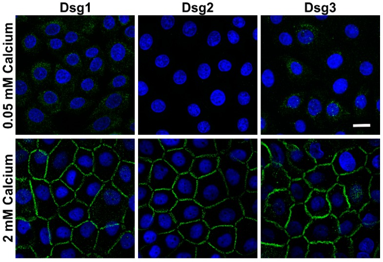
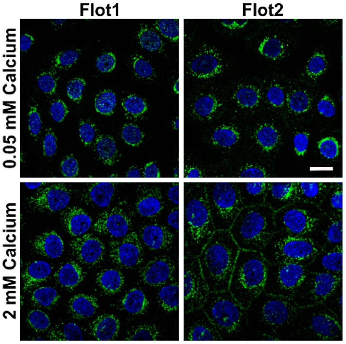
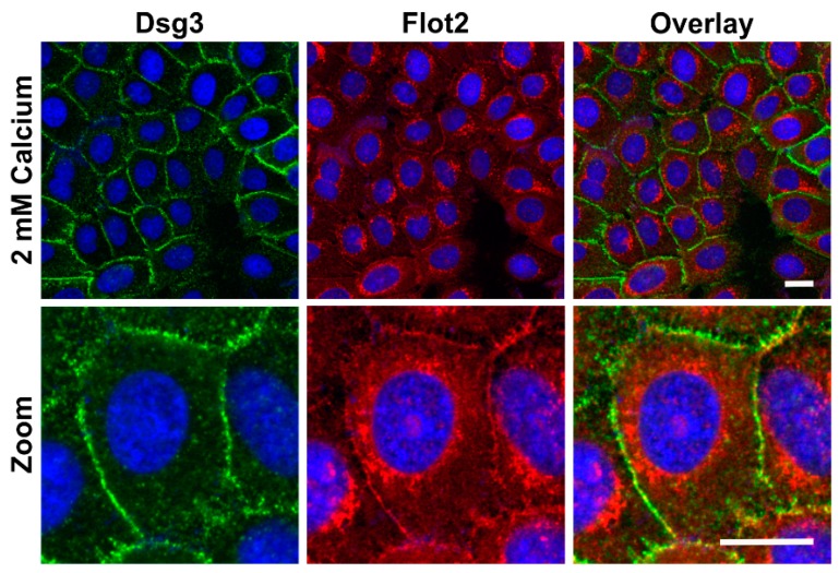
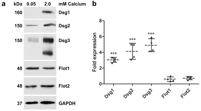
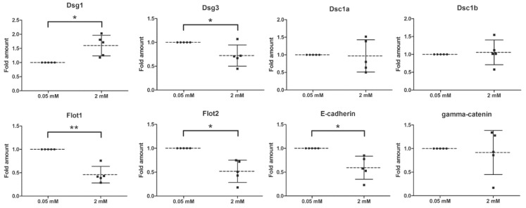
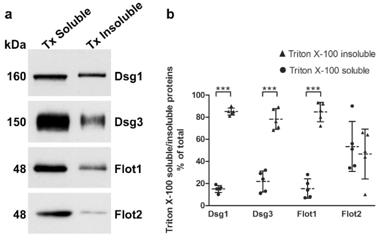
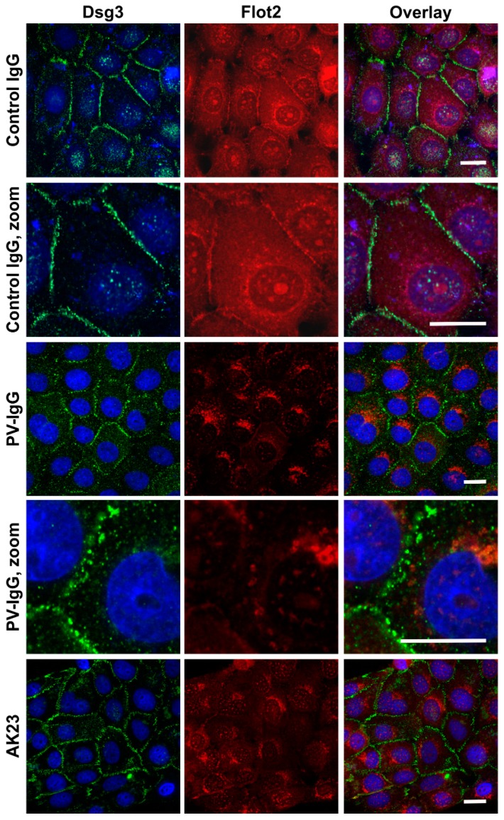
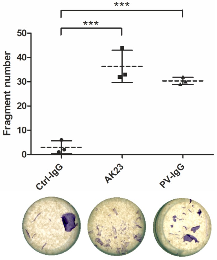
Pemphigus Vulgaris is an autoimmune disease that results in blister formation in the epidermis and in mucosal tissues due to antibodies recognizing desmosomal cadherins, mainly desmoglein-3 and -1. Studies on the molecular mechanisms of Pemphigus have mainly been carried out using the spontaneously immortalized human keratinocyte cell line HaCaT or in primary keratinocytes. However, both cell systems have suboptimal features, with HaCaT cells exhibiting a large number of chromosomal aberrations and mutated p53 tumor suppressor, whereas primary keratinocytes are short-lived, heterogeneous and not susceptible to genetic modifications due to their restricted life-span. We have here tested the suitability of the commercially available human keratinocyte cell line hTert/KER-CT as a model system for research on epidermal cell adhesion and Pemphigus pathomechanisms. We here show that hTert cells exhibit a calcium dependent expression of desmosomal cadherins and are well suitable for typical assays used for studies on Pemphigus, such as sequential detergent extraction and Dispase-based dissociation assay. Treatment with Pemphigus auto-antibodies results in loss of monolayer integrity and altered localization of desmoglein-3, as well as loss of colocalization with flotillin-2. Our findings demonstrate that hTert cells are well suitable for studies on epidermal cell adhesion and Pemphigus pathomechanisms.

Keywords: Pemphigus vulgaris; blistering disease; cell-cell adhesion; dermatology; desmoglein; desmosome; epidermis; flotillin.


Conflict of interest statement

The authors declare no conflict of interest. The funders had no role in the design of the study; in the collection, analyses, or interpretation of data; in the writing of the manuscript, or in the decision to publish the results.

Figures

Figure 1
Figure 1
Effect of calcium concentration on the localization of desmogleins in human telomerase reverse transcriptase (hTert) cells. The cells were grown on coverslips in Keratinocyte growth medium (KGM) with 0.05 mM calcium for at least four days, and then shifted to 2 mM calcium for 24 h. After methanol (MeOH) fixation, the cells were stained with anti-desmoglein (Dsg) antibodies and fluorochrome coupled secondary antibodies (anti-mouse Alexa488, green). The coverslips were mounted in a mounting medium with 4′,6-diamidino-2-phenylindole (DAPI, blue) to visualize nuclei. n = 4 independent experiments, scale bar 20 µm.
Figure 2
Figure 2
Effect of calcium concentration on the localization of flotillins in hTert cells. The cells were grown on coverslips in KGM with 0.05 mM calcium for at least four days, and then shifted to 2 mM calcium for 24 h. After MeOH fixation, the cells were stained with anti-flotillin antibodies and fluorochrome coupled secondary antibodies (anti-mouse Alexa488, green). The coverslips were mounted in a mounting medium with DAPI (blue) to visualize nuclei. n = 4 independent experiments, scale bar 20 µm.
Figure 3
Figure 3
Colocalization of desmogleins and flotillin-2 upon 2 mM calcium in hTert cells. The cells were grown on coverslips in KGM with 0.05 mM calcium for at least four days, and then shifted to 2 mM calcium for 24 h. After MeOH fixation, the cells were stained with anti-Dsg3 (green) and anti-Flot2 (red) antibodies and fluorochrome coupled secondary antibodies (anti-mouse Alexa488 and anti-rabbit Alexa546). The coverslips were mounted in a mounting medium with DAPI. n = 3 independent experiments, scale bar 20 µm.
Figure 4
Figure 4
Effect of calcium on the expression of desmogleins and flotillins in hTert cells. The cells were grown in KGM with 0.05 mM calcium, and one plate was treated with 2 mM calcium for 24 h. (a) After cell lysis, equal protein amounts of the lysates were loaded onto gel and the expression of the indicated proteins was analyzed by Western blot. Glyceraldehyde 3-phosphate dehydrogenase (GAPDH) was used as a loading control; (b) quantification of the bands was performed with Quantity-One software. The expression signal in the 0.05 mM sample was set to one, and the relative fold expression levels in the 2 mM samples are shown as a scatter plot. Statistical analysis was done using one-way analysis of variance (ANOVA) with Bonferroni’s multiple comparison test. Statistically significant differences, as compared to the respective 0.05 mM sample, are indicated by *** p < 0.001. n = 4 independent experiments. Dotted line: Mean of samples, solid lines: SD.
Figure 5
Figure 5
Effect of calcium concentration on the mRNA levels of adhesion proteins in hTert cells. The cells were grown in KGM with 0.05 mM calcium, then treated or not with 2 mM calcium for 24 h. RNA was isolated from the cells and quantitative real-time PCR was performed using the primers shown in Table 1. The ∆Ct-method was used to quantify the PCR products. The mean of the reference genes RPL13a, GAPDH, Ywhaz and B2M was used for normalization. The signal in the 0.05 mM calcium sample was set to one, and the relative fold amount in the 2 mM sample was calculated. For statistical analysis, the unpaired Student’s t-test was used. Statistically significant differences are indicated by * p < 0.05, and ** p < 0.01. n = 5 independent experiments. Dotted line: Mean of samples, solid lines: SD.
Figure 6
Figure 6
Detergent solubility of desmogleins and flotillins in hTert cells. The cells were grown in KGM with 2 mM calcium for 24 h and a sequential detergent extraction was performed, resulting in two pools with detergent soluble and insoluble proteins. (a) Western blot analysis of these fractions was used to detect Dsg1, Dsg3, Flot1 and Flot2; (b) quantification of the gel bands was performed with the Quantity-One software, and the percentage of the total cellular protein was calculated taking into account that triton X-100 (TX) soluble bands represent a higher relative percentage of the respective fraction than the TX insoluble bands. For statistical analysis, unpaired Student’s t-test was used. Statistically significant differences are indicated by *** p < 0.001. n = 5 independent experiments. Dotted line: Mean of samples, solid lines: SD.
Figure 7
Figure 7
Effect of purified Pemphigus auto-antibodies (PV-IgG) and anti-Dsg3 antibodies on the localization of Dsg3 and Flot2 in hTert cells. The cells were grown on coverslips in KGM with 0.05 mM calcium for at least four days, and then shifted to 2 mM calcium for 24 h. Thereafter, the cells were treated with either purified control Immunoglobulin G (IgG), PV-IgG or pathogenic monoclonal anti-Dsg3 (AK23) antibody for further 24 h. After MeOH fixation, the cells were stained with anti-Dsg3 (green) and anti-Flot2 (red) antibodies and fluorochrome coupled secondary antibodies (anti-mouse Alexa488 and anti-rabbit Alexa546). The coverslips were mounted in a mounting medium with DAPI. n = 3 independent experiments, scale bar 20 µm.
Figure 8
Figure 8
Effect of PV-IgG and anti-Dsg3 antibodies on the monolayer integrity in hTert cells. The cells were grown in KGM with 0.05 mM calcium until confluent and then shifted to 2 mM calcium for 24 h. Thereafter, the cells were treated with either purified control IgG, PV-IgG or AK23 antibody for further 24 h. Dispase was used to break the cell-matrix adhesion, and resulting monolayers were pipetted up and down to induce fragmentation. The resulting fragments were counted using ImageJ. Statistical analysis was done using ANOVA with Bonferroni’s multiple comparison test. Statistically significant differences are indicated by *** p < 0.001. n = 3 independent experiments. Dotted line: Mean of samples, solid lines: SD.

References

    1. Pollmann R., Schmidt T., Eming R., Hertl M. Pemphigus: A Comprehensive Review on Pathogenesis, Clinical Presentation and Novel Therapeutic Approaches. Clin. Rev. Allergy. Immunol. 2018;54:1–25. doi: 10.1007/s12016-017-8662-z. - DOI - PubMed
    1. Spindler V., Eming R., Schmidt E., Amagai M., Grando S., Jonkman M.F., Kowalczyk A.P., Muller E.J., Payne A.S., Pincelli C., et al. Mechanisms Causing Loss of Keratinocyte Cohesion in Pemphigus. J. Invest. Dermatol. 2018;138:32–37. doi: 10.1016/j.jid.2017.06.022. - DOI - PubMed
    1. Spindler V., Waschke J. Pemphigus-A Disease of Desmosome Dysfunction Caused by Multiple Mechanisms. Front. Immunol. 2018;9:136. doi: 10.3389/fimmu.2018.00136. - DOI - PMC - PubMed
    1. Boukamp P., Petrussevska R.T., Breitkreutz D., Hornung J., Markham A., Fusenig N.E. Normal keratinization in a spontaneously immortalized aneuploid human keratinocyte cell line. J. Cell. Biol. 1988;106:761–771. doi: 10.1083/jcb.106.3.761. - DOI - PMC - PubMed
    1. Boukamp P., Popp S., Altmeyer S., Hulsen A., Fasching C., Cremer T., Fusenig N.E. Sustained nontumorigenic phenotype correlates with a largely stable chromosome content during long-term culture of the human keratinocyte line HaCaT. Genes Chromosomes Cancer. 1997;19:201–214. doi: 10.1002/(SICI)1098-2264(199708)19:4<201::AID-GCC1>3.0.CO;2-0. - DOI - PubMed