. 2019 Jun 19;17(6):363.
doi: 10.3390/md17060363.

Oncolytic Vaccinia Virus Expressing Aphrocallistes vastus Lectin as a Cancer Therapeutic Agent

Affiliations

Oncolytic Vaccinia Virus Expressing Aphrocallistes vastus Lectin as a Cancer Therapeutic Agent

Tao Wu et al. Mar Drugs. .

Abstract

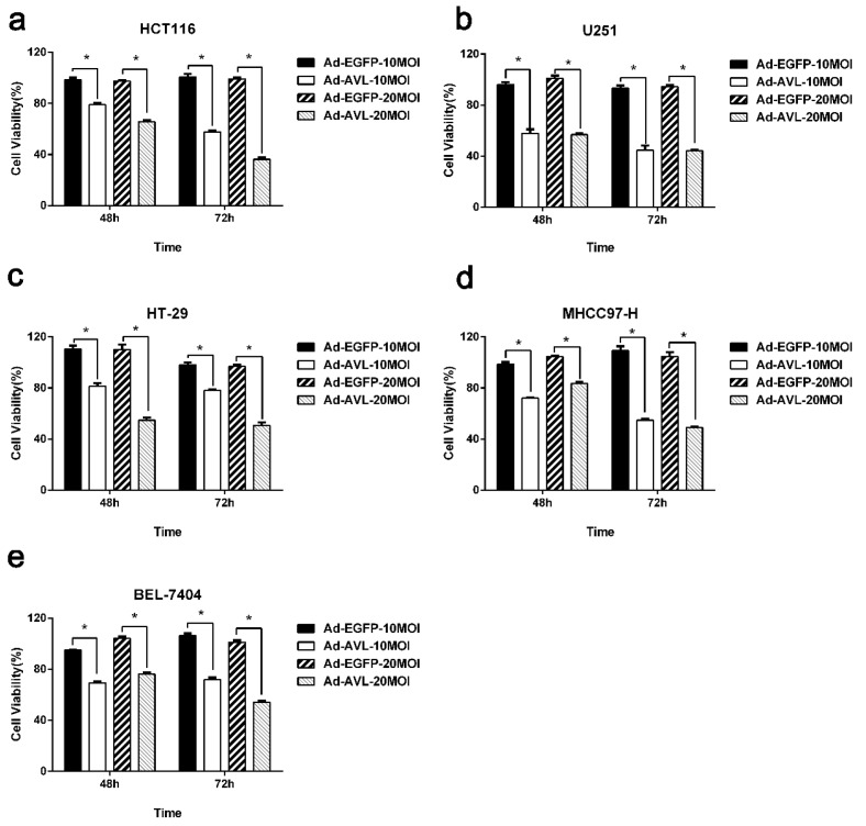
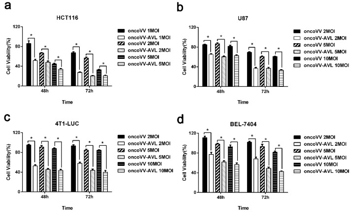
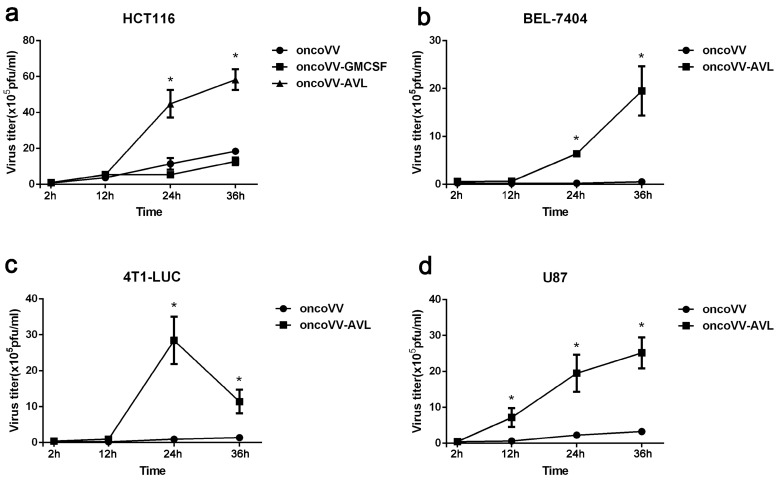
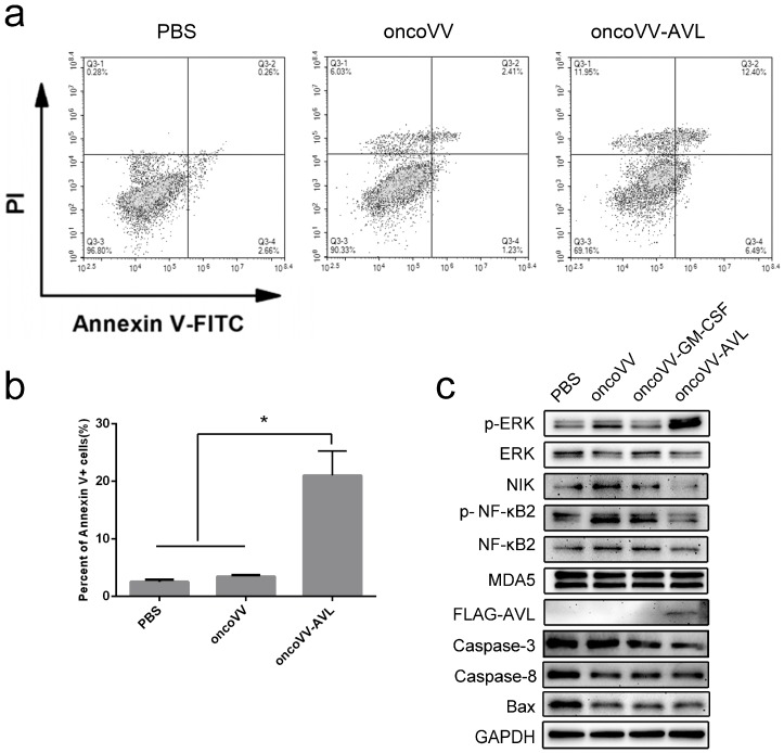
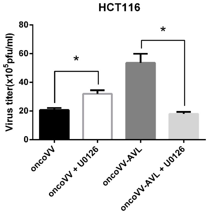
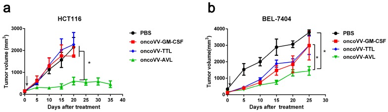
Lectins display a variety of biological functions including insecticidal, antimicrobial, as well as antitumor activities. In this report, a gene encoding Aphrocallistes vastus lectin (AVL), a C-type lectin, was inserted into an oncolytic vaccinia virus vector (oncoVV) to form a recombinant virus oncoVV-AVL, which showed significant in vitro antiproliferative activity in a variety of cancer cell lines. Further investigations revealed that oncoVV-AVL replicated faster than oncoVV significantly in cancer cells. Intracellular signaling elements including NF-κB2, NIK, as well as ERK were determined to be altered by oncoVV-AVL. Virus replication upregulated by AVL was completely dependent on ERK activity. Furthermore, in vivo studies showed that oncoVV-AVL elicited significant antitumor effect in colorectal cancer and liver cancer mouse models. Our study might provide insights into a novel way of the utilization of marine lectin AVL in oncolytic viral therapies.

Keywords: Aphrocallistes vastus lectin; ERK; oncolytic vaccinia virus.


Conflict of interest statement

The authors declare no conflict of interest.

Figures

Figure 1
Figure 1
The cytotoxicity of Ad-AVL in various tumor cells. The cytotoxicity of Ad-AVL was measured by MTT assay in HCT116 cells (a), U251 cells (b), HT-29 cells (c), MHCC-97-H cells (d), and BEL-7404 cells (e). Ad-EGFP served as a control. Data were expressed as the mean ± SEM from at least three separate experiments. (* p < 0.05).
Figure 2
Figure 2
The antiproliferative effect of oncoVV-AVL in cancer cells. The cell viability was measured by MTT assay in HCT116 cells (a), U87 cells (b), 4T1-LUC (c), and BEL-7404 cells (d). OncoVV was used as a control virus. Data were expressed as the mean ± SEM from at least three separate experiments. (* p < 0.05).
Figure 3
Figure 3
oncoVV-AVL replication in multiple tumor cell lines. The replication of oncoVV-AVL in HCT116 cells (a), BEL-7404 cells (b), 4T1-LUC cells (c), and U87 cells (d). Viral replication was determined by TCID50 assay. Data were expressed as the mean ± SEM from at least three separate experiments. (* p < 0.05).
Figure 4
Figure 4
oncoVV-AVL induced apoptosis and altered various intracellular signaling pathways in HCT116 cells. (a) HCT116 cells were treated with oncoVV or oncoVV-AVL at 2 MOI as well as PBS control for 24 h. Cells were stained with Annexin V-FITC and PI followed by analysis under a flow cytometer; (b) the percent of Annexin V-positive cells from three repeats was shown as mean ± SEM (* p < 0.05). (c) The levels of p-ERK, ERK, MDA5, NF-κB2, p-NF-κB2, NIK, Caspase-3, Caspase8, Bax and FLAG tagged AVL was detected by Western blot. GAPDH served as a loading control.
Figure 5
Figure 5
The virus titers of oncoVV and oncoVV-AVL affected by U0126 in HCT116 cells. OncoVV or oncoVV-AVL at 5MOI was used to treat HCT116 cells for 24 h in combination with 10 μM of U0126. Virus titers were measured by TCID50 assay. Data were expressed as the mean ± SEM from at least three separate experiment (* p < 0.05).
Figure 6
Figure 6
The antitumor effect of oncoVV-AVL on BEL-7404 and HCT116 tumors. (a) HCT116 cells or (b) BEL7404 cells were injected into the Balb/c nude mice on the back. Tumors were then injected with PBS, oncoVV-GM-CSF, oncoVV-TTL, or oncoVV-AVL. Arrows indicate injections. Data were expressed as the mean ± SEM. (* p < 0.05).

Similar articles

Cited by

References

    1. Neth O., Jack D.L., Dodds A.W., Holzel H., Klein N.J., Turner M.W. Mannose-binding lectin binds to a range of clinically relevant microorganisms and promotes complement deposition. Infect. Immun. 2000;68:688–693. doi: 10.1128/IAI.68.2.688-693.2000. - DOI - PMC - PubMed
    1. Weis W.I., Drickamer K. Structural basis of lectin-carbohydrate recognition. Annu. Rev. Biochem. 1996;65:441–473. doi: 10.1146/annurev.bi.65.070196.002301. - DOI - PubMed
    1. Worbs S., Köhler K., Pauly D., Avondet M.A., Schaer M., Dorner M.B., Dorner B.G. Ricinus communis intoxications in human and veterinary medicine-a summary of real cases. Toxins. 2011;3:1332–1372. doi: 10.3390/toxins3101332. - DOI - PMC - PubMed
    1. Fujita T. Evolution of the lectin—Complement pathway and its role in innate immunity. Nat. Rev. Immunol. 2002;2:346–353. doi: 10.1038/nri800. - DOI - PubMed
    1. Lillie B.N., Brooks A.S., Keirstead N.D., Hayes M.A. Comparative genetics and innate immune functions of collagenous lectins in animals. Vet. Immunol. Immunopathol. 2005;108:97–110. doi: 10.1016/j.vetimm.2005.07.001. - DOI - PubMed

MeSH terms

LinkOut - more resources