. 2019 Jun 27;20(1):17.
doi: 10.1186/s12867-019-0133-z.

MicroRNA-325-3p protects the heart after myocardial infarction by inhibiting RIPK3 and programmed necrosis in mice

Affiliations

MicroRNA-325-3p protects the heart after myocardial infarction by inhibiting RIPK3 and programmed necrosis in mice

Dong-Ying Zhang et al. BMC Mol Biol. .

Erratum in

Abstract

Background: Receptor-interacting serine-threonine kinase 3 (RIPK3)-mediated necroptosis has been implicated in the progression of myocardial infarction (MI), but the underlying mechanisms, particularly whether microRNAs (miRNAs) are involved, remain largely unknown.

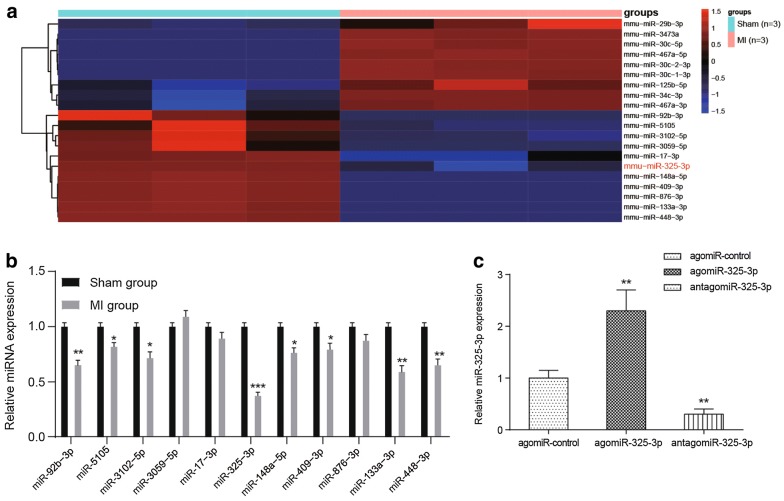
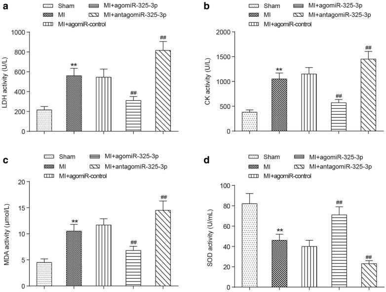
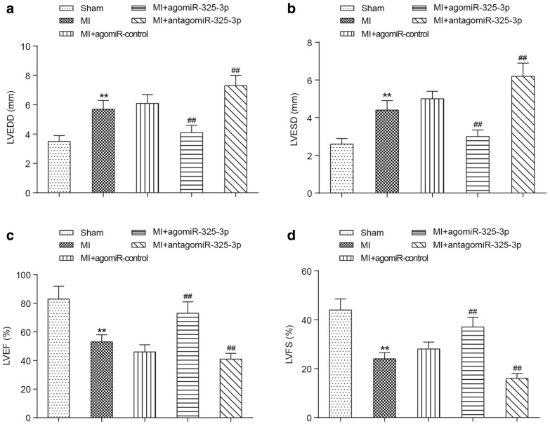
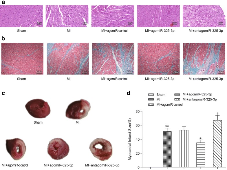
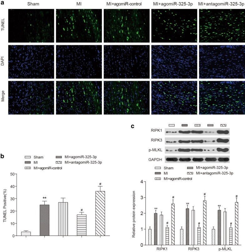
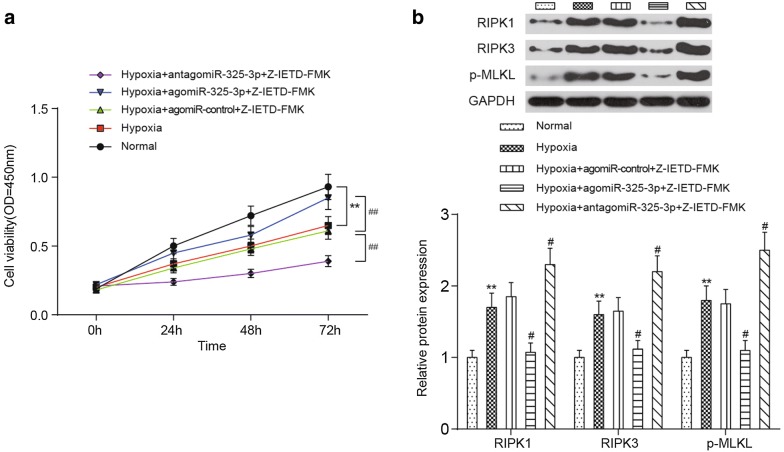
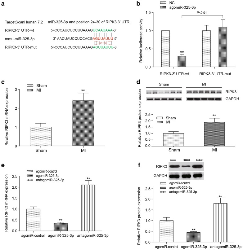
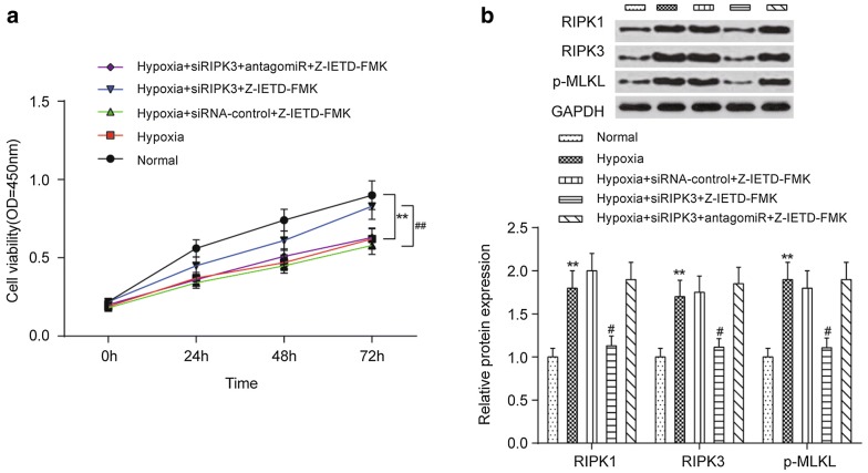
Results: A microarray analysis was used to screen for miR-325-3p expression in myocardial tissues from MI mice, and the expression was confirmed with qRT-PCR. The levels of myocardial enzymes were measured using commercial kits, and an echocardiography system was utilized for the detection of cardiac function parameters. The pathological features and infarction sizes of cardiac tissues were examined using H&E, TCC and Masson's trichrome staining, and the amount of cell apoptosis was determined using an in situ TUNEL assay. Cardiomyocytes were isolated and then subjected to hypoxia induction in vitro. The expression of the RIPK1, RIPK3 and phosphorylated MLKL (p-MLKL) proteins was measured using a Western blot. The mouse cardiomyocyte cell viability was analyzed by an MTT assay. The mRNA target of miR-325-3p was predicted using TargetScan v7.2 and then validated using a dual-luciferase reporter assay. The overexpression of miR-325-3p evidently decreased the expression levels of lactate dehydrogenase (LDH), phosphocreatine kinase (CK), superoxide dismutase (SOD) and malondialdehyde (MDA), inhibited left ventricular end-diastolic diameter (LVEDD) and left ventricular end-systolic diameter (LVESD), and promoted left ventricular ejection fraction (LVEF) and left ventricular fractional shortening (LVES). In addition, miR-325-3p overexpression attenuated the degree of injury to the cardiac tissue, decreased the infarct sizes and downregulated the expression of the necrosis-related proteins RIPK1, RIPK3 and p-MLKL.

Conclusions: The RIPK1/RIPK3/p-MLKL axis-induced necroptosis that occurred during MI was mediated by a miRNA module, miR-325-3p, which can effectively ameliorate the symptoms of MI by suppressing the expression of RIPK3.

Keywords: MiR-325-3p; Myocardial infarction; Necroptosis; RIPK3.


Conflict of interest statement

The authors declare that they have no competing interests.

Figures

Fig. 1
Fig. 1
Expression features of miR-325-3p in MI mice. a The expression profiles of miRNAs in MI mice. Twenty miRNAs with the greatest changes in MI mice are shown. The miRNAs that have high expression or low expression are colored red and blue. b The expression of the downregulated miRNAs in MI tissues were analyzed by qRT-PCR analysis. *P < 0.05, **P < 0.01, ***P < 0.001 compared to the mice that received the sham operation. c The influence of agomiR-325-3p or antagomiR-325-3p on the expression of miR-325-3p in MI mice. **P < 0.01 compared to MI mice treated with agomiR-control. MI, myocardial infarction; agomiR-325-3p, miR-325-3p agomir; antagomiR-325-3p, miR-325-3p antagomir; agomiR-control, scrambled agomir or antagomir control
Fig. 2
Fig. 2
The change of myocardial enzyme activity in MI mice with miR-325-3p dysregulation. The adult male mice were randomly divided into the sham group, MI group, MI + agomiR-control group, MI + agomiR-325-3p group and MI + antagomiR-325-3p group. a The serum concentrations of LDH in different groups. b The serum concentration of CK in different groups. c The serum concentration of MDA in different groups. d The serum concentration of SOD in different groups. **P < 0.01 compared to the mice that received the sham operation, ##P < 0.01 compared to MI mice. MI, myocardial infarction; agomiR-325-3p, miR-325-3p agomir; antagomiR-325-3p, miR-325-3p antagomir; agomiR-control, scrambled agomir or antagomir control; LDH, lactate dehydrogenase; CK, phosphocreatine kinase; SOD, superoxide dismutase; MDA, malondialdehyde
Fig. 3
Fig. 3
Influences of miR-325-3p dysregulation on the cardiac function parameters in MI mice. a, b The comparison of LVEDD and LVESD between the sham group, MI group, MI + agomiR-control group, MI + agomiR-325-3p group and MI + antagomiR-325-3p group mice. c, d The comparison of LVEF and LVFS between the sham group, MI group, MI + agomiR-control group, MI + agomiR-325-3p group and MI + antagomiR-325-3p group mice. **P < 0.01 compared to the mice that received the sham operation, ##P < 0.01 compared to MI mice. MI, myocardial infarction; agomiR-325-3p, miR-325-3p agomir; antagomiR-325-3p, miR-325-3p antagomir; agomiR-control, scrambled agomir or antagomir control; LVEDD, left ventricular end-diastolic diameter; LVESD, left ventricular end-systolic diameter; LVEF, left ventricular ejection fraction; LVES, left ventricular fractional shortening
Fig. 4
Fig. 4
Pathological injury and the infarct size of cardiac tissues in MI mice with miR-325-3p dysregulation. a Representative images of cardiac tissues stained with H&E. b Representative images of cardiac tissues stained with Masson’s trichrome. The myocardial fibers and collagen are colored blue. c Representative images of cardiac tissues that were stained with TCC. d The comparison of infarct size among the groups. **P < 0.01 compared to the mice that received the sham operation, #P < 0.05 compared to MI mice. MI, myocardial infarction; agomiR-325-3p, miR-325-3p agomir; antagomiR-325-3p, miR-325-3p antagomir; agomiR-control, scrambled agomir or antagomir control; H&E, hematoxylin and eosin; TCC, tetrazolium chloride
Fig. 5
Fig. 5
Influences of miR-325-3p dysregulation on the apoptosis of cardiomyocytes in MI mice. a Representative images of cardiac tissues that were stained with TUNEL (green), DAPI (blue) and the merge images of TUNEL and DAPI are shown. b The quantification of TUNEL-positive cells. c The Western blot results of RIPK1, RIPK3 and p-MLKL. Representative images (top) and quantitative comparison (bottom) are shown. **P < 0.01 compared to the mice that received the sham operation, #P < 0.05 compared to MI mice. MI, myocardial infarction; agomiR-325-3p, miR-325-3p agomir; antagomiR-325-3p, miR-325-3p antagomir; agomiR-control, scrambled agomir or antagomir control; TUNEL, terminal deoxynucleotidyl transferase dUTP nick-end labeling; DAPI, 4-6-diamidino-2-phenylindole; RIPK1, receptor-interacting serine/threonine protein kinase 1; RIPK3, receptor-interacting serine/threonine protein kinase 3; p-MLKL, phosphorylated mixed-lineage kinase domain-like protein
Fig. 6
Fig. 6
Influences of miR-325-3p dysregulation on the viability of cardiomyocytes in vitro. a Both the up- and downregulation of miR-325-3p affect the cell viability of hypoxia-induced cardiomyocytes. b The protein expression of RIPK1, RIPK3 and p-MLKL in cardiomyocytes is shown. Representative images (top) and quantitative comparisons (bottom) are shown. **P < 0.01 compared to cells cultured under normal conditions, ##P < 0.01 compared to cells treated with hypoxia + agomiR-control + Z-IETD-FMK. MI, myocardial infarction; agomiR-325-3p, miR-325-3p agomir; antagomiR-325-3p, miR-325-3p antagomir; agomiR-control, scrambled agomir or antagomir control; Z-IETD-FMK, a caspase inhibitor, benzyloxycarbonyl (Cbz)-Ile-Glu (OMe)-Thr-Asp (OMe)-FMK; RIPK1, receptor-interacting serine/threonine protein kinase 1; RIPK3, receptor-interacting serine/threonine protein kinase 3; p-MLKL, phosphorylated mixed-lineage kinase domain-like protein
Fig. 7
Fig. 7
Target relationship between miR-325-3p and RIPK3. a The binding sites between the 3′UTR of RIPK3 and miR-325-3p predicted by TargetScanHuman 7.2. b A dual-luciferase reporter assay validated the target relationship between miR-325-3p and the 3′UTR of RIPK3. **P < 0.01 between the wild-type and mutated 3′UTR of RIPK3. c, d The differential expression of RIPK3 mRNA (c) or protein (d) in the sham-operated mice and the MI mice. **P < 0.01 compared to the mice that received the sham operation. e, f The influence of miR-325-3p dysregulation on the expression of RIPK3 mRNA (e) and protein (f) in MI mice. **P < 0.01 compared to MI mice treated with agomiR-control. MI, myocardial infarction; agomiR-325-3p, miR-325-3p agomir; antagomiR-325-3p, miR-325-3p antagomir; agomiR-control, scrambled agomir or antagomir control; RIPK3, receptor-interacting serine/threonine protein kinase 3
Fig. 8
Fig. 8
Influences of RIPK3 dysregulation on the viability of cardiomyocytes in vitro. a RIPK3-targeting siRNA increased the cell viability of cardiomyocytes under hypoxic conditions. b The protein expression of RIPK1, RIPK3 and p-MLKL in cardiomyocytes affected by RIPK3-targeting siRNA. Representative images (top) and quantitative comparison (bottom) are shown. **P < 0.01 compared to cells cultured under normal conditions, #P < 0.05 and ##P < 0.01 compared to cells treated with hypoxia + siRNA-control + Z-IETD-FMK. MI, myocardial infarction; antagomiR-325-3p, miR-325-3p antagomir; siRIPK3, a RIPK3-targeting small interfering RNA; siRNA-control, scrambled siRNA control; Z-IETD-FMK, a caspase inhibitor, benzyloxycarbonyl (Cbz)-Ile-Glu (OMe)-Thr-Asp (OMe)-FMK; RIPK1, receptor-interacting serine/threonine protein kinase 1; RIPK3, receptor-interacting serine/threonine protein kinase 3; p-MLKL, phosphorylated mixed-lineage kinase domain-like protein

References

    1. Li C, Ren H, Chen H, Song J, Li S, Lee C, Liu J, Cui Y. Prothrombin G20210A (rs1799963) polymorphism increases myocardial infarction risk in an age-related manner: a systematic review and meta-analysis. Sci Rep. 2017;7(1):13550. doi: 10.1038/s41598-017-13623-6. - DOI - PMC - PubMed
    1. Sim DS, Jeong MH. Differences in the Korea acute myocardial infarction registry compared with western registries. Korean Circ J. 2017;47(6):811–822. doi: 10.4070/kcj.2017.0027. - DOI - PMC - PubMed
    1. Lee ES, Vedanthan R, Jeemon P, Kamano JH, Kudesia P, Rajan V, Engelgau M, Moran AE. Quality improvement in cardiovascular disease care. In: Prabhakaran D, Anand S, Gaziano TA, Mbanya JC, Wu Y, Nugent R, editors. Cardiovascular, respiratory, and related disorders. Washington (DC); 2017. - PubMed
    1. Raygan F, Karimian M, Rezaeian A, Bahmani B, Behjati M. Angiotensinogen-M235T as a risk factor for myocardial infarction in Asian populations: a genetic association study and a bioinformatics approach. Croat Med J. 2016;57(4):351–362. doi: 10.3325/cmj.2016.57.351. - DOI - PMC - PubMed
    1. Fiorito G, Guarrera S, Valle C, Ricceri F, Russo A, Grioni S, Mattiello A, Di Gaetano C, Rosa F, Modica F, et al. B-vitamins intake, DNA-methylation of one carbon metabolism and homocysteine pathway genes and myocardial infarction risk: the EPICOR study. Nutr Metab Cardiovasc Dis. 2014;24(5):483–488. doi: 10.1016/j.numecd.2013.10.026. - DOI - PubMed

Publication types

Substances