. 2019 Aug;96(2):320-326.
doi: 10.1016/j.kint.2019.04.014. Epub 2019 May 8.

Roscovitine blocks collecting duct cyst growth in Cep164-deficient kidneys

Affiliations

Roscovitine blocks collecting duct cyst growth in Cep164-deficient kidneys

Rannar Airik et al. Kidney Int. 2019 Aug.

Abstract

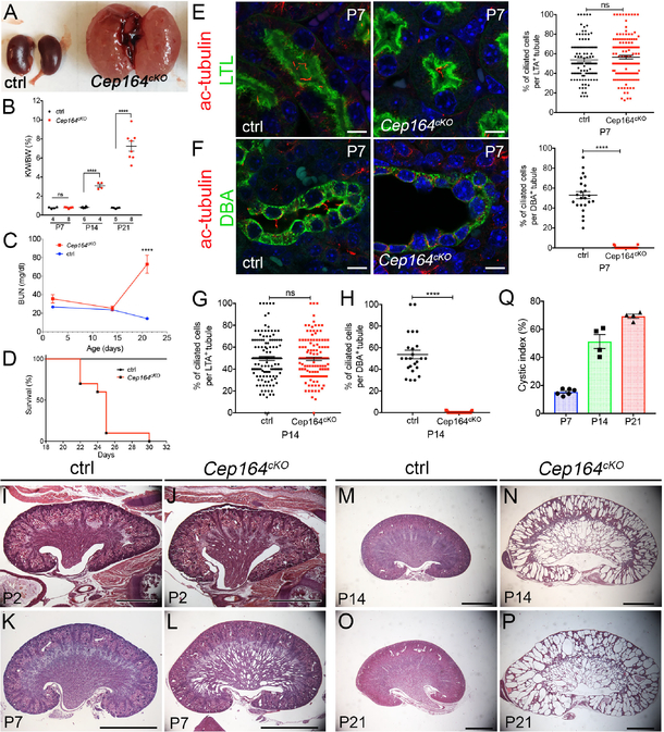
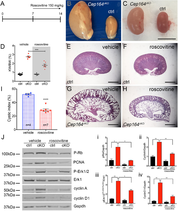
Nephronophthisis is an autosomal recessive kidney disease with high genetic heterogeneity. Understanding the functions of the individual genes contributing to this disease is critical for delineating the pathomechanisms of this disorder. Here, we investigated kidney function of a novel gene associated with nephronophthisis, CEP164, coding a centriolar distal appendage protein, using a Cep164 knockout mouse model. Collecting duct-specific deletion of Cep164 abolished primary cilia from the collecting duct epithelium and led to rapid postnatal cyst growth in the kidneys. Cell cycle and biochemical studies revealed that tubular hyperproliferation is the primary mechanism that drives cystogenesis in the kidneys of these mice. Administration of roscovitine, a cell cycle inhibitor, blocked cyst growth in the cortical collecting ducts and preserved kidney parenchyma in Cep164 knockout mice. Thus, our findings provide evidence that therapeutic modulation of cell cycle activity can be an effective approach to prevent cyst progression in the kidney.

Keywords: CEP164; centrosome; cilia; nephronophthisis; polycystic kidney disease.


Conflict of interest statement

DISCLOSURE

F.H. is a cofounder of Goldfinch-Bio. The other authors have no competing financial interests.

Figures

Figure 1.
Figure 1.. Deletion of Cep164 from collecting duct epithelium leads to postnatal cystogenesis.
(A) Overview of P21 kidneys. Cep164cKO mice develop polycystic kidney disease by P21. (B) Quantification of kidney weight to body weight ratio of Cep164cKO mice at postnatal days 7, 14 and 21. P7 ctrl 0.75 ± 0.04, n=4 vs. Cep164cKO 0.77 ± 0.03, n=5, ns; P14 ctrl 0.82 ± 0.03, n=6 vs. Cep164cKO 3.09 ± 0.18, n=4, ****p<0.0001; P21 difference ctrl 0.74 ± 0.02, n=5 vs. Cep164cKO 7.2 ± 0.53, n=8, ****p<0.0001. (C) Blood urea nitrogen levels are significantly increased in Cep164cKO animals by P21 and indicate kidney failure. BUN values are comparable in control and Cep164cKO mice at P2 and P14. At P21 the BUN levels in Cep164cKO mice indicated kidney failure (ctrl 14.31 ± 1.09, n=7 vs. Cep164cKO 72.87 ± 9.69, n=5, ****p<0.0001). (D) Mantel-Cox logrank test shows a statistically significant difference between control and Cep164cKO mouse survival (**p<0.001). Median survival time of Cep164cKO mice is 25 days (n = 10). (E) P7 kidney sections stained with antibodies against acetylated α-tubulin to mark primary cilia (red) and Lotus tetragonolobus lectin (LTL) to mark proximal tubules (green). Both, control and Cep164cKO mice have primary cilia in the proximal tubular epithelium. DAPI nuclei stained in blue. Scale bar 7.5 μm. There is no change in the fraction of ciliated cells in the proximal tubule of control vs. Cep164cKO kidneys at P7; ctrl (53.87 % ± 2.00, n=105 tubules, n=699 cells) vs. Cep164cKO (56.55 % ± 1.89, n=131 tubules, n=707 cells), p=0.3336. (F) P7 kidney sections stained with antibodies against acetylated α-tubulin to mark primary cilia (red) and Dolichos biflorus agglutinin (DBA) lectin to mark collecting ducts (green). Primary cilia are visible in the lumen of the collecting ducts in control kidneys, while no cilia are present in the Cep164cKO collecting duct epithelium. DAPI nuclei stained in blue. Scale bar 7.5 μm. Cep164cKO kidneys lack primary cilia in the collecting duct at P7; ctrl (52.95 % ± 3.58, n=23 tubules, n=292 cells) vs. Cep164cKO (0.36 % ± 0.20, n=25 tubules, n=643 cells), ****p<0.0001. (G) There is no change in the fraction of ciliated cells in the proximal tubule of control vs. Cep164cKO kidneys at P14; ctrl (49.64 % ± 1.811, n=130 tubules, n=909 cells) vs. Cep164cKO (49.39 % ± 1.617, n=133 tubules, n=887 cells), p=0.9180. (H) Cep164cKO kidneys lack primary cilia in the collecting duct at P14; ctrl (53.77 % ± 3.96, n=24 tubules, n=266 cells) vs. Cep164cKO (0.24 % ± 0.11, n=37 tubules, n=562 cells), ****p<0.0001. (I–P) Comparison of hematoxylin and eosin-stained mid-sagittal sections from control (I, K, M, O) and Cep164cKO kidneys (J, L, M, P) at various postnatal stages, reveals the formation of collecting duct dilations in the medullary region of the mutant kidneys at P7 (K, L). By P14 the medullary cysts have greatly expanded in size, and cystogenesis has occurred in the cortical collecting ducts (M, N). By P21, corticomedullary border and kidney parenchyma has been destroyed by the expanding cysts in Cep164cKO kidneys (O, P). Scale bar, 1 mm (I, J), 2 mm (K – P). (Q) Quantification of the cystic area (%) in Cep164cKO kidneys at postnatal stages P7, P14, P21. There is a sudden increase in the cystic index from P7 to P14, coinciding with the appearance of cortical collecting duct cysts.
Figure 2.
Figure 2.. Roscovitine slows cyst growth in Cep164cKO kidneys.
(A) Schematic of timeline of roscovitin injections and kidney analysis. (B) Overview of kidney morphology of vehicle treated control (ctrl) and Cep164cKO kidneys at P14. Scale bar 5mm. (C) Overview of kidney morphology of roscovitine (150 mg/kg) treated control (ctrl) and Cep164cKO kidneys at P14. Scale bar 5mm. (D) Roscovitine treatment reduces the kidney weight to body weight ratio in P14 Cep164cKO mice. Vehicle 2.76 ± 0.10, n=5, vs. roscovitine 1.79 ± 0.15, n=6, ***p<0.001. (E – H) Comparison of hematoxylin and eosin-stained mid-sagittal sections of vehicle treated (E,G) and roscovitine-treated kidneys (F,H) demonstrate a significant reduction in the size of the cortical collecting duct cysts in roscovitine-treated P14 Cep164cKO kidneys. (I) Cystic index is significantly reduced in P14 Cep164cKO kidneys after roscovitin treatment. Vehicle 52.25 % ± 1.15 %, n=4 vs. roscovitine 28.92 % ± 1.68 %, n=7, **** p<0.0001. (J) Roscovitine treatment blocks cell cycle activity in Cep164cKO kidneys. Western blotting against markers of cell cycle activity pRb, PCNA, pERK1/2, cyclin A and cyclin D1 are significantly decreased in roscovitine-treated Cep164cKO kidneys at P14 compared to vehicle-treated kidneys. Erk1 and Gapdh are used as loading controls. Phospho-proteins were compared with total protein or GAPDH as loading control for densitometric analysis which is presented graphically in i, ii, iii and iv. i) pRb ctrl vs cKOveh (1 ± 0.2, n=3 vs. 3.6 ± 0.7, n=3), cKOveh vs. cKOros (3.4 ± 0.7, n=3 vs. 0.9 ± 0.0, n=3, *p<0.05); ii) cyclinA ctrl vs. cKOveh (1 ± 0.1, n=3 vs. 2.6 ± 0.1, n=3, **p<0.01), cKOveh vs. cKOros (2.6 ± 0.1, n=3 vs. 1.3 ± 0.1, n=3, **p<0.01); iii) pErk1 ctrl vs. cKOveh (1 ± 0.1, n=3 vs. 4.8 ± 0.6, n=3, **p<0.01), cKOveh vs. cKOros (4.8 ± 0.6, n=3 vs. 2.8 ± 0.2, n=3, *p<0.05); iv) cyclinD1 ctrl vs. cKOveh (1 ± 0.2, n=3 vs. 7.8 ± 0.9, n=3, **p<0.01), cKOveh vs. cKOros (7.8 ± 0.9, n=3 vs. 2.7 ± 0.4, n=3, **p<0.01). Data from 3 individual animals in a given group were used for densitometric analysis.

References

    1. Hildebrandt F, Benzing T, Katsanis N: Ciliopathies. The New England journal of medicine, 364: 1533–1543, 2011. - PMC - PubMed
    1. Igarashi P, Somlo S: Genetics and pathogenesis of polycystic kidney disease. Journal of the American Society of Nephrology : JASN, 13: 2384–2398, 2002. - PubMed
    1. Ward CJ, Hogan MC, Rossetti S, Walker D, Sneddon T, Wang X, Kubly V, Cunningham JM, Bacallao R, Ishibashi M, Milliner DS, Torres VE, Harris PC: The gene mutated in autosomal recessive polycystic kidney disease encodes a large, receptor-like protein. Nat Genet, 30: 259–269, 2002. - PubMed
    1. Yokoyama T: Ciliary subcompartments and cysto-proteins. Anat Sci Int, 92: 207–214, 2017. - PubMed
    1. Sorokin SP: Centriole formation and ciliogenesis. Aspen Emphysema Conf, 11: 213–216, 1968. - PubMed

Publication types

MeSH terms

LinkOut - more resources