Clinical Trial
. 2019 Jul:45:341-350.
doi: 10.1016/j.ebiom.2019.06.016. Epub 2019 Jun 24.

Clearance of apoptotic cells by mesenchymal stem cells contributes to immunosuppression via PGE2

Affiliations
Clinical Trial

Clearance of apoptotic cells by mesenchymal stem cells contributes to immunosuppression via PGE2

Zhuoya Zhang et al. EBioMedicine. 2019 Jul.

Abstract

Background: Defective clearance of apoptotic cells (ACs) has been suggested to be involved in the pathogenesis of systemic lupus erythematosus (SLE). Mesenchymal stem cells (MSCs) exhibit promising therapeutic effects on SLE, but whether MSCs phagocytose ACs and contributes to the underlying mechanism in the treatment of SLE remain unknown.

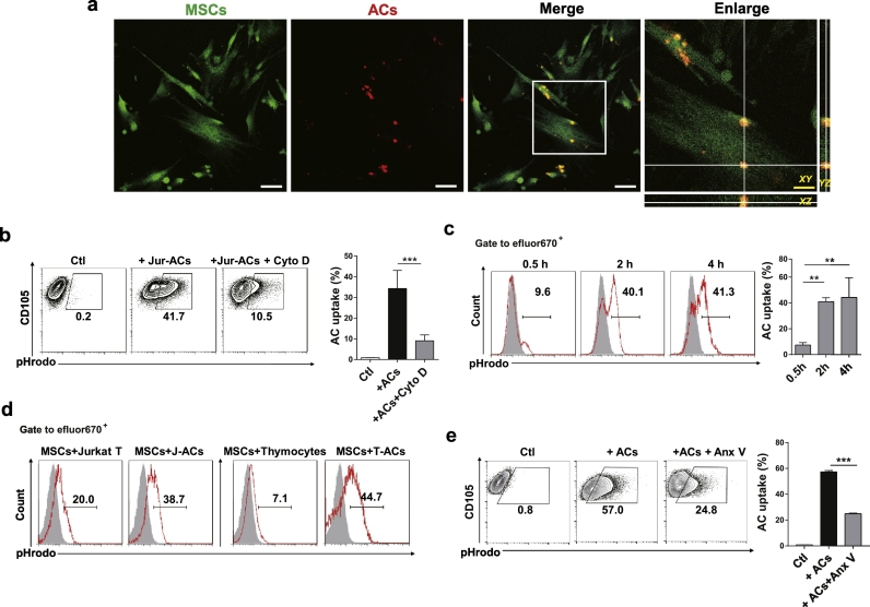
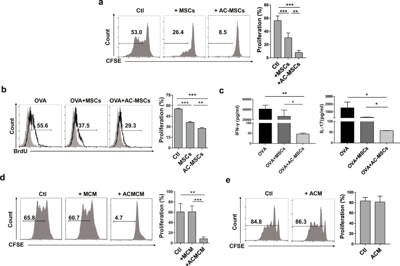
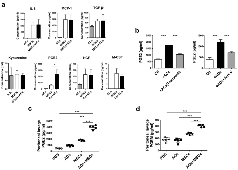
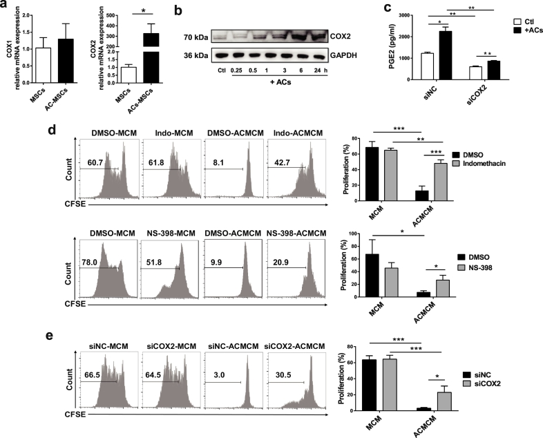
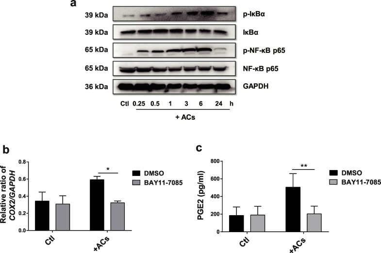
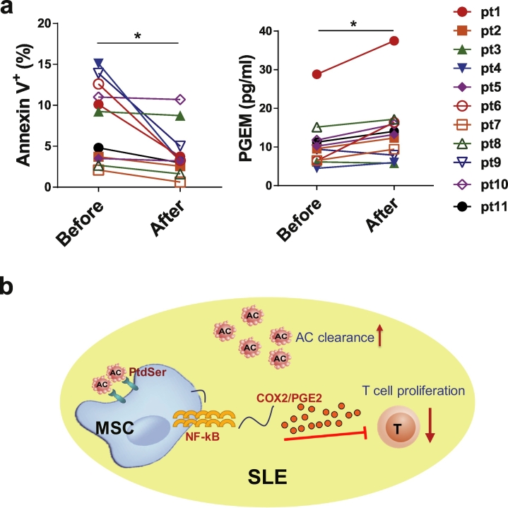
Methods: Human umbilical cord (UC) MSCs were co-cultured with ACs, and the engulfment of ACs by MSCs was either detected by flow cytometry or observed under confocal laser scanning microscope. Peripheral blood mononuclear cells (PBMCs) from healthy controls (HCs) were cultured in MSC conditioned medium (MCM) or MSC exposed to ACs (AC-MSC) conditioned medium (ACMCM), and then CD4+ T cell proliferation was detected. Soluble factors including prostaglandin (PG)E2 in the supernatants of MSCs and AC-MSCs, as well as in the mouse peritoneal lavage fluids (PLF) were determined by enzyme-linked immunosorbent assay (ELISA). Cyclooxygenase (COX)2 inhibitors and siRNA transfection were utilized to determine the function of COX2/PGE2 in AC-MSC-mediated immunosuppression. PGE2 metabolites (PGEM) in the plasma of SLE patients were measured before and 24 h after MSC transplantation respectively.

Findings: Human UC MSCs possessed the ability to engulf ACs. AC-MSCs increased MSC-mediated suppression of CD4+ T cell proliferation compared to MSCs alone. Mechanistically, ACs stimulated MSCs to express COX2 and consequently produced PGE2 that inhibited T cell responses. NF-κB signalling pathway mediated the activation of COX2/PGE2 in AC-MSCs. Importantly, in patients with SLE, the plasma PGEM levels increased significantly in those with reduced apoptotic mononuclear cells in peripheral blood after MSC transplantation.

Interpretation: Clearance of ACs by MSCs contributes to immunosuppressive function via increasing PGE2 production. These findings reveal a previously unrecognized role of MSC-mediated phagocytosis of ACs in MSC-based immunotherapy. FUND: This study was supported by grants from the Chinese Major International (Regional) Joint Research Project (No. 81720108020), the Jiangsu Province Major Research and Development Program (No. BE2015602) and the Jiangsu Province 333 Talent Grant (BRA2016001). WJ. Chen was supported by the Intramural Research Program of NIH, NIDCR.


Figures

Fig. 1
Fig. 1
UC MSCs Engulf ACs in vitro and in vivo. (a) Engulfment of apoptotic cells (ACs) by UC MSCs observed under laser scanning confocal microscopy in all three axes. White scale bar: 75 μm;Yellow scale bar: 30 μm. (b) In vitro AC uptake of UC MSCs (n = 4) by flow cytometry. (c-d) pHrodo labelled living cells or ACs were injected into B6 mice intraperitoneally with eFluor670 labelled UC MSCs. Mice were sacrificed and peritoneal exudate cells were collected. (c) The internalization of ACs by UC MSCs (n = 3) were scored 0.5 h, 2 h and 4 h after injection. (d) The internalization of living cells and ACs by UC MSCs were determined 2 h after injection. (e) Annexin V (Anx V) blocked the engulfment of ACs by UC MSCs (n = 4). Bar graphs show means ± SD. **p < .01, ***p < .001 (One-way ANOVA with Tukey's multiple comparisons test). (For interpretation of the references to colour in this figure legend, the reader is referred to the web version of this article.)
Fig. 2
Fig. 2
Phagocytosis of ACs Increases Immunosuppressive Function of MSCs. (a) MSCs exposed to ACs (AC-MSCs) showed enhanced suppression on CD4+ T cell proliferation (n = 5). (b-c) Lymph node cells from the inguinal draining lymph node of OVA-immunized mice (n = 3) were cocultured with MSCs or AC-MSCs at 10:1 ratio, in the presence of OVA, for three days. (b) OVA-specific CD4+ T cell proliferation. (c) IFN-γ and IL-17 levels in the coculture supernatants. (d) Conditioned media of AC-MSCs (ACMCM) significantly inhibited CD4+ T cell proliferation (n = 6). (e) Conditioned media of ACs (ACM) could not suppress CD4+ T cell proliferation (n = 7, student's t-test). Bar graphs show means ± SD. *p < .05, **p < .01, ***p < .001 (One-way ANOVA with Tukey's multiple comparisons test).
Fig. 3
Fig. 3
Phagocytosis of ACs by MSCs Releases PGE2. (a) ACs and MSCs were cultured alone or cocultured together at a ratio of 20:1 for 24 h (n = 4, **P < .01, student's t-test). Related soluble factor levels in the culture supernatants. (b) ACs were cultured in the upper chamber of the transwell system (with 0.4 μm pores), with MSCs in the lower (n = 4). Alternatively, ACs were pre-treated with Annexin V and cocultured with MSCs at a ratio of 20:1 for 24 h (n = 4). PGE2 levels in the culture supernatants were measured. (c-d) B6 mice (n = 5/group) were pre-treated with clodronate liposomes to deplete peritoneal macrophages one day before ACs, MSCs, or ACs plus MSCs (10:1) injection. After 4 h, mice were sacrificed and peritoneal lavage fluid (PLF) was harvested for PGE2 (c) and PGEM (d) concentration assessment. Data were presented as means ± SD. *p < .05, ***p < .001 (One-way ANOVA with Tukey's multiple comparisons test).
Fig. 4
Fig. 4
PGE2 Contributes to Immunosuppression by MSC Uptake of ACs. (a) Increased COX2 mRNA expression in AC-MSCs 24 h after MSCs and ACs coculture (n = 3). (b) Protein levels of COX2 in MSCs at the indicated time points after coculturing with ACs. (c) COX2 knockdown by siRNA transfection in MSCs decreased PGE2 releasing (n = 3). (d) Indomethacin and NS-398 (dissolved in DMSO) were added into the MSC and AC cocultures and then the conditioned media were prepared. Indomethacin and NS-398 partly reversed the inhibition of CD4+ T cells (n = 4) by the conditioned media of AC-MSCs (ACMCM). (e) MSCs were transfected with siRNA targeted to COX2 (siCOX2) or negative control siRNA (siNC) and then the conditioned media were prepared. COX2 knockdown partially reversed CD4+ T cell suppression (n = 4) by the ACMCM. Bar graphs show means ± SD. *p < .05, **p < .01, ***p < .001 (Student's t-test).
Fig. 5
Fig. 5
PGE2 Production in AC-MSCs is Dependent on NF-κB Signalling. (a) MSCs were cocultured with ACs at a ratio of 1:20 for the indicated time. The expressions of IκBα, p65 and their phosphorylated forms in MSCs were analysed by western blot. (b-c) MSCs were cocultured with ACs for 6 h in the presence or absence of BAY 11–7085. (b) The relative ratio of COX2/GAPDH expression in MSCs by western blot analysis (n = 3). (c) PGE2 levels in the culture supernatants (n = 4). Bar graphs show means ± SD. *p < .05, **p < .01 (Student's t-test).
Fig. 6
Fig. 6
MSCs Transplantation Increases PGE2 in patients with SLE. (a) Apoptotic levels of PBMCs and plasma levels of PGE2 metabolites (PGEM) in eleven patients with SLE before and after MSC transplantation. (b) Scheme of MSCs promote AC clearance and induced immunosuppression in SLE. *p < .05 (Student's t-test).

Similar articles

Cited by

References

    1. Tsokos G.C., Lo M.S., Costa Reis P., Sullivan K.E. New insights into the immunopathogenesis of systemic lupus erythematosus. Nat Rev Rheumatol. 2016;12:716–730. - PubMed
    1. Mahajan A., Herrmann M., Munoz L.E. Clearance deficiency and cell death pathways: A model for the pathogenesis of SLE. Front Immunol. 2016;7:35. - PMC - PubMed
    1. Arandjelovic S., Ravichandran K.S. Phagocytosis of apoptotic cells in homeostasis. Nat Immunol. 2015;16:907–917. - PMC - PubMed
    1. Munoz L.E., Lauber K., Schiller M., Manfredi A.A., Herrmann M. The role of defective clearance of apoptotic cells in systemic autoimmunity. Nat Rev Rheumatol. 2010;6:280–289. - PubMed
    1. Shoshan Y., Mevorach D. Accelerated autoimmune disease in MRL/MpJ-Faslpr but not in MRL/MpJ following immunization with high load of syngeneic late apoptotic cells. Autoimmunity. 2004;37:103–109. - PubMed

Publication types

MeSH terms