

Primary angiosarcoma of thyroid
- PMID: 31248893
- PMCID: PMC6605889
- DOI: 10.1136/bcr-2018-228862
Primary angiosarcoma of thyroid
Abstract
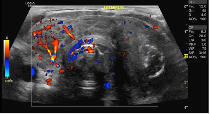
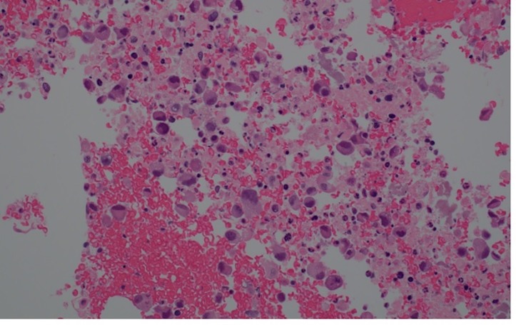
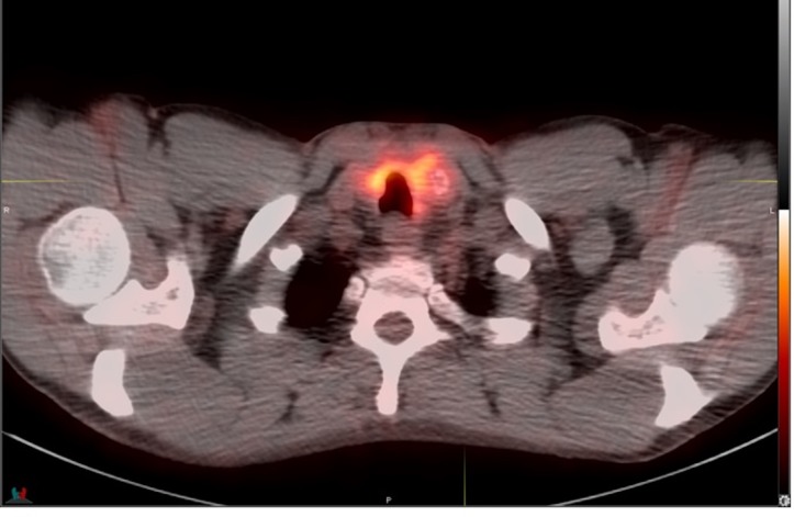
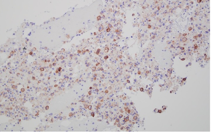
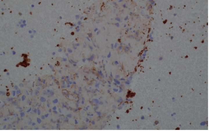
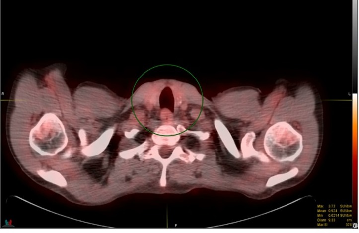
Mesenchymal origin of primary thyroid angiosarcomas (TAS) is extremely rare and comprises less than 1% of primary thyroid cancer worldwide. While TAS are most commonly occurring in the Alpine region, there are multiple reported cases of TAS in non-Alpine regions. Diagnosis of TAS is commonly made after thyroidectomy as cytologic diagnosis can be challenging due to paucity of cells, presence of necrosis and unawareness of the disease due to rarity. We report a case of primary TAS diagnosed by cytology in a 56-year-old man who presented with a sudden onset of left neck pain, swelling and haemoptysis. He was later noted to have suspicious nodules on both lobes of thyroid on ultrasound. Fine needle aspiration of thyroid nodules showed malignant epithelioid cells. The diagnosis of TAS was made based on positive endothelial markers such as thrombomodulin and CD31, with many pertinent negatives, including negative cytokeratins,thyroid transcription factor (TTF1), thyroglobulin, calcitonin and carcinoembryonic antigen (CEA).
Keywords: cancer intervention; endocrine cancer; thyroid disease.
© BMJ Publishing Group Limited 2019. No commercial re-use. See rights and permissions. Published by BMJ.
Conflict of interest statement
Competing interests: None declared.
Figures
References
-
- Weiss SW GJ. Soft tissue tumors. St Louis: Mosby, 2001.
-
- Surov A, Gottschling S, Wienke A, et al. . Primary Thyroid Sarcoma: a systematic review. Anticancer Res 2015;35:5185–91. - PubMed
Publication types
MeSH terms
Substances
LinkOut - more resources
Full Text Sources
Medical