. 2019 Aug 16;294(33):12521-12533.
doi: 10.1074/jbc.RA119.009666. Epub 2019 Jun 27.

Ca2+ flux through splice variants of the ATP-gated ionotropic receptor P2X7 is regulated by its cytoplasmic N terminus

Affiliations

Ca2+ flux through splice variants of the ATP-gated ionotropic receptor P2X7 is regulated by its cytoplasmic N terminus

Xin Liang et al. J Biol Chem. .

Abstract

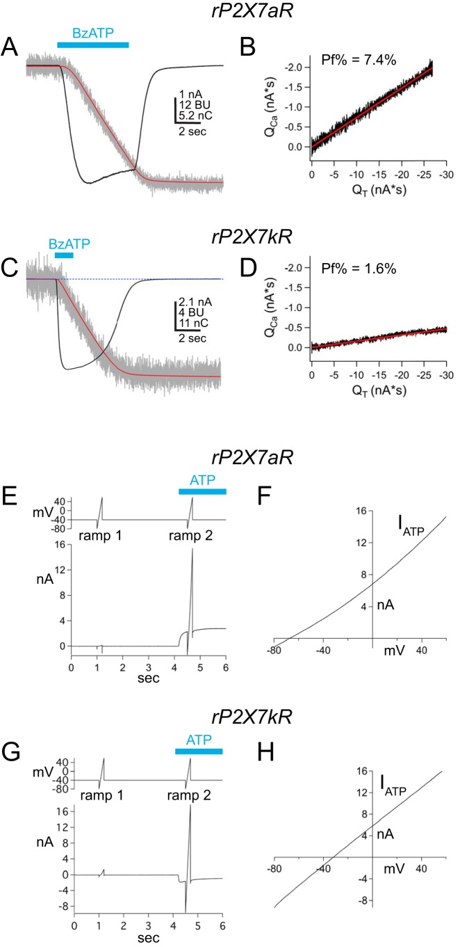
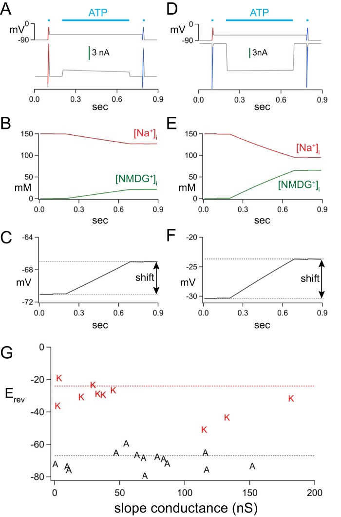
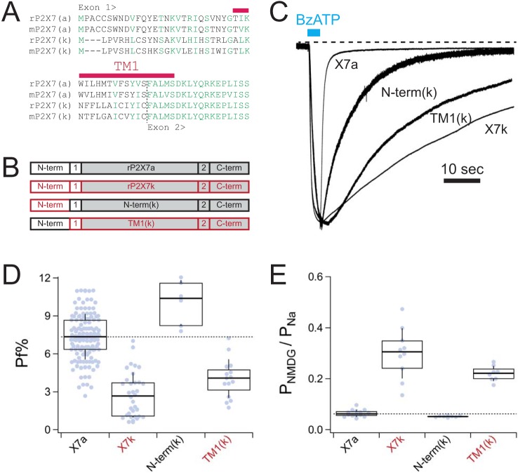
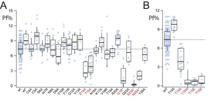
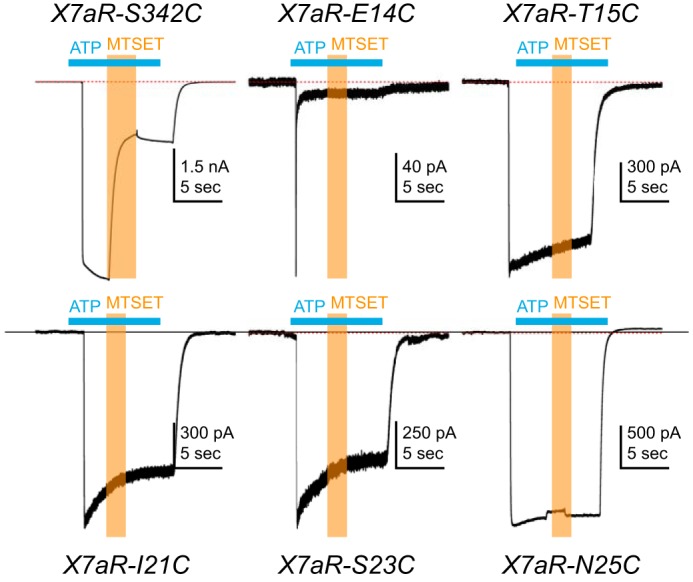
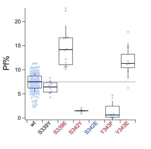
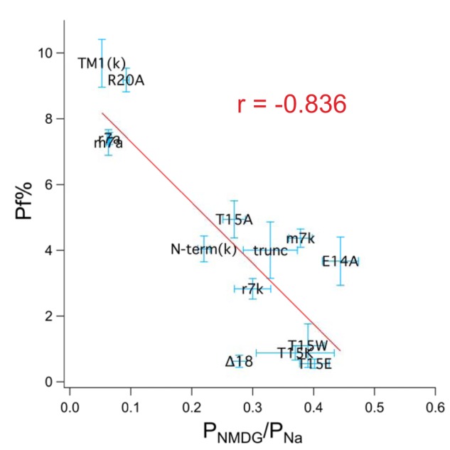
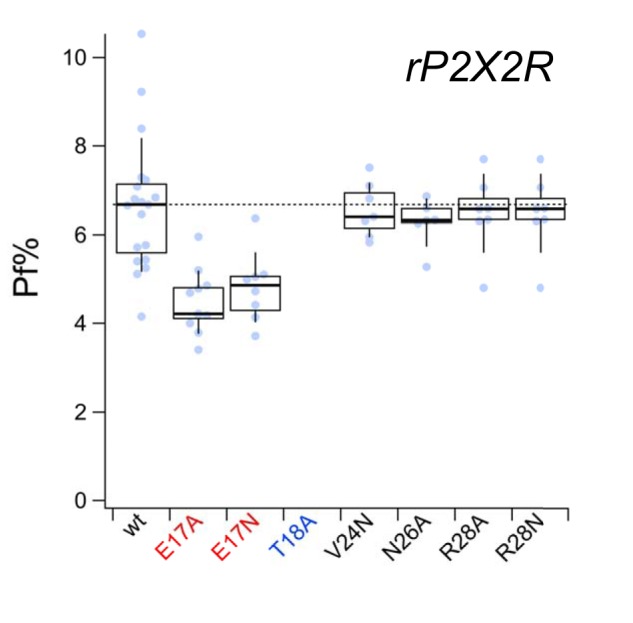
Activation of ionotropic P2X receptors increases free intracellular Ca2+ ([Ca2+] i ) by initiating a transmembrane cation flux. We studied the "a" and "k" splice variants of the rat purinergic P2X7 receptor (rP2X7aR and rP2X7kR) to exhibit a significant difference in Ca2+ flux through this channel. This difference is surprising because the variants share absolute sequence identity in the area of the pore that defines ionic selectivity. Here, we used patch-clamp fluorometry and chimeric receptors to show that the fraction of the total current carried by Ca2+ is a function of the primary sequence of the cytoplasmic N terminus. Using scanning mutagenesis, we identified five sites within the N terminus that respond to mutagenesis with a decrease in fractional calcium current and an increase in permeability to the polyatomic cation, N-methyl-d-glucamine (NMDG+), relative to Na+ (PNMDG/PNa). We tested the hypothesis that these sites line the permeation pathway by measuring the ability of thiol-reactive MTSET+ to alter the current of cysteine-substituted variants, but we detected no effect. Finally, we studied the homologous sites of the rat P2X2 receptor (rP2X2R) and observed that substitutions at Glu17 significantly reduced the fractional calcium current. Taken together, our results suggest that a change in the structure of the N terminus alters the ability of an intra-pore Ca2+ selectivity filter to discriminate among permeating cations. These results are noteworthy for two reasons: they identify a previously unknown outcome of mutagenesis of the N-terminal domain, and they suggest caution when assigning structure to function for truncated P2X receptors that lack a part of the N terminus.

Keywords: P2X 7 (P2X7) P2RX7; calcium channel; calcium transport; fractional calcium current; ion channel; ligand-gated; patch clamp; patch-clamp fluorometry; permeability; purinergic receptor; splice variant.


Conflict of interest statement

The authors declare that they have no conflicts of interest with the contents of this article

Figures

Figure 1.
Figure 1.
Fractional calcium current and PNMDG/PNa of two rP2X7R splice variants. A and C show raw and integrated data collected from voltage-clamped HEK293T cells transiently expressing either rP2X7aRs (A) or rP2X7kRs (C). Holding voltage = −60 mV. Membrane currents (nA) are shown as black traces, integrated currents (nC) as red traces, and fura-2 fluorescence (BU; 380-nm excitation and 510-nm emission) as gray traces. Applications of 100 μm BzATP (cyan bars) generated inward membrane current and decreases in fura-2 fluorescence. These data were used to plot integrated currents (QT) versus calibrated fluorescent signals (QCa) in B and D. The fractional calcium currents (Pf%) were calculated from the slopes of the linear fits to the plotted data. The fractional calcium current of the rP2X7kR is smaller than that of the rP2X7aR despite the fact that the two splice variants share sequence identity in the pore-forming domains. E and G show raw data of Erev measurements from the two WT splice variants bathed in the extracellular NMDG+ solution. Holding voltage = −40 mV, and voltage ramps ranged from −80 to 60 mV. ATP (500 μm) evoked outward current through the rP2X7aR (E), and inward current through the rP2X7kR (G). Ramp1 was used to measure leak current, and Ramp2 was used to measure total current in the presence of ATP. F and H, plot ATP-gated current, equal to Ramp2 − Ramp1, versus ramp voltage. PNMDG/PNa values were calculated from the values of the X-intercept (i.e. Erev) as described under “Experimental procedures.”
Figure 2.
Figure 2.
In silico modeling of ATP-gated responses. A and D show the voltage protocols (top traces) and resulting currents (bottom traces) for rP2X7aRs or rP2X7kRs, respectively. ATP causes an immediate inward current through the rP2X7aR (A) and an immediate outward current through the rP2X7kR (D) at a holding voltage of −60 mV. B and E show the expected changes in intracellular concentrations of ions, and C and F show the expected shift in Erev caused by the ionic changes. The shifts are modest for both splice variants. G plots the results of empirical measurements of the slope conductances of the ATP-gated currents obtained near the Erev of the rP2X7aR (“A”) and rP2X7kR (“K”) for cells bathed in the NMDG+ bath solution.
Figure 3.
Figure 3.
Chimeric receptors. A, exon 1 of rat and mouse P2X7aRs and P2X7kRs shows differences in primary sequence. All other parts are identical (see also Fig. S1). B, cartoon representations of the different chimeric rP2X7Rs. Domains contributed by rP2X7aRs are shown in black. Domains contributed by rP2X7kRs are shown in red. The shaded boxes are common to both splice variants. C, raw data traces of representative currents through WT and chimeric rP2X7Rs recorded at a holding voltage of −60 mV and normalized to their peak currents. D, box-and-whisker plots of fractional calcium currents of the two splice variants. The thick solid black lines demarcate median values; boxes are interquartile ranges, and whiskers are equal to standard deviations. Constructs that are significantly different from the WT rP2X7aR are denoted by red lettering in the x axis label. E, box-and-whisker plots of intrinsic PNMDG/PNa of the WT and chimeric receptors. Note that lower fractional calcium current (D) correlates with higher intrinsic PNMDG/PNa (E).
Figure 4.
Figure 4.
Fractional calcium current of N-terminal P2X7aR mutants. A, amino acid sequence of the N terminus of the rP2X7aR was changed one residue at a time to yield 23 mutants. Nineteen of these gave measurable currents in response to 100 μm BzATP. Only E14A and T15A had fractional calcium currents that were significantly different (marked with red lettering the x axis label) from the WT rP2X7aR (wt). The four unresponsive mutants were regenerated in a rP2X7aR-YFP background (I21R*, S23A*, V24R*, and N25A*); of these, only V24A* failed to respond to BzATP. The fractional calcium currents (Pf%) of the other three were significantly smaller than the WT rP2X7aR-YFP (wt*). B, fractional calcium current was measured from a range of rP2X7aR mutants involving Thr15. Again, results significantly different from the WT rP2X7aR are denoted with red lettering.
Figure 5.
Figure 5.
Cysteine-scanning mutagenesis. HEK293T cells expressing cysteine-substituted mutant rP2X7aRs were voltage-clamped at a holding potential of −60 mV. ATP (2 mm) was applied at times indicated by the horizontal cyan bars, and MTSET (1 mm) was co-applied at times indicated by the vertical orange bars. Co-application of MTSET caused a significant and irreversible inhibition of rP2X7aR-S342C mutant receptors but had no effect on any of the other five cysteine-substituted rP2X7aRs. Note that the rP2X7aR-E14C differed from the WT rP2X7aR and the other mutant receptors in showing a biphasic current. Although MTSET had no effect on the plateau phase of the rP2X7aR-E14C, the altered phenotype of the ATP-gated current suggests that caution be used in drawing firm conclusions from this mutant receptor.
Figure 6.
Figure 6.
Pore-lining residues form the intra-pore Ca2+-binding site. Fractional calcium current (Pf%) was measured from HEK293T cells expressing rP2X7Rs with single mutations at sites in TM2 that are thought to line the pore (31). Mutants with fractional calcium currents that significantly differ from the WT P2X7aR (wt) are labeled with red lettering on the x axis. Only the S342E mutant (blue lettering) failed to respond to BzATP with measurable inward current. Other mutants (S339E, S342Y, Y343F, and Y343E) showed changes in fractional calcium current that suggest their involvement with permeating Ca2+ in the pore of the channel.
Figure 7.
Figure 7.
Fractional calcium current negatively correlates with PNMDG/PNa. The graph shows data obtained from rat, mouse, and human P2X7Rs. Fractional calcium current (Pf%) is plotted against PNMDG/PNa. The red line is the best linear fit to the data using the resident curve fitting algorithm of Igor Pro.
Figure 8.
Figure 8.
Fractional calcium current of N-terminal rP2X2R mutants. Fractional calcium current (Pf%) was measured from WT (wt) and N-terminal mutant rP2X2Rs as described under “Experimental procedures.” Although T18A (labeled with blue lettering on the x axis) showed an ATP-gated current, it desensitized too quickly to give a measurable change in the fractional calcium current. Mutants with fractional calcium currents that were significantly different from the WT rP2X2R are labeled with red lettering.

Similar articles

Cited by

References

    1. Nicke A., Grutter T., and Egan T. M. (2018) in The Oxford Handbook of Neuronal Ion Channels (Bhattacharjee A., ed) pp. 1–32, Oxford University Press, Oxford, UK
    1. Schmid R., and Evans R. J. (2019) ATP-Gated P2X receptor channels: molecular insights into functional roles. Annu. Rev. Physiol. 81, 43–62 10.1146/annurev-physiol-020518-114259 - DOI - PubMed
    1. Skaper S. D., Debetto P., and Giusti P. (2010) The P2X7 purinergic receptor: from physiology to neurological disorders. FASEB J. 24, 337–345 10.1096/fj.09-138883 - DOI - PubMed
    1. Wiley J. S., Sluyter R., Gu B. J., Stokes L., and Fuller S. J. (2011) The human P2X7 receptor and its role in innate immunity. Tissue Antigens 78, 321–332 10.1111/j.1399-0039.2011.01780.x - DOI - PubMed
    1. Di Virgilio F. (2015) P2X receptors and inflammation. Curr. Med. Chem. 22, 866–877 10.2174/0929867322666141210155311 - DOI - PubMed

Publication types

LinkOut - more resources