. 2019 Aug 16;294(33):12507-12520.
doi: 10.1074/jbc.RA119.008255. Epub 2019 Jun 27.

An extracellular acidic cleft confers profound H+-sensitivity to epithelial sodium channels containing the δ-subunit in Xenopus laevis

Affiliations

An extracellular acidic cleft confers profound H+-sensitivity to epithelial sodium channels containing the δ-subunit in Xenopus laevis

Lukas Wichmann et al. J Biol Chem. .

Abstract

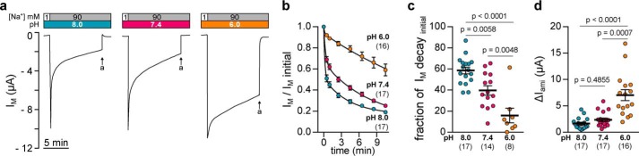
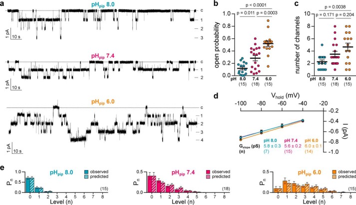
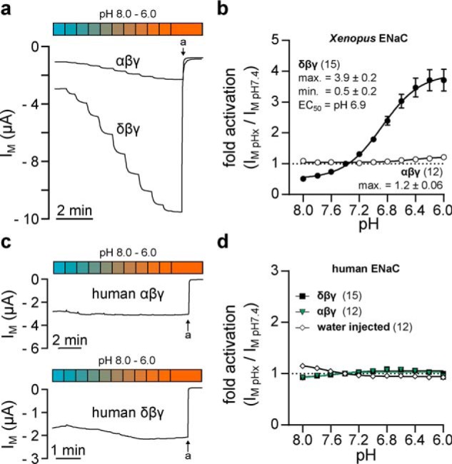
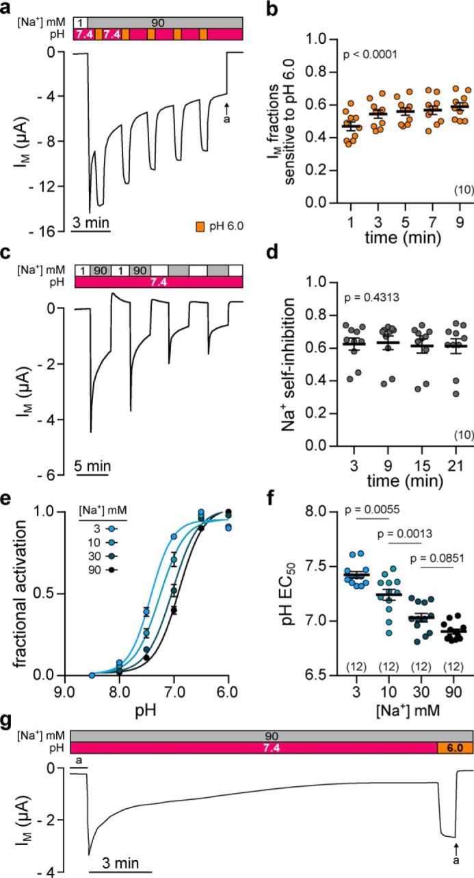
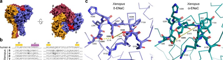
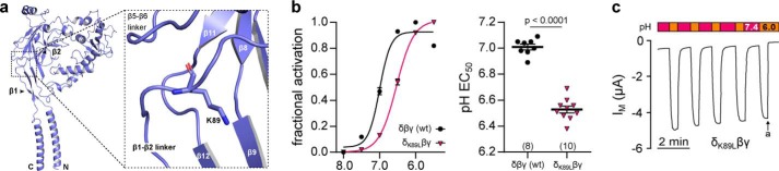
The limited sodium availability of freshwater and terrestrial environments was a major physiological challenge during vertebrate evolution. The epithelial sodium channel (ENaC) is present in the apical membrane of sodium-absorbing vertebrate epithelia and evolved as part of a machinery for efficient sodium conservation. ENaC belongs to the degenerin/ENaC protein family and is the only member that opens without an external stimulus. We hypothesized that ENaC evolved from a proton-activated sodium channel present in ionocytes of freshwater vertebrates and therefore investigated whether such ancestral traits are present in ENaC isoforms of the aquatic pipid frog Xenopus laevis Using whole-cell and single-channel electrophysiology of Xenopus oocytes expressing ENaC isoforms assembled from αβγ- or δβγ-subunit combinations, we demonstrate that Xenopus δβγ-ENaC is profoundly activated by extracellular acidification within biologically relevant ranges (pH 8.0-6.0). This effect was not observed in Xenopus αβγ-ENaC or human ENaC orthologs. We show that protons interfere with allosteric ENaC inhibition by extracellular sodium ions, thereby increasing the probability of channel opening. Using homology modeling of ENaC structure and site-directed mutagenesis, we identified a cleft region within the extracellular loop of the δ-subunit that contains several acidic amino acid residues that confer proton-sensitivity and enable allosteric inhibition by extracellular sodium ions. We propose that Xenopus δβγ-ENaC can serve as a model for investigating ENaC transformation from a proton-activated toward a constitutively-active ion channel. Such transformation might have occurred during the evolution of tetrapod vertebrates to enable bulk sodium absorption during the water-to-land transition.

Keywords: Xenopus; allosteric regulation; delta-subunit; epithelial sodium channel (ENaC); evolution; molecular evolution; pH; sodium self-inhibition; tetrapod; water–to–land transition.


Conflict of interest statement

The authors declare that they have no conflicts of interest with the contents of this article.

Figures

Figure 1.
Figure 1.
Extracellular pH modifies Xenopus δβγ-ENaC currents. a, representative recordings of transmembrane currents (IM) in oocytes expressing Xenopus δβγ-ENaC. The extracellular pH alters transient current kinetics of Xenopus δβγ-ENaC. Arrows indicate application of amiloride (a, 100 μm). b, current decays normalized to the initial peak values (IM, initial) from recordings as depicted in a follow a two-phase decay consisting of a fast, initial decay as well as a slow, continuous current rundown. c, fractions of the fast, initial current decay (IM decayinitial) are reduced with decreasing extracellular pH (one-way ANOVA, F = 20.31, p < 0.0001; Tukey's multiple comparisons test). d, amiloride-sensitive current fractions (ΔIami) derived from recordings as shown in a are markedly increased at pH 6.0 (Kruskal-Wallis test, p < 0.0001; Dunn's multiple comparisons test).
Figure 2.
Figure 2.
Extracellular pH affects gating of Xenopus-δβγ ENaC. a, representative current traces from cell-attached patch-clamp recordings of oocytes expressing Xenopus δβγ-ENaC at a holding potential of −100 mV. Recordings were performed using pipette solutions at pH 8.0, 7.4, or 6.0 (pHpip). Dashed lines indicate the number of individual open channel levels or the current baseline (c). b, open probability of individual channels increases with decreasing pHpip (one-way ANOVA, F = 24.93, p < 0.0001; Tukey's multiple comparisons test). c, number of visible channels in cell-attached recordings as depicted in a (one-way ANOVA, F = 5.83, p = 0.0056; Tukey's multiple comparisons test). d, current (I)/voltage (Vhold) plots derived from cell-attached patch-clamp recordings of Xenopus δβγ-ENaC at different pHpip. The pHpip does not affect the channel's slope conductance (Gslope) (one-way ANOVA, F = 1.854, p = 0.1725), which was calculated from the linear regression of unitary channel conductance (mean of at least three single channel amplitudes per n) at −40 to −100 mV. e, estimation of the number of channels in the patch. Pn is the probability of n channels out of the total number of channels within the patch being opened. The observed probability for each n (i.e. observed current levels) was compared with a theoretical distribution of Pn as predicted by a binomial distribution, assuming n channels present in the patch. There is no significant difference between observed and predicted Pn values under the employed pHpip conditions (Kruskal-Wallis test with Dunn's multiple comparisons test in each panel. p > 0.9999 between each pair of Pn observed and Pn predicted.). Statistical evaluation is based on individual recordings lasting for 120–180 s with a maximum of eight channels per patch. Please note that single-channel characteristics at pHpip 7.4 include data, which have been reported earlier (18). Patch-clamp data for both studies were collected simultaneously using the same oocyte batches for all pH conditions and αβγ- as well as δβγ-ENaC.
Figure 3.
Figure 3.
Xenopus δβγ-ENaC is activated by extracellular acidification. a, recordings of IM in oocytes expressing Xenopus αβγ- or δβγ-ENaC. Amiloride-sensitive (a, amiloride, 100 μm) currents mediated by δβγ- but not αβγ ENaC are dose-dependently increased by a stepwise acidification of the extracellular solution (pH 8.0–6.0; pH 0.2 increments). b, dose-response curves of acid-mediated activation of Xenopus ENaC isoforms. Normalized (IM, pHx/IM, pH 7.4) current levels were fit to a sigmoidal dose-response function with variable slope (Hill-slope of Xenopus δβγ-ENaC: 1.6 ± 0.4). Compared with pH 7.4, alkaline conditions decrease channel activity to a minimal factor (min.) of 0.5 ± 0.2, whereas acidification enhances activity of δβγ-ENaC by a maximal factor (max.) of 3.9 ± 0.2. c and d, maximal acid-induced activation of currents mediated by human αβγ-ENaC (max.: 1.03 ± 0.01) or δβγ-ENaC (max.: 1.05 ± 0.01) is considerably reduced with respect to Xenopus δβγ-ENaC.
Figure 4.
Figure 4.
Acidic pH elicits a reversible, consistent, and Na+-dependent activation of Xenopus δβγ-ENaC. a, representative current trace of an oocyte expressing Xenopus δβγ-ENaC. Repetitive acidification of the extracellular milieu (from pH 7.4 to pH 6.0 for 30 s) reversibly increases IM (a = amiloride, 100 μm). b, normalized IM fractions sensitive to pH 6.0 ((IM, pH 6.0IM, pH 7.4)/IM, pH 6.0) from recordings as depicted in a. Acid-sensitive current fractions do not decline but rather increase over time (repeated-measures one-way ANOVA, F = 57.8, p < 0.0001; Tukey's multiple comparisons test). c, assessment of repetitive SSI in an oocyte expressing Xenopus δβγ-ENaC. SSI was defined as the decline in IM at 3 min after increasing extracellular [Na+] from 1 to 90 mm. d, Na+ self-inhibition values from recordings as depicted in c. Current fractions lost to SSI ((IM, peakIM, 3 min)/IM, peak) are constant over time (repeated-measures one-way ANOVA, F = 0.8537, p = 0.4313; Tukey's multiple comparisons test). e, Na+ dependence of acid-induced ENaC activation was examined through determination of the pH EC50 (pH 8.5–6.0; pH 0.5 increments) under different Na+ concentrations (3–90 mm). Fractional channel activation was calculated as the difference between IM at a given pH (IM, x) and the minimal IM (IM, min) in relation to the maximally observed difference in IMmax IM) in the respective recording ((IM, xIM, min)/Δmax IM)). f, alkaline-shift of the pH EC50 in the presence of reduced [Na+] indicates that less protons are needed for acid-induced channel activation under low [Na+] (one-way ANOVA, F = 38.51, p < 0.0001; Tukey's multiple comparisons test). g, extracellular acidification (from pH 7.4 to pH 6.0) still activates Xenopus δβγ-ENaC after maximal rundown of currents.
Figure 5.
Figure 5.
Extracellular acidification antagonizes sodium self-inhibition of Xenopus δβγ-ENaC. a, representative recording of IM in oocytes expressing Xenopus δβγ-ENaC. SSI was determined by rapidly changing extracellular [Na+] from 1 to 3–120 mm at pH 8.0. The magnitude of Na+ self-inhibition ((IM, peakIM, 3 min)/IM, peak) was plotted against the respective [Na+] and fitted to the Michaelis-Menten equation, allowing estimation of maximal inhibition (Vmax) and apparent affinity for Na+ (Km). b–d, current recordings and Michaelis-Menten plots of SSI in Xenopus δβγ-ENaC at pH 7.4, 7.0, and 6.0. At pH 8.0, Vmax of SSI is similar to values at pH 7.4 (b), but SSI displays an enhanced Na+ affinity (Kruskal-Wallis test, p = 0.035; Dunn's multiple comparisons test). Compared with values at pH 7.4, further acidification (pH 7.0, c) decreases Vmax (one-way ANOVA, F = 5.508, Tukey's multiple comparisons test, p = 0.017), whereas the apparent Na+ affinity of SSI is not significantly changed (Kruskal-Wallis test, Dunn's multiple comparisons test, p > 0.999). Irrespective of the extracellular [Na+], SSI of Xenopus δβγ-ENaC is markedly reduced at pH 6.0 (d) and does not converge to a Michaelis-Menten fit. e, interaction landscape for Na+-dependent SSI at different extracellular H+. The Bliss score indicates deviations of the measured combinational responses from a reference model that assumes independence between Na+- and H+-mediated responses. Negative Bliss values across the interaction landscape indicate antagonism of Na+ dependent SSI by increasing H+.
Figure 6.
Figure 6.
Comparison of acidic cleft residues in Xenopus δ- and α-ENaC subunits. a, structural arrangement of a Xenopus δβγ-ENaC trimer with the position of the δ-ENaC acidic cleft highlighted in red. b, sequence alignments of human and Xenopus ENaC subunits depicting the β2–α1 and β6–β7 loops that constitute parts of the δ-ENaC acidic cleft. Bold letters indicate acidic cleft residues that have been mutated in this study. c, models of the acidic cleft within Xenopus δ- and α-ENaC. Residues in the acidic cleft are shown as stick representations with the rest of the protein chain depicted as ribbon representations. Contacts between residues in the acidic cleft are highlighted with distances shown.
Figure 7.
Figure 7.
Three aspartates in the δ-ENaC acidic cleft distinctly affect ENaC sensitivity to pH and Na+. a, representative IM recordings of Xenopus oocytes expressing WT δβγ-ENaC or channels containing a single aspartate to asparagine (δD296Nβγ) mutation. Channel sensitivity to pH was assessed by a stepwise reduction of extracellular pH from 8.0 to 6.0 (pH 0.2 increments). b, pH-dependent activation of δβγ-ENaC containing none (wt), one (δD105Kβγ, δD293Nβγ, and δD296Nβγ), two (δD293N,D296Nβγ and δD105K,D296Nβγ) or three (δD3βγ: δD105K,D293N,D296Nβγ) mutations leading to substitution of single aspartates. Maximum acid-induced channel activation is reduced in ENaC-containing single mutations (δD105Kβγ, δD293Nβγ, and δD296Nβγ) when compared with WT δβγ-ENaC. There is no cumulative effect in channels containing two mutated aspartates, but substitution of three aspartates (δD3βγ) further decreases maximum acid-induced activation as well as alkaline channel inhibition (Kruskal-Wallis test, p < 0.0001; Dunn's multiple comparisons test). c, assessment of SSI at [Na+] from 3 to 120 mm in oocytes expressing WT (δβγ) or mutant (δD296Nβγ) ENaC. d, Na+ self-inhibition ((IM, peakIM, 3 min)/IM, peak) of WT, single, double, and triple mutant channels. Introduction of δD293N or δD105K moderately decreases maximal SSI when compared with WT δβγ-ENaC, whereas ENaC containing δD296N has a profoundly reduced SSI irrespective of the extracellular [Na+]. Substitution of additional aspartates in double or triple mutant channels does not further decrease ENaC SSI (one-way ANOVA, F = 10.26, p = 0.0003; Tukey's multiple comparisons test). e, proton-sensitivity of δβγ ENaC mutants was assessed by determining fractional channel activation ((IM, xIM, min)/Δmax IM) resulting from a stepwise reduction of the extracellular pH (pH 8.5–6.0; pH 0.5 increments) in the presence of 90 or 3 mm extracellular Na+. f, reduction of extracellular [Na+] evokes an alkaline-shift of the pH EC50 in WT δβγ-ENaC but not in channels containing the δD296Nβγ or δD3βγ mutations. However, in the presence of 90 mm Na+, introduction of these mutations shifts the pH EC50 to more alkaline values, when compared with WT ENaC (one-way ANOVA, F = 48.02, p < 0.0001; Tukey's multiple comparisons test). g, pH-sensitivity of Xenopus αβγ-ENaC (n = 11) is not enhanced by partial (αK105Dβγ; n = 13) or full (αK105D,E296D,Q297Lβγ; n = 12) reconstitution of the δ-ENaC acidic cleft in this subunit. Individual values for pH-mediated regulation and SSI of WT and mutant ENaC (a–d), including statistical analyses, are listed in Table 1.
Figure 8.
Figure 8.
Proton sensitivity of Xenopus δβγ ENaC is further modulated by a lysine in the δ-ENaC β1–β2 linker. a, homology model of the Xenopus δ-ENaC subunit indicating the location of δLys-89 in the linker region between the β1 and β2 sheets in the palm domain. b, fractional channel activation ((IM, xIM, min)/Δmax IM) resulting from a stepwise reduction of the extracellular pH (pH 8.0–5.5; pH 0.5 increments) in WT and δK89Lβγ ENaC. Introduction of the δK89L mutation leads to a significant acidic-shift of the channel's pH EC50 (Student's unpaired t test). c, representative IM recording of an oocyte expressing δK89Lβγ-ENaC. Macroscopic currents mediated by mutant channels display “stimulus-activated” characteristics as they are low at neutral pH 7.4 and reversibly increased by extracellular acidification (pH 6.0, 30 s; a = amiloride, 100 μm).
Figure 9.
Figure 9.
Conservation of motifs involved in pH-sensitivity and proteolytic maturation of α- and δ-ENaC orthologs. Multiple sequence alignment inferred from amino acid sequences of α- and δ-ENaC orthologs (Table S1) showing sites involved in proton-mediated activation of X. laevis δβγ-ENaC that have been identified in this study (β1–β2, β2–α1, and β1–β2 linker). Note that only one α-like subunit ortholog has been identified in hagfish Eptatretus burgeri and lamprey Petromyzon marinus, which therefore are displayed in a separate group. Rats do not have a functional gene for δ-ENaC. Colored backgrounds illustrate the presence of residues with hydrophobic (blue) or polar (red) side chains in the β1–β2 linker or negatively-charged residues (yellow) in the acidic cleft. The Asp-296 position, which is essential for Na+ coordination in Xenopus δβγ-ENaC, is highlighted in red. Conservation of the minimal consensus sequence for proteolytic maturation of ENaC subunits by furin (sequence RXXR) is indicated by green check marks, and red crosses represent the absence of this consensus sequence. Gray checkmarks indicate the presence of an RXXR motif dislocated from the conserved location of the respective furin cleavage site.

Similar articles

Cited by

References

    1. Bayley M., Damsgaard C., Thomsen M., Malte H., and Wang T. (2019) Learning to air-breathe: the first steps. Physiology 34, 14–29 10.1152/physiol.00028.2018 - DOI - PubMed
    1. Hanukoglu I., and Hanukoglu A. (2016) Epithelial sodium channel (ENaC) family: phylogeny, structure–function, tissue distribution, and associated inherited diseases. Gene 579, 95–132 10.1016/j.gene.2015.12.061 - DOI - PMC - PubMed
    1. Studer R. A., Person E., Robinson-Rechavi M., and Rossier B. C. (2011) Evolution of the epithelial sodium channel and the sodium pump as limiting factors of aldosterone action on sodium transport. Physiol. Genomics 43, 844–854 10.1152/physiolgenomics.00002.2011 - DOI - PubMed
    1. Kerem E., Bistritzer T., Hanukoglu A., Hofmann T., Zhou Z., Bennett W., MacLaughlin E., Barker P., Nash M., Quittell L., Boucher R., and Knowles M. R. (1999) Pulmonary epithelial sodium-channel dysfunction and excess airway liquid in pseudohypoaldosteronism. N. Engl. J. Med. 341, 156–162 10.1056/NEJM199907153410304 - DOI - PubMed
    1. Cogswell M. E., Zhang Z., Carriquiry A. L., Gunn J. P., Kuklina E. V., Saydah S. H., Yang Q., and Moshfegh A. J. (2012) Sodium and potassium intakes among US adults: NHANES 2003–2008. Am. J. Clin. Nutr. 96, 647–657 10.3945/ajcn.112.034413 - DOI - PMC - PubMed

Associated data

LinkOut - more resources