. 2019 Aug 16;294(33):12444-12458.
doi: 10.1074/jbc.RA119.009471. Epub 2019 Jun 27.

Direct observation of intermediates in the SufS cysteine desulfurase reaction reveals functional roles of conserved active-site residues

Affiliations

Direct observation of intermediates in the SufS cysteine desulfurase reaction reveals functional roles of conserved active-site residues

Matthew Blahut et al. J Biol Chem. .

Abstract

Iron-sulfur (Fe-S) clusters are necessary for the proper functioning of numerous metalloproteins. Fe-S cluster (Isc) and sulfur utilization factor (Suf) pathways are the key biosynthetic routes responsible for generating these Fe-S cluster prosthetic groups in Escherichia coli Although Isc dominates under normal conditions, Suf takes over during periods of iron depletion and oxidative stress. Sulfur acquisition via these systems relies on the ability to remove sulfur from free cysteine using a cysteine desulfurase mechanism. In the Suf pathway, the dimeric SufS protein uses the cofactor pyridoxal 5'-phosphate (PLP) to abstract sulfur from free cysteine, resulting in the production of alanine and persulfide. Despite much progress, the stepwise mechanism by which this PLP-dependent enzyme operates remains unclear. Here, using rapid-mixing kinetics in conjunction with X-ray crystallography, we analyzed the pre-steady-state kinetics of this process while assigning early intermediates of the mechanism. We employed H123A and C364A SufS variants to trap Cys-aldimine and Cys-ketimine intermediates of the cysteine desulfurase reaction, enabling direct observations of these intermediates and associated conformational changes of the SufS active site. Of note, we propose that Cys-364 is essential for positioning the Cys-aldimine for Cα deprotonation, His-123 acts to protonate the Ala-enamine intermediate, and Arg-56 facilitates catalysis by hydrogen bonding with the sulfhydryl of Cys-aldimine. Our results, along with previous SufS structural findings, suggest a detailed model of the SufS-catalyzed reaction from Cys binding to C-S bond cleavage and indicate that Arg-56, His-123, and Cys-364 are critical SufS residues in this C-S bond cleavage pathway.

Keywords: PLP; PLP-dependent sulfur abstraction; SufS; X-ray crystallography; cysteine desulfurase; cysteine sulfur bond cleavage; enzyme catalysis; enzyme mechanism; iron-sulfur protein; pre-steady-state kinetics.


Conflict of interest statement

The authors declare that they have no conflicts of interest with the contents of this article

Figures

SCHEME 1.
SCHEME 1.
General cysteine desulfurase reaction scheme for SufS prior to this study. l-Cysteine substrate is shown in blue (C, H, O, and N) and orange (S). PLP and SufS active-site residues are in black.
Figure 1.
Figure 1.
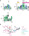
A, a sequence logo indicates active-site residues of interest that are highly conserved across SufS proteins. The sequence alignment was made from 29 members of the InterPro SufS family (IPR010970) chosen from distinct nodes of a sequence similarity network to model sequence diversity in the family. B, a structural superposition of six SufS family members from the PDB reveals that several residues, such as His-123 and His-124, adopt an identical position in all structures, while the location of Arg-56 (indicated by an arc) varies.
Figure 2.
Figure 2.
A, UV-visible absorption spectra for fractions containing WT or H123A SufS from the initial purification step using an anion exchange column. B, UV-visible absorption spectra for fractions containing WT or H123A SufS from the second purification step using a hydrophobic interaction column. Presence of SufS was confirmed in all fractions by SDS-PAGE. C, an image of the SufS homodimer including a box denoting the active-site region shown below. D, a superposition of WT SufS and SufS H123A reveals that the mutation does not affect the position of other critical active-site components such as PLP or Cys-364.
Figure 3.
Figure 3.
UV-visible absorption spectra during 150 min treatment with 500 μml-cysteine for (A) WT, (B) H123A, and (C) C364A SufS.
Figure 4.
Figure 4.
Stopped-flow analysis of 37.5 μm (post-mix) WT (left), H123A (center), and C364A SufS (right) with various cysteine concentrations. A, absorbance spectrum at various time points for the reaction of 400 μm cysteine with SufS. B, absorbance versus time traces monitored at 340 and 420 nm for 400 μm cysteine mixed with SufS. C, fast phase (1/τ1) at different cysteine concentrations monitored at 340 nm (in which 1/τ1 is the reciprocal relaxation time).
Figure 5.
Figure 5.
UV-visible absorption features of 37.5 μm H123A SufS (post-mix) with 400 μml-cysteine fit to an A to B to C transition, where A, B, and C represent distinct species (see text for details). A, spectra obtained from the indicated time points after mixing of H123A SufS and cysteine. B, pure component spectra of H123A SufS determined using global fitting SVD analysis of PDA data. B, inset, fractional concentration of each species in the H123A SufS spectra over time after l-cysteine addition.
Figure 6.
Figure 6.
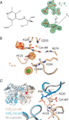
A, unbiased FoFc difference electron density maps of C364A crystals incubated with l-cysteine possess electron density for a Cys-aldimine enzymatic intermediate covalently bound to PLP. A tetrahedral arrangement of the atoms bonded to Cα indicates sp3 hybridization consistent with assignment of the intermediate as Cys-aldimine. A line drawing of Cys-aldimine is also shown. B, anomalous electron density maps of the active-site of SufS C364A with Cys-aldimine, contoured at 3 σ, possess a strong signal indicating the presence of the Cys-aldimine S atom. Anomalous signals are also observed corresponding to the P atom of PLP and a solvent molecule that is most likely Cl. C, a superposition of SufS Cys-364 with Cys-aldimine in the active-site versus SufS WT with no substrate is shown. A sharp kink in the region of Arg-56 in SufS monomer B unfurls to allow hydrogen bonding between Arg-56 and the sulfhydryl of Cys-aldimine.
Figure 7.
Figure 7.
A, crystals of SufS H123A lose 420 nm absorbance (loss of yellow color) upon incubation with l-cysteine. B, unbiased FoFc difference electron density maps of H123A crystals incubated with l-cysteine possess electron density for a Cys-ketimine enzymatic intermediate covalently bound to PLP. A planar arrangement of the atoms bonded to Cα indicates sp2 hybridization consistent with assignment of the intermediate as Cys-ketimine. A line drawing of Cys-ketimine is also shown. C, anomalous difference electron density maps of the active-site of SufS H123A with Cys-ketimine, contoured at 3 σ, possess a strong signal indicating the position of the Cys-ketimine S atom. Anomalous signals are also observed corresponding to the S atom of Cys-364 and a solvent atom adjacent to Cys-364. The solvent atom has been previously observed in WT SufS structures and is most likely Cl. A weak anomalous signal is observed for the P atom of PLP. D, a superposition of SufS H123A without substrate and H123A in the Cys-ketimine state reveals rotation of the SufS monomers occurs in the Cys-ketimine state. Subtle changes to the electrostatic interactions at the SufS dimer interface occur as part of the monomer rotation.
Figure 8.
Figure 8.
The roles of active-site residues in the SufS cysteine desulfurase mechanism. Active-site residues implicated in type II cysteine desulfurase function are labeled. Arrows show residues that change conformation during reaction progression. Dotted lines represent hydrogen bonds or electrostatic interactions. SufS-S-Sin indicates the persulfide covalently linked to SufS Cys-364 is positioned toward PLP. Coordinate super-positions used to generate the model are described under “Experimental procedures.” SufS bound to l-cysteine is derived from PDB 5db5. The Cys-aldimine and Cys-ketimine structures were created by modeling Cys-364 or His-123 into the corresponding mutant structures (PDB codes 6O11 and 6O13). The Ala-aldimine structure was modeled by combining PDB 1i29 (external aldimine) and PDB 6mr2 (S-Sin and Arg-56).
SCHEME 2.
SCHEME 2.
Updated cysteine desulfurase reaction scheme for SufS based on results presented in this work. l-Cysteine substrate is shown in blue (C, H, O, and N) and orange (S). PLP and SufS active-site residues are in black.

References

    1. Fontecave M. (2006) Iron-sulfur clusters: ever-expanding roles. Nat. Chem. Biol. 2, 171–174 10.1038/nchembio0406-171 - DOI - PubMed
    1. Volbeda A., Charon M. H., Piras C., Hatchikian E. C., Frey M., and Fontecilla-Camps J. C. (1995) Crystal structure of the nickel-iron hydrogenase from Desulfovibrio gigas. Nature 373, 580–587 10.1038/373580a0 - DOI - PubMed
    1. Roche B., Aussel L., Ezraty B., Mandin P., Py B., and Barras F. (2013) Iron/sulfur proteins biogenesis in prokaryotes: formation, regulation and diversity. Biochim. Biophys. Acta 1827, 455–469 10.1016/j.bbabio.2012.12.010 - DOI - PubMed
    1. Tokumoto U., and Takahashi Y. (2001) Genetic analysis of the isc operon in Escherichia coli involved in the biogenesis of cellular iron-sulfur proteins. J. Biochem. 130, 63–71 10.1093/oxfordjournals.jbchem.a002963 - DOI - PubMed
    1. Fontecave M., Choudens S. O., Py B., and Barras F. (2005) Mechanisms of iron-sulfur cluster assembly: the SUF machinery. J. Biol. Inorg. Chem. 10, 713–721 10.1007/s00775-005-0025-1 - DOI - PubMed

Publication types

LinkOut - more resources