Review
. 2019 Jun 27;146(12):dev169854.
doi: 10.1242/dev.169854.

A periodic table of cell types

Affiliations
Review

A periodic table of cell types

Bo Xia et al. Development. .

Abstract

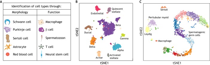
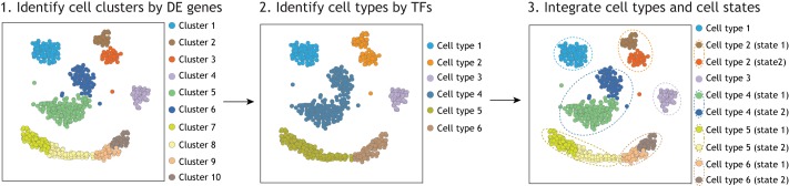
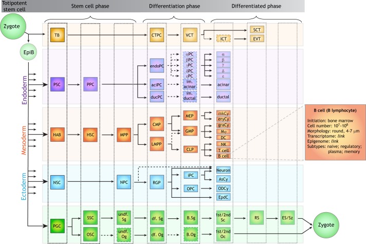
Single cell biology is currently revolutionizing developmental and evolutionary biology, revealing new cell types and states in an impressive range of biological systems. With the accumulation of data, however, the field is grappling with a central unanswered question: what exactly is a cell type? This question is further complicated by the inherently dynamic nature of developmental processes. In this Hypothesis article, we propose that a 'periodic table of cell types' can be used as a framework for distinguishing cell types from cell states, in which the periods and groups correspond to developmental trajectories and stages along differentiation, respectively. The different states of the same cell type are further analogous to 'isotopes'. We also highlight how the concept of a periodic table of cell types could be useful for predicting new cell types and states, and for recognizing relationships between cell types throughout development and evolution.

Keywords: Cell atlas; Cell states; Cell types; Periodic table; Single cell RNA-seq.


Conflict of interest statement

Competing interestsThe authors declare no competing or financial interests.

Figures

Fig. 1.
Fig. 1.
Historical and scRNA-seq approaches to identifying cell types. (A) Examples of historical identification of cell types through morphologies and cell functions. (B,C) tSNE plots of scRNA-seq data from adult human pancreatic cell populations (B) and adult human testicular cell populations (C). These data highlight the existence of discrete cell populations in the mature pancreas tissue (B) and a continuum of germ cell populations in the testicular tissue (C). The pancreas data is adapted from Baron et al. (2016) and the testis data is adapted from Xia et al. (2018 preprint).
Fig. 2.
Fig. 2.
A framework for distinguishing cell types from cell states using scRNA-seq. Individual cell transcriptomes can be clustered using typical methods, for example based on DE genes (left), as performed by most scRNA-seq projects. At this point, the cell clusters may not be equivalent to cell types. However, a second step of cell clustering, using only informative transcription factors (TFs), can be used as an additional criterion to distinguish cell types (middle). Thus, by integrating different cell clustering tools (right), we can distinguish cell types from cell states in silico, which can be further tested by molecular and cell biology validation.
Fig. 3.
Fig. 3.
A draft periodic table of cell types. Using the mouse as an example, a periodic table of cell types can be assembled based on current knowledge. The table is formatted from left to right based on development and/or differentiation trajectories, in which each element represents a cell type in the developmental system. Each column represents a similar developmental stage of cell types across different developing trajectories. As a proof-of-principle, each embryonic layer is arranged as a single period of cell types. The dashed cell type squares and arrows indicate unclear cell identities. Each cell type can be further annotated to provide information relating to its morphology, abundance, transcriptome, epigenome and states. Note that this draft periodic table of cell types is just a summary of a limited set of cell types and is based on our current understanding of developmental programs, despite some ongoing debates. αPC, alpha progenitor cell; βPC, beta progenitor cell; γPC, gamma progenitor cell; δPC, delta progenitor cell; εPC, epsilon progenitor cell; aciPC, acinar progenitor cells; AsCy, astrocyte; B.Og, B oogonia; B.Sg, B spermatogonia; CLP, common lymphoid progenitors; CMP, common myeloid progenitors; CTPC, cytotrophoblast progenitor cell; DC, dendritic cell; d.f.Og, differentiated oogonia; d.f.Sg, differentiated spermatogonia; ducPC, ductal progenitor cells; endoPC, endocrine progenitor cells; EpdC, ependymal cell; EpiB, epiblast; eryCy, erythrocyte; ES, elongating spermatids; EVT, extravillous cytotrophoblasts. GMP, granulocyte/macrophage progenitors; graCy, granulocytes; HAB, hemangioblast; HSC, hematopoietic stem cell; ICT, interstitial cytotrophoblast; im.acinar, immature acinar cell; im.ductal, immature ductal cell; iPC, intermediate progenitor cells; LMPP, lymphoid-primed multipotent progenitors; MEP, megakaryocyte/erythrocyte progenitors; mkCy, megakaryocyte; Mφ, macrophage; MPP, multipotent progenitors; NK, natural killer cell; NPC, neural progenitor cells; NSC, neural stem cells; ODCy, oligodendrocytes; OPC, oligodendrocyte progenitor cells; OSC, oogonial stem cell; PGC, primordial germ cell; PPC, pancreatic progenitor cells; PSC, pancreatic stem cells; RGP, radial glial progenitor cells; RS, round spermatids; SCT, syncytiotrophoblast; SSC, spermtogonial stem cell; Sz, spermatozoon; TB, trophoblast; undiff.Og, undifferentiated oogonia; undiff.Sg, undifferentiated spermatogonia; VCT, villous cytotrophoblast; 1st/2nd Sc, primary/secondary spermatocytes; 1st/2nd Oc, primary/secondary oocytes.

References

    1. Alberts B., Johnson A., Lewis J., Morgan D., Raff M., Roberts K. and Walter P. (2014). The innate and adaptive immune systems. In Molecular Biology of the Cell, 6th edn, pp. 1297-1342. Garland Science.
    1. Arendt D. (2008). The evolution of cell types in animals: emerging principles from molecular studies. Nat. Rev. Genet. 9, 868-882. 10.1038/nrg2416 - DOI - PubMed
    1. Arendt D., Musser J. M., Baker C. V. H., Bergman A., Cepko C., Erwin D. H., Pavlicev M., Schlosser G., Widder S., Laubichler M. D. et al. (2016). The origin and evolution of cell types. Nat. Rev. Genet. 17, 744-757. 10.1038/nrg.2016.127 - DOI - PubMed
    1. Artegiani B., Lyubimova A., Muraro M., van Es J. H., van Oudenaarden A. and Clevers H. (2017). A single-cell RNA sequencing study reveals cellular and molecular dynamics of the hippocampal neurogenic niche. Cell Rep. 21, 3271-3284. 10.1016/j.celrep.2017.11.050 - DOI - PubMed
    1. Ashburner M., Ball C. A., Blake J. A., Botstein D., Butler H., Cherry J. M., Davis A. P., Dolinski K., Dwight S. S., Eppig J. T. et al. (2000). Gene ontology: tool for the unification of biology. The Gene Ontology Consortium. Nat. Genet. 25, 25-29. 10.1038/75556 - DOI - PMC - PubMed

Publication types