. 2019 Jun 28;364(6447):1283-1287.
doi: 10.1126/science.aaw8981. Epub 2019 Jun 27.

Mechanism of β2AR regulation by an intracellular positive allosteric modulator

Affiliations

Mechanism of β2AR regulation by an intracellular positive allosteric modulator

Xiangyu Liu et al. Science. .

Abstract

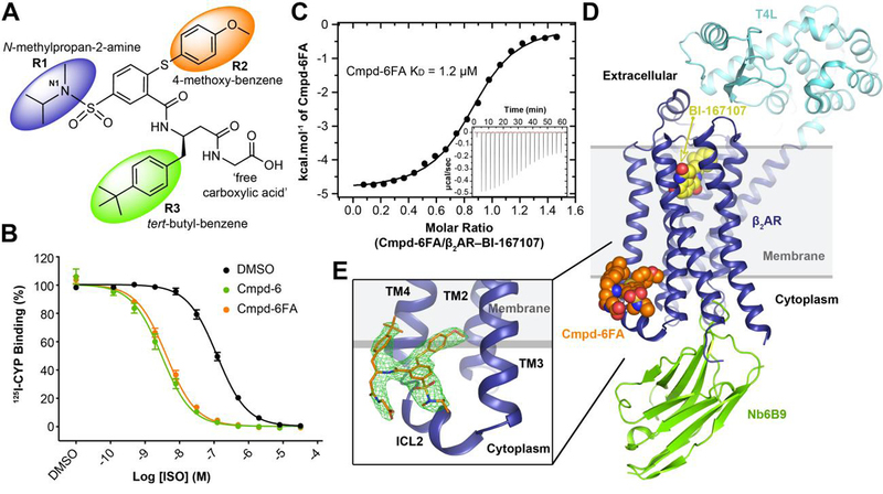
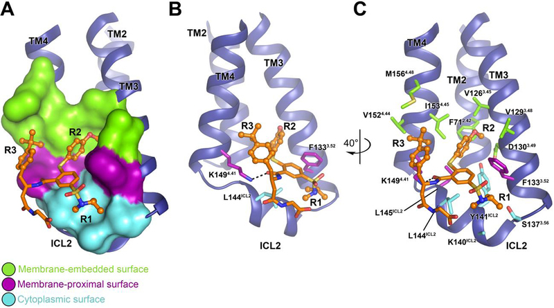
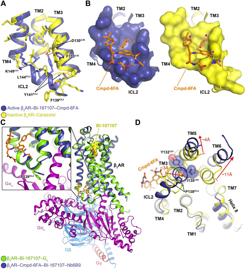
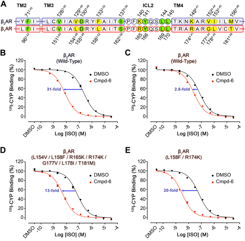
Drugs targeting the orthosteric, primary binding site of G protein-coupled receptors are the most common therapeutics. Allosteric binding sites, elsewhere on the receptors, are less well-defined, and so less exploited clinically. We report the crystal structure of the prototypic β2-adrenergic receptor in complex with an orthosteric agonist and compound-6FA, a positive allosteric modulator of this receptor. It binds on the receptor's inner surface in a pocket created by intracellular loop 2 and transmembrane segments 3 and 4, stabilizing the loop in an α-helical conformation required to engage the G protein. Structural comparison explains the selectivity of the compound for β2- over the β1-adrenergic receptor. Diversity in location, mechanism, and selectivity of allosteric ligands provides potential to expand the range of receptor drugs.


Conflict of interest statement

Competing interests: Authors declare no competing interests.

Figures

Fig. 1.
Fig. 1.. Structure of the active-state T4L-β2AR in complex with the orthosteric agonist BI-167107, nanobody 6B9, and Compound-6FA.
A, The chemical structure of Compound-6FA (Cmpd-6FA). B, Isoproterenol (ISO) competition binding with 125I-cyanopindolol (CYP) to the β2AR reconstituted in nanodiscs in the presence of vehicle (0.32 % dimethylsulfoxide; DMSO), Cmpd-6, or Cmpd-6FA at 32 μM. Values were normalized to percentages of the maximal 125I-CYP binding level obtained from a one-site competition binding-Log IC50 curve fit. Binding curves were generated by GraphPad Prism. Points on curves represent mean ± SEM obtained from five independent experiments performed in duplicate. C, Analysis of Cmpd-6FA interaction with the BI-167107-bound β2AR by isothermal titration calorimetry (ITC). Representative thermogram (insert) and binding isotherm, of three independent experiments, with the best titration curve fit are shown. Summary of thermodynamic parameters obtained by ITC: binding affinity (Kd = 1.2 ± 0.1 µM), stoichiometry (N = 0.9 ± 0.1 sites), enthalpy (ΔH = 5.0 ± 1.2 kcal/mol), and entropy (ΔS =13 ± 2.0 cal/mol/deg). D, Side-view of T4L-β2AR bound to the orthosteric agonist BI-167107, nanobody 6B9 (Nb6B9), and Cmpd-6FA. The gray box indicates the membrane layer as defined by the OPM database. e, Close-up view of Cmpd-6FA binding site. Covering Cmpd-6FA is 2FoFc electron density contoured at 1.0 σ (green mesh).
Fig. 2.
Fig. 2.. Intermolecular interaction between the β2AR and Cmpd-6FA.
A, Overview of the Cmpd-6FA binding site. The molecular surface of the β2AR bound to Cmpd-6FA is subdivided into three surface regions: membrane embedded (green), membrane-proximal (purple), and cytoplasmic (cyan). B, C, Detailed interactions of the β2AR bound to Cmpd-6FA. B, The side-chains of those residues that interact with the core scaffold of Cmpd-6FA are shown. The hydrogen bond is indicated by a black dashed line. C, Rotated view showing all β2AR residues that contribute at the β2AR–Cmpd-6FA interface.
Fig. 3.
Fig. 3.. Mechanism of allosteric activation of the β2AR by Cmpd-6FA.
A, Superposition of the inactive β2AR bound to the antagonist carazolol (PDB code: 2RH1) and the active β2AR bound to the agonist BI-167107, Cmpd-6FA, and Nb6B9. Close-up view of the Cmpd-6FA binding site is shown. The residues of the inactive (yellow) and active (blue) β2AR are depicted and the hydrogen bond formed between Asp1303.49 and Tyr141ICL2 in the active-state is indicated by a black dashed line. B, Topography of Cmpd-6FA binding surface on the active β2AR (left, blue) and the corresponding surface of the inactive β2AR (right, yellow) with Cmpd-6FA (orange sticks) docked on top. Molecular surfaces are of only those residues involved in interaction with Cmpd-6FA. Steric clash between Cmpd-6FA and the surface of inactive β2AR is represented by a purple asterisk. C, Overlay of the β2AR bound to BI-167107, Nb6B9, and Cmpd-6FA with the β2AR–Gs complex (PDB code: 3SN6). The inset shows the position of Phe139ICL2 relative to the α-subunit of Gs. D, Superposition of the active β2AR bound to the agonist BI-167107, Nb6B9, and Cmpd-6FA (blue) with the inactive β2AR bound to carazolol (yellow) (PDB code: 2RH1) as viewed from the cytoplasm. For clarity, Nb6B9 and the orthosteric ligands are omitted. The arrows indicate shifts in the intracellular ends of the TMs 3, 5, and 6 upon activation and their relative distances.
Fig. 4.
Fig. 4.. Gain-of-function of Cmpd-6 on the human β1AR mutants as compared to wild-type.
A, Partial protein sequence alignment of the human β2AR (blue highlight) and β1AR (red highlight) that includes only the relevant locations that engage Cmpd-6FA. Those residues that interact with Cmpd-6FA and are conserved are highlighted in green. Residues that are not conserved, but also interact with Cmpd-6FA are highlighted in yellow. Residue numbers for the β2AR and β1AR are shown above and below the alignment, respectively. B-E, Cmpd-6 positive allosteric effect on agonist binding of B, wild-type β2AR C, wild-type β1AR D, L154V / L158F / R165K / R174K / G177V / L178I / T181M β1AR mutant and E, L158F / R174K β1AR mutant. 125I-CYP vs. ISO competition binding was done using membrane preparations from HEK293 cells expressing the indicated receptor in the absence or presence of Cmpd-6 at 25 μM. Values were normalized to percentages of the maximal 125I-CYP binding level obtained from a one-site competition binding-Log IC50 curve fit (GraphPad Prism) of the vehicle (0.25% dimethylsulfoxide; DMSO) control data. Points on curves represent mean ± SEM obtained from four independent experiments performed in duplicate.

References

    1. Pierce KL, Premont RT, Lefkowitz RJ, Seven-transmembrane receptors. Nat Rev Mol Cell Biol 3, 639–650 (2002). - PubMed
    1. Hauser AS, Attwood MM, Rask-Andersen M, Schioth HB, Gloriam DE, Trends in GPCR drug discovery: new agents, targets and indications. Nat Rev Drug Discov 16, 829–842 (2017). - PMC - PubMed
    1. Thal DM, Glukhova A, Sexton PM, Christopoulos A, Structural insights into G-protein-coupled receptor allostery. Nature 559, 45–53 (2018). - PubMed
    1. Wacker D, Stevens RC, Roth BL, How Ligands Illuminate GPCR Molecular Pharmacology. Cell 170, 414–427 (2017). - PMC - PubMed
    1. Staus DP et al., Allosteric nanobodies reveal the dynamic range and diverse mechanisms of G-protein-coupled receptor activation. Nature 535, 448–452 (2016). - PMC - PubMed

Publication types

MeSH terms

Substances