. 2019 Aug 2;365(6452):eaax1033.
doi: 10.1126/science.aax1033. Epub 2019 Jun 27.

Substrate processing by the Cdc48 ATPase complex is initiated by ubiquitin unfolding

Affiliations

Substrate processing by the Cdc48 ATPase complex is initiated by ubiquitin unfolding

Edward C Twomey et al. Science. .

Abstract

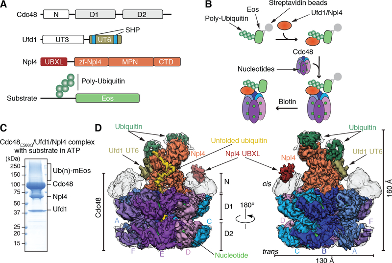
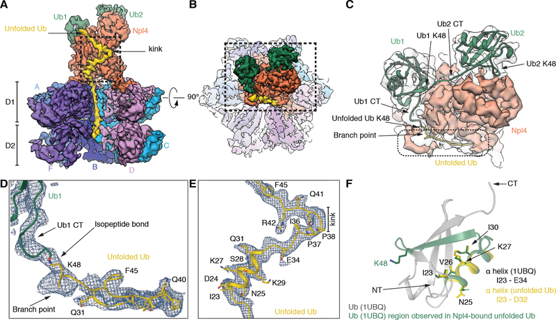
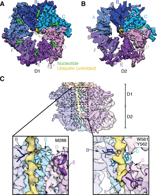
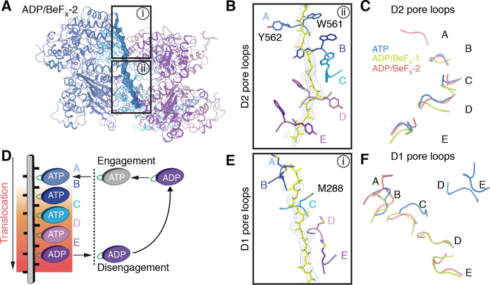
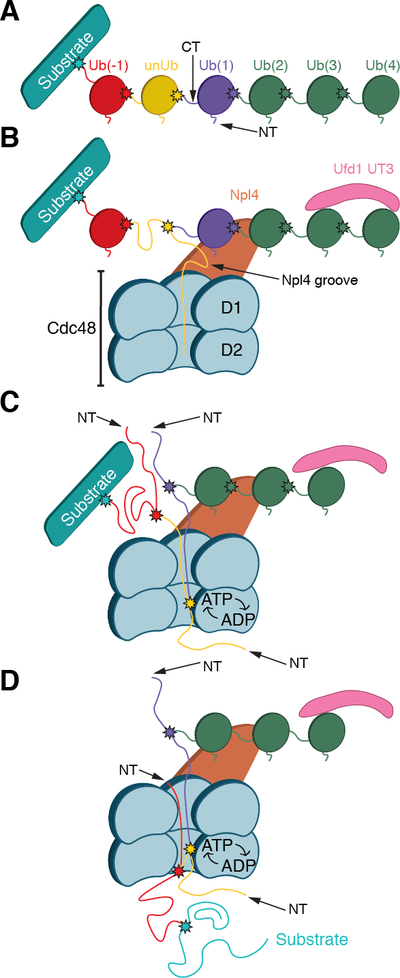
The Cdc48 adenosine triphosphatase (ATPase) (p97 or valosin-containing protein in mammals) and its cofactor Ufd1/Npl4 extract polyubiquitinated proteins from membranes or macromolecular complexes for subsequent degradation by the proteasome. How Cdc48 processes its diverse and often well-folded substrates is unclear. Here, we report cryo-electron microscopy structures of the Cdc48 ATPase in complex with Ufd1/Npl4 and polyubiquitinated substrate. The structures show that the Cdc48 complex initiates substrate processing by unfolding a ubiquitin molecule. The unfolded ubiquitin molecule binds to Npl4 and projects its N-terminal segment through both hexameric ATPase rings. Pore loops of the second ring form a staircase that acts as a conveyer belt to move the polypeptide through the central pore. Inducing the unfolding of ubiquitin allows the Cdc48 ATPase complex to process a broad range of substrates.


Conflict of interest statement

Competing interests: The authors declare no competing financial interests.

Figures

Fig. 1.
Fig. 1.. Generation and cryo-EM analysis of a substrate-engaged Cdc48 ATPase complex in ATP.
(A) Domain structures of Cdc48, Ufd1, and Npl4. (B) Scheme showing the step-wise assembly of the ATPase complex. The poly-ubiquitinated Eos substrate was generated as in fig. S1A, bound via a SBP tag to streptavidin beads, and incubated sequentially with Ufd1/Npl4 cofactor and Cdc48. After incubation with different nucleotides, the complex was eluted with biotin. (C) Complex generated with ATP and Cdc48 carrying a Walker B mutation in D2 (E588Q). The eluted complex was analyzed by SDS-PAGE and Coomassie blue staining. (Ub)n, poly-ubiquitin. (D) Cryo-EM reconstruction of the complex in two different views. The domains of Cdc48, cofactor, and ubiquitin molecules attached to substrate are shown in different colors. The subunits in the hexameric ATPase ring are labeled A-F. The refined, unsharpened map is shown in transparent grey over the final composite map sharpened with a B-factor of −150. The densities for the N domains, Npl4’s UBXL domain, and Ufd1’s UT6 domain are from the unsharpened map.
Fig. 2.
Fig. 2.. Ubiquitin molecules bound to the Npl4 cofactor of Cdc48.
(A) Side view of the density map of the Cdc48 complex, with subunit E removed to show the interior of the central pore. Two folded ubiquitin molecules (Ub1 and Ub2) are shown in green and an unfolded ubiquitin molecule (unUb) in yellow. (B) Top view of the map, with the Npl4 tower in the foreground. (C) Magnified view of the boxed area in (B). Density for the folded ubiquitins is shown in transparent grey. Models for folded ubiquitin models (PDB 1UBQ), fit as rigid bodies into the map, are in green ribbon presentation, and unfolded ubiquitin in yellow ribbon presentation. The C-terminal flexible tails of folded ubiquitins were fit into the map. The position of Lys48 in each ubiquitin is indicated. (D) Magnified view of the density enclosed by a broken line in (C), together with a model of unfolded ubiquitin. Prominent features used for modeling are indicated. (E) Magnified view of the kink in unfolded ubiquitin indicated in the boxed area in (A), together with a model. (F) Superposition of the helix I23-D32 in unfolded ubiquitin (yellow) with the corresponding helix in folded ubiquitin, both in ribbon presentation. The segment bound to the Npl4 groove is shown in green. Some residues in the helix face Npl4 in the complex (Ile23, Val26, Ile30), while others face away (Lys27, Asn25).
Fig. 3.
Fig. 3.. Binding of unfolded ubiquitin to a conserved Npl4 groove.
(A) Surface model of the Npl4 tower with residues colored according to the degree of their conservation, as calculated by the ConSurf Server. The conserved groove to which unfolded ubiquitin binds is enclosed by a broken line. The positions of the b-strand finger and Zn2+-finger 1 (ZF-1) are indicated. (B) The highlighted conserved residues in the Npl4 groove, colored according to their position, were mutated to test their effect on substrate unfolding by the Cdc48 ATPase complex. (C) UN cofactor containing wild-type Ufd1 and either wild-type (WT) or the indicated Npl4 mutant was purified (fig. S4) and tested together with Cdc48 for unfolding of poly-ubiquitinated, photo-converted Eos, measured as loss of fluorescence. The curves show the mean and standard deviation of three replicates. (D) Experiments as in (C) were done with selected Npl4 mutants and controls. The initial unfolding rates were determined and normalized to that of WT. Shown are the mean and standard deviation of three replicates. (E) Regions of Npl4 protected by substrate against HDX in the Cdc48/UN complex (in blue). HDX MS was performed with Cdc48/UN/ADP/BeFx in the absence or presence of poly-ubiquitinated substrate at different time points (fig. S5). (F) As in (E), but in the absence of Cdc48.
Fig. 4.
Fig. 4.. Insertion of the N-terminus of ubiquitin into the D2 ring of Cdc48.
(A) Side view of the model of the Cdc48 complex with the unfolded ubiquitin molecule in yellow. The boxed region is magnified to show the N-terminus unfolded (Ub NT; in yellow) in proximity of Asp602 residues (sticks) of several D2 subunits (in ribbon presentation). (B) Cdc48-FLAG containing Bpa at position 602 was incubated with UN and dye-labeled, poly-ubiquitinated sfGFP (Dy-Ub(n)-sfGFP) in the presence of ADP or ATP. The sample was irradiated, as indicated, Cdc48-FLAG and crosslinked products were isolated with FLAG-antibody beads, and analyzed by SDS-PAGE and fluorescence scanning. Where indicated, the samples were treated with the DUB Usp21 before analysis. The stars indicate crosslinks between Dy-Ub(n)-sfGFP and one or more Cdc48 molecules. The intensity of the crosslinks was quantitated and normalized to that of the sample in ATP (numbers underneath the lanes). The lower panel shows an immunoblot using Cdc48 antibodies. (C) As in (B), but in the presence of ADP, and analysis of the crosslinked products by nano-liquid chromatography/mass spectrometry after trypsin digestion. The indicated peptides of ubiquitin and Cdc48 were crosslinked through Bpa (B). Colored glyphs highlight fragment ions detected in the MS/MS spectrum, and indicate that the Bpa was crosslinked to either Met1 or Gln2 of ubiquitin (fig. S8).
Fig. 5.
Fig. 5.. The N-terminal segment of unfolded ubiquitin in the central pore of the ATPase rings.
(A) Top view of the density map of the substrate-engaged Cdc48 complex in ATP, cut to the surface of the D1 ring. Substrate density is shown in yellow. The D1 ATPases are colored individually and labeled A-F. Density for bound nucleotides is shown in green. (B) As in (A), but cut to the surface of the D2 ring. (C) The upper panel shows a cut-away side view of the map, with unUb in yellow. The boxed areas in D1 and D2 are magnified in the lower panels. Amino acids of the pore loops contacting the polypeptide chain are indicated.
Fig. 6.
Fig. 6.. Structures of substrate-engaged Cdc48 complex in ADP/BeFx reveal translocation mechanism.
(A) Structures of the substrate-engaged Cdc48 complex were determined in the presence of ADP and BeFx as in Fig. 1. Shown is a side view of a ribbon-diagram model built into the higher resolution map (ADP/BeFx-2), together with density for the substrate shown in blue mesh. (B) Magnified, 90°-rotated view of the boxed region ii in (A), showing the D2 pore loop residues that contact the polypeptide. The pore loops are labeled by the ATPase subunits to which they belong. (C) Comparison of the positions of the substrate-engaged D2 pore loops in the three structures, aligned on the basis of subunit B. Note that subunit A is only engaged in ADP/BeFx-2. (D) Model for polypeptide translocation. Subunit A binds ATP and moves to the top of the staircase. The previously engaged subunits move downwards, dragging the polypeptide with them by interacting with every other peptide bond of the substrate. Subunit E hydrolyzes ATP before its disengagement. The disengaged subunit, F, then moves back to the top and starts a new cycle. (E) Magnified view of the boxed region i in (A), with Met288 in the D1 pore loops highlighted. (F) Comparison of the position of the D1 pore loops in the three structures. Note that the pore loops are almost planar in ATP (blue) and arranged as a staircase in ADP/BeFx (green and red).
Fig. 7.
Fig. 7.. Model for substrate processing by the Cdc48 ATPase complex.
(A) Scheme of a substrate with an attached Lys48-linked poly-ubiquitin chain. In the example chosen, the ubiquitin molecule shown in yellow will be unfolded; it is separated from the substrate by one ubiquitin molecule (Ub(−1)). Ubiquitins distal to the one being unfolded are numbered (Ub(1)…Ub(4)). Lys48 branch points are indicated by stars. (B) Binding of the substrate to the Cdc48 complex. The distal folded ubiquitins Ub(1) and Ub(2) bind to the top of Npl4, and Ub(3) and possibly Ub(4) bind to the UT3 domain of Ufd1. The unfolded ubiquitin (unUb) binds to the groove in Npl4 and projects its N-terminus across both ATPase rings. (C) ATP hydrolysis in the D2 ring pulls the N-terminus of the unfolded ubiquitin through the central pore, moving the branch point into the ATPase rings and causing unfolding of attached Ub(1). Pulling on the proximal side of the ubiquitin chain results in unfolding of Ub(−1). (D) Ultimately the substrate is moved through the central pore and unfolded. Substrate release from the ATPase complex requires the removal of the distal ubiquitins Ub(2)-Ub(4) by a DUB.

Similar articles

Cited by

References

    1. Xia D, Tang WK, Ye Y, Structure and function of the AAA+ ATPase p97/Cdc48p. Gene. 583, 64–77 (2016). - PMC - PubMed
    1. van den Boom J, Meyer H, VCP/p97-Mediated Unfolding as a Principle in Protein Homeostasis and Signaling. Mol. Cell 69, 182–194 (2018). - PubMed
    1. Bodnar N, Rapoport T, Toward an understanding of the Cdc48/p97 ATPase. F1000Research. 6, 1318 (2017). - PMC - PubMed
    1. Watts GDJ et al., Inclusion body myopathy associated with Paget disease of bone and frontotemporal dementia is caused by mutant valosin-containing protein. Nat. Genet 36, 377–381 (2004). - PubMed
    1. Kimonis VE, Fulchiero E, Vesa J, Watts G, VCP disease associated with myopathy, Paget disease of bone and frontotemporal dementia: Review of a unique disorder. Biochim. Biophys. Acta - Mol. Basis Dis 1782, 744–748 (2008). - PubMed

Publication types

MeSH terms

Substances