

Interface-mediated Kirkendall effect and nanoscale void migration in bimetallic nanoparticles during interdiffusion
- PMID: 31249286
- PMCID: PMC6597554
- DOI: 10.1038/s41467-019-10623-0
Interface-mediated Kirkendall effect and nanoscale void migration in bimetallic nanoparticles during interdiffusion
Abstract
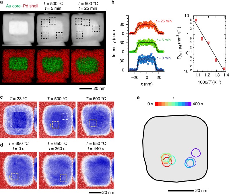
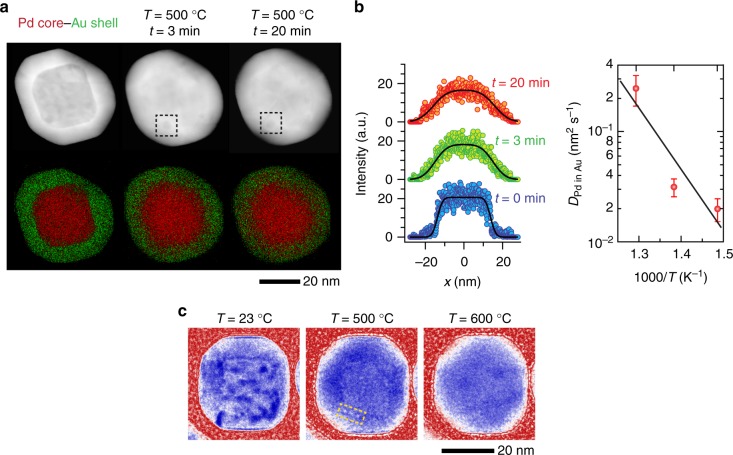
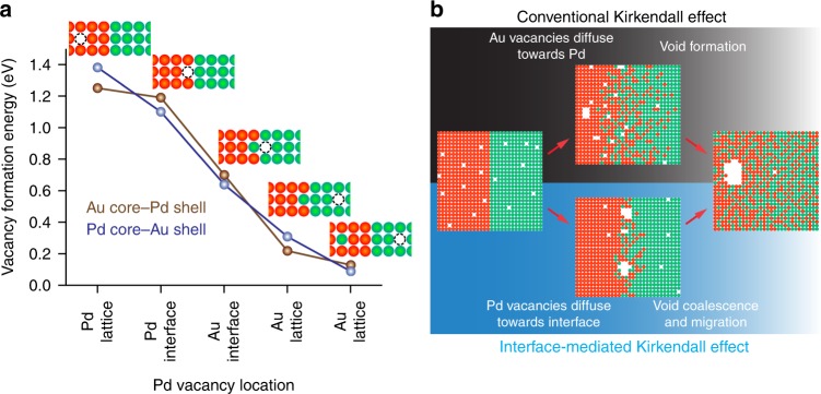
At elevated temperatures, bimetallic nanomaterials change their morphologies because of the interdiffusion of atomic species, which also alters their properties. The Kirkendall effect (KE) is a well-known phenomenon associated with such interdiffusion. Here, we show how KE can manifest in bimetallic nanoparticles (NPs) by following core-shell NPs of Au and Pd during heat treatment with in situ transmission electron microscopy. Unlike monometallic NPs, these core-shell NPs did not evolve into hollow core NPs. Instead, nanoscale voids formed at the bimetallic interface and then, migrated to the NP surface. Our results show that: (1) the direction of vacancy flow during interdiffusion reverses due to the higher vacancy formation energy of Pd compared to Au, and (2) nanoscale voids migrate during heating, contrary to conventional assumptions of immobile voids and void shrinkage through vacancy emission. Our results illustrate how void behavior in bimetallic NPs can differ from an idealized picture based on atomic fluxes and have important implications for the design of these materials for high-temperature applications.
Conflict of interest statement
The authors declare no competing interests.
Figures

Similar articles
-
Size-dependent nanoscale kirkendall effect during the oxidation of nickel nanoparticles.ACS Nano. 2010 Apr 27;4(4):1913-20. doi: 10.1021/nn901736y. ACS Nano. 2010. PMID: 20361781
-
Synthesis and exceptional thermal stability of Mg-based bimetallic nanoparticles during hydrogenation.Nanoscale. 2014 Oct 21;6(20):11963-70. doi: 10.1039/c4nr03885a. Nanoscale. 2014. PMID: 25178019
-
Atomic structure of Au-Pd bimetallic alloyed nanoparticles.J Am Chem Soc. 2010 Sep 8;132(35):12480-6. doi: 10.1021/ja105614q. J Am Chem Soc. 2010. PMID: 20712315
-
Melting Behavior of Bimetallic and Trimetallic Nanoparticles: A Review of MD Simulation Studies.Top Curr Chem (Cham). 2021 Apr 22;379(3):22. doi: 10.1007/s41061-021-00332-y. Top Curr Chem (Cham). 2021. PMID: 33890199 Review.
-
Formation of nanotubes and hollow nanoparticles based on Kirkendall and diffusion processes: a review.Small. 2007 Oct;3(10):1660-71. doi: 10.1002/smll.200700382. Small. 2007. PMID: 17890644 Review.
Cited by
-
From Multi- to Single-Hollow Trimetallic Nanocrystals by Ultrafast Heating.Chem Mater. 2023 Nov 6;35(22):9603-9612. doi: 10.1021/acs.chemmater.3c01698. eCollection 2023 Nov 28. Chem Mater. 2023. PMID: 38047181 Free PMC article.
-
A multiple Kirkendall strategy for converting nanosized zero-valent iron to highly active Fenton-like catalyst for organics degradation.Proc Natl Acad Sci U S A. 2023 Sep 26;120(39):e2304552120. doi: 10.1073/pnas.2304552120. Epub 2023 Sep 19. Proc Natl Acad Sci U S A. 2023. PMID: 37725641 Free PMC article.
-
Unraveling Interdiffusion Phenomena and the Role of Nanoscale Diffusion Barriers in the Copper-Gold System.ACS Nano. 2024 Oct 29;18(43):29658-29666. doi: 10.1021/acsnano.4c08502. Epub 2024 Oct 16. ACS Nano. 2024. PMID: 39414568 Free PMC article.
-
Facile aqueous synthesis of hollow dual plasmonic hetero-nanostructures with tunable optical responses through nanoscale Kirkendall effects.Nanoscale Adv. 2022 Nov 10;5(1):88-95. doi: 10.1039/d2na00606e. eCollection 2022 Dec 20. Nanoscale Adv. 2022. PMID: 36605812 Free PMC article.
-
Time-resolved transmission electron microscopy for nanoscale chemical dynamics.Nat Rev Chem. 2023 Apr;7(4):256-272. doi: 10.1038/s41570-023-00469-y. Epub 2023 Feb 22. Nat Rev Chem. 2023. PMID: 37117417 Review.
References
-
- Erdélyi Z, Beke DL. Nanoscale volume diffusion: diffusion in thin films, multilayers and nanoobjects (hollow nanoparticles) J. Mater. Sci. 2011;46:6465–6483. doi: 10.1007/s10853-011-5720-4. - DOI
-
- Smigelskas AD, Kirkendall EO. Zinc diffusion in alpha brass. Trans. AIME. 1947;171:130–142.
Grants and funding
LinkOut - more resources
Full Text Sources
Miscellaneous
