Biosynthesis and Extracellular Concentrations of N,N-dimethyltryptamine (DMT) in Mammalian Brain
- PMID: 31249368
- PMCID: PMC6597727
- DOI: 10.1038/s41598-019-45812-w
Biosynthesis and Extracellular Concentrations of N,N-dimethyltryptamine (DMT) in Mammalian Brain
Abstract
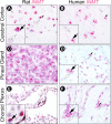
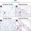
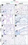
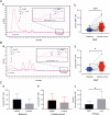
N,N-dimethyltryptamine (DMT), a psychedelic compound identified endogenously in mammals, is biosynthesized by aromatic-L-amino acid decarboxylase (AADC) and indolethylamine-N-methyltransferase (INMT). Whether DMT is biosynthesized in the mammalian brain is unknown. We investigated brain expression of INMT transcript in rats and humans, co-expression of INMT and AADC mRNA in rat brain and periphery, and brain concentrations of DMT in rats. INMT transcripts were identified in the cerebral cortex, pineal gland, and choroid plexus of both rats and humans via in situ hybridization. Notably, INMT mRNA was colocalized with AADC transcript in rat brain tissues, in contrast to rat peripheral tissues where there existed little overlapping expression of INMT with AADC transcripts. Additionally, extracellular concentrations of DMT in the cerebral cortex of normal behaving rats, with or without the pineal gland, were similar to those of canonical monoamine neurotransmitters including serotonin. A significant increase of DMT levels in the rat visual cortex was observed following induction of experimental cardiac arrest, a finding independent of an intact pineal gland. These results show for the first time that the rat brain is capable of synthesizing and releasing DMT at concentrations comparable to known monoamine neurotransmitters and raise the possibility that this phenomenon may occur similarly in human brains.
Conflict of interest statement
The authors declare no competing interests.
Figures


Similar articles
-
Indolethylamine N-methyltransferase (INMT) is not essential for endogenous tryptamine-dependent methylation activity in rats.Sci Rep. 2023 Jan 6;13(1):280. doi: 10.1038/s41598-023-27538-y. Sci Rep. 2023. PMID: 36609666 Free PMC article.
-
Indolethylamine-N-methyltransferase Polymorphisms: Genetic and Biochemical Approaches for Study of Endogenous N,N,-dimethyltryptamine.Front Neurosci. 2018 Apr 23;12:232. doi: 10.3389/fnins.2018.00232. eCollection 2018. Front Neurosci. 2018. PMID: 29740267 Free PMC article. Review.
-
Noncompetitive inhibition of indolethylamine-N-methyltransferase by N,N-dimethyltryptamine and N,N-dimethylaminopropyltryptamine.Biochemistry. 2014 May 13;53(18):2956-65. doi: 10.1021/bi500175p. Epub 2014 Apr 28. Biochemistry. 2014. PMID: 24730580 Free PMC article.
-
Exploring DMT: Endogenous role and therapeutic potential.Neuropharmacology. 2025 May 1;268:110314. doi: 10.1016/j.neuropharm.2025.110314. Epub 2025 Jan 18. Neuropharmacology. 2025. PMID: 39832530 Review.
-
Significance of mammalian N, N-dimethyltryptamine (DMT): A 60-year-old debate.J Psychopharmacol. 2022 Aug;36(8):905-919. doi: 10.1177/02698811221104054. Epub 2022 Jun 13. J Psychopharmacol. 2022. PMID: 35695604 Review.
Cited by
-
Why N,N-dimethyltryptamine matters: unique features and therapeutic potential beyond classical psychedelics.Front Psychiatry. 2024 Nov 6;15:1485337. doi: 10.3389/fpsyt.2024.1485337. eCollection 2024. Front Psychiatry. 2024. PMID: 39568756 Free PMC article.
-
Psychedelics for Brain Injury: A Mini-Review.Front Neurol. 2021 Jul 29;12:685085. doi: 10.3389/fneur.2021.685085. eCollection 2021. Front Neurol. 2021. PMID: 34393973 Free PMC article. Review.
-
DMT alters cortical travelling waves.Elife. 2020 Oct 12;9:e59784. doi: 10.7554/eLife.59784. Elife. 2020. PMID: 33043883 Free PMC article.
-
Three Naturally-Occurring Psychedelics and Their Significance in the Treatment of Mental Health Disorders.Front Pharmacol. 2022 Jun 28;13:927984. doi: 10.3389/fphar.2022.927984. eCollection 2022. Front Pharmacol. 2022. PMID: 35837277 Free PMC article. Review.
-
An experimental platform for stochastic analyses of single serotonergic fibers in the mouse brain.Front Neurosci. 2023 Oct 6;17:1241919. doi: 10.3389/fnins.2023.1241919. eCollection 2023. Front Neurosci. 2023. PMID: 37869509 Free PMC article.
References
-
- The Hallucinogens by Hoffer, A. & Osmondm, H.: Academic Press Hardcover, 1st Edition - bookseller e.g. Wolfgang Risch. Available at, https://www.abebooks.com/first-edition/Hallucinogens-A-Hoffer-H-Osmond-A....
Publication types
MeSH terms
Substances
LinkOut - more resources
Full Text Sources
Other Literature Sources

