. 2019 Nov;99(11):1636-1649.
doi: 10.1038/s41374-019-0281-2. Epub 2019 Jun 27.

Thy-1 depletion and integrin β3 upregulation-mediated PI3K-Akt-mTOR pathway activation inhibits lung fibroblast autophagy in lipopolysaccharide-induced pulmonary fibrosis

Affiliations

Thy-1 depletion and integrin β3 upregulation-mediated PI3K-Akt-mTOR pathway activation inhibits lung fibroblast autophagy in lipopolysaccharide-induced pulmonary fibrosis

Hanxi Wan et al. Lab Invest. 2019 Nov.

Abstract

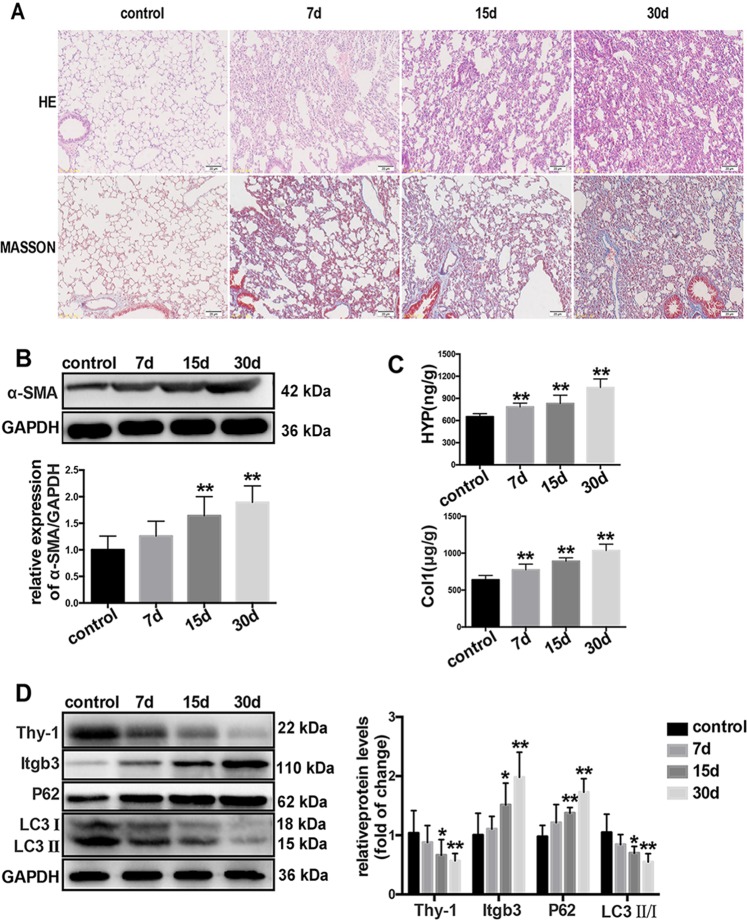
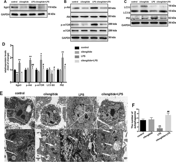
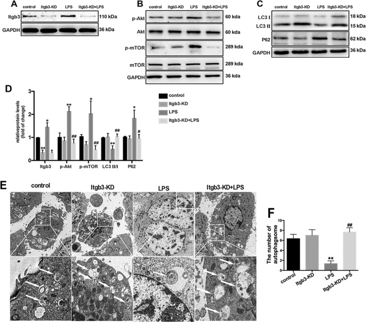
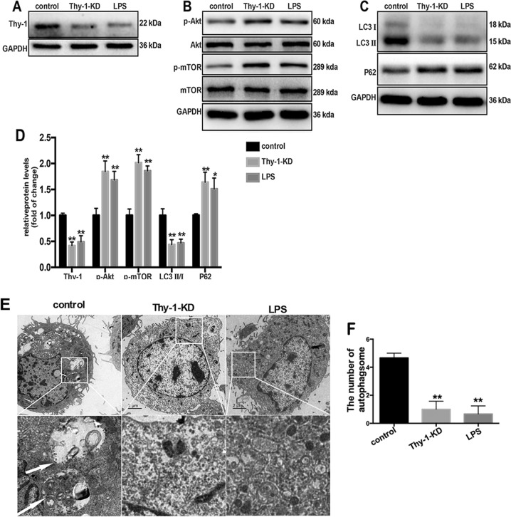
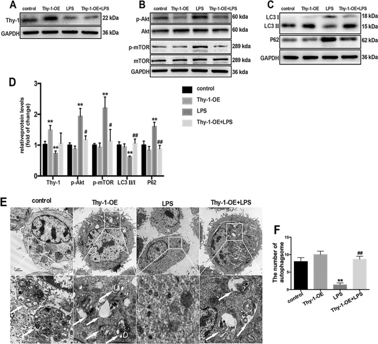
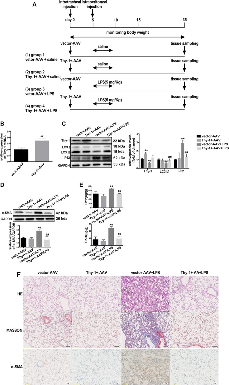
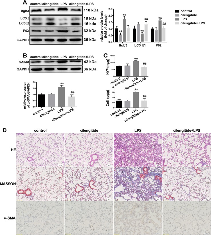
Lipopolysaccharide (LPS)-induced autophagy inhibition in lung fibroblasts is closely associated with the activation of the phosphatidylinositol 3-kinase/protein kinase B/mammalian target of rapamycin (PI3K-Akt-mTOR) pathway. However, the underlying mechanism remains unknown. In this study, we demonstrated that LPS activated the PI3K-Akt-mTOR pathway and inhibited lung fibroblast autophagy by depleting thymocyte differentiation antigen-1 (Thy-1) and upregulating integrin β3 (Itgb3). Challenge of the human lung fibroblast MRC-5 cell line with LPS resulted in significant upregulation of integrin β3, activation of the PI3K-Akt-mTOR pathway and inhibition of autophagy, which could be abolished by integrin β3 silencing by specific shRNA or treatment with the integrin β3 inhibitor cilengitide. Meanwhile, LPS could inhibit Thy-1 expression accompanied with PI3K-Akt-mTOR pathway activation and lung fibroblast autophagy inhibition; these effects could be prevented by Thy-1 overexpression. Meanwhile, Thy-1 downregulation with Thy-1 shRNA could mimic the effects of LPS, inducing the activation of PI3K-Akt-mTOR pathway and inhibiting lung fibroblast autophagy. Furthermore, protein immunoprecipitation analysis demonstrated that LPS reduced the binding of Thy-1 to integrin β3. Thy-1 downregulation, integrin β3 upregulation and autophagy inhibition were also detected in a mouse model of LPS-induced pulmonary fibrosis, which could be prohibited by intratracheal injection of Thy-1 overexpressing adeno-associated virus (AAV) or intraperitoneal injection of the integrin β3 inhibitor cilengitide. In conclusion, this study demonstrated that Thy-1 depletion and integrin β3 upregulation are involved in LPS-induced pulmonary fibrosis, and may serve as potential therapeutic targets for pulmonary fibrosis.


Conflict of interest statement

The authors declare that they have no conflict of interest.

Figures

Fig. 1
Fig. 1
LPS-induced autophagy inhibition is accompanied with Thy-1 downregulation and integrin β3 upregulation in LPS-induced pulmonary fibrosis. The severity of pulmonary fibrosis was determined by hematoxylin-eosin (H&E) staining; collagen deposition was revealed by MASSON staining (magnification, ×200) (a). The expression of α-SMA in lung tissues was also measured by Western blot (b). Collagen deposition was measured by hydroxyproline content and collagen level assessments (c). Representative Western blot images showing expression of Thy-1, integrin β3, LC3 and P62 in the lung tissue (d). Values are mean ± SD (n = 6). *p < 0.05 vs control group; **P < 0.01 vs control group
Fig. 2
Fig. 2
LPS-induced autophagy inhibition is accompanied with Thy-1 downregulation and integrin β3 upregulation and activation in lung fibroblasts. Western blot was performed to detect Thy-1 and integrin β3 in lung fibroblasts challenged with 1 μg/ml LPS for 6 h, and P62 and LC3 in lung fibroblasts challenged with 1 μg/ml LPS for 24 h (a). Immunofluorescent staining of the nucleus (blue), integrin β3 (green) and Thy-1 (red) in lung fibroblasts cultured in the absence or presence of 1 μg/ml LPS for 6 h (b). Lung fibroblasts were observed by transmission electron microscopy. White arrows indicate autophagosomes (c), the semi-quantification of autophagosomes of per cell were also shown (d). Co-IP was used to detect the interaction between Thy-1 and integrin β3 in MRC-5 cells after LPS challenge. Western blot for co-immunoprecipitates of IgG control, input, Thy-1 and integrin β3 (e). Flow cytometry was used to detect the expression of PAC-1, an integrin β3 activation marker (f).Values are mean ± SD from triplicate experiments. *p < 0.05, **p < 0.01
Fig. 3
Fig. 3
Integrin β3 inhibition precludes LPS-induced PI3K-Akt-mTOR activation and lung fibroblast autophagy blockade. Western blot was used to detect the expression of integrin β3 (a), phospho-AKT (p-AKT), total AKT, phospho-mTOR (p-mTOR) and total mTOR (b). Representative images showing protein expression of LC3 I, LC3 II and P62 in MRC-5 cells challenged without or with 1 μg/ml LPS in the absence or presence of the inhibitor cilengitide (2 mM) for 24 h (c). Quantitation of p-AKT/total AKT, p-mTOR/total mTOR, LC3 II/ I, integrin β3 and P62 protein levels normalized to GAPDH (d). Lung fibroblasts were assessed by transmission electron microscopy. White arrows indicate autophagosomes (e), the semi-quantification of autophagosomes of per cell were also shown (f). Values are mean ± SD from triplicate experiments. *p < 0.05 vs control group; **p < 0.01 vs control group; #p < 0.05 vs LPS group; ##p < 0.05 vs. LPS group
Fig. 4
Fig. 4
Genetic integrin β3 inhibition prevents LPS-induced PI3K-Akt-mTOR activation and lung fibroblast autophagy inhibition. Western blot was used to detect the expression levels of integrin β3 (a), phospho-AKT (p-AKT), total AKT, phospho-mTOR (p-mTOR) and total mTOR (b) in lung fibroblasts after 6 h of 1 μg/ml LPS challenge after transfection with integrin β3 shRNA or the empty vector. Representative images showing expression of LC3 I, LC3 II and P62 in lung fibroblasts after 24 h of 1 μg/ml LPS challenge after transfection with integrin β3 shRNA or the empty vector (c). Quantification of p-AKT/total AKT, p-mTOR/total mTOR, LC3 II/ I, integrin β3and P62 protein levels normalized to GAPDH (d). Lung fibroblasts were observed by transmission electron microscopy. White arrows indicate autophagosomes (e), the semi-quantification of autophagosomes of per cell were also shown (f). Values are mean ± SD from triplicate experiments. *p < 0.05 vs. control group; **p < 0.01 vs. control group; #p < 0.05 vs. LPS group; ##p < 0.05 vs. LPS group
Fig. 5
Fig. 5
Thy-1 downregulation induces PI3K-Akt-mTOR activation and lung fibroblast autophagy inhibition. Western blot was performed to detect the expression levels of Thy-1 (a), phospho-AKT (p-AKT), total AKT, phospho-mTOR (p-mTOR) and total mTOR (b) in lung fibroblasts after 6 h of 1 μg/ml LPS challenge after transfection with Thy-1 shRNA or the empty vector. Representative images showing expression of LC3 I, LC3 II and P62 in lung fibroblasts after 24 h of 1 μg/ml LPS challenge after transfection with Thy shRNA or the empty vector (c). Lung fibroblasts were observed by transmission electron microscopy. White arrows indicate autophagosomes (e), the semi-quantification of autophagosomes of per cell were also shown (f). Values are mean ± SD from triplicate experiments. *p < 0.05 vs. control group; **p < 0.01 vs. control group
Fig. 6
Fig. 6
Thy-1 overexpression precludes LPS-induced PI3K-Akt-mTOR activation and lung fibroblast autophagy inhibition. Western blot was performed to detect the expression levels of Thy-1(a), phospho-AKT (p-AKT), total AKT, phospho-mTOR (p-mTOR) and total mTOR (b) in lung fibroblasts after 6 h of 1 μg/ml LPS challenge after transfection with Thy-1-OE or the empty vector. Representative images showing expression of LC3 I, LC3 II and P62 in lung fibroblasts after 24 h of 1 μg/ml LPS challenge after transfection with Thy-1-OE or empty vector (c). Lung fibroblasts were observed by transmission electron microscopy. White arrows indicate autophagosomes (e), the semi-quantification of autophagosomes of per cell were also shown (f). Values are mean ± SD (n = 3). *p < 0.05 vs. control group; **p < 0.01 vs. control group; #p < 0.05 vs. LPS group; ##p < 0.05 vs. LPS group
Fig. 7
Fig. 7
Thy-1 overexpression prevents LPS-induced autophagy inhibition and LPS-induced pulmonary fibrosis. Male C57BL/6J mice aged 6–8 weeks (n = 6/group) were treated intratracheally with AAV6-CMV-Thy-1 (5 × 1010 vg/mouse) or vector AAV, as shown in (a). Mice were treated with vector-AAV or Thy-1+-AAV, and Thy-1 mRNA levels were assessed (b). Western blot was performed to detect the expression levels of Thy-1, LC3, P62 (c) and α-SMA (d) in the lung tissue. The severity of collagen deposition was measured by assessing hydroxyproline and collagen amounts (e). The severity of pulmonary fibrosis was determined by hematoxylin-eosin (H&E) staining; collagen deposition was assessed by Masson’s trichrome staining, and α-SMA expression in the lung tissue was detected by immunohistochemistry (magnification, ×200) (f). Values are mean ± SD (n = 6). *p < 0.05 vs. control group; **p < 0.01 vs. control group; #p < 0.05 vs. LPS group; ##p < 0.05 vs. LPS group
Fig. 8
Fig. 8
Integrin β3 inhibition prevents LPS-induced autophagy inhibition and LPS-induced pulmonary fibrosis. Male C57BL/6J mice aged 6–8 weeks (n = 6/group) were pretreated with cilengitide (2 mg/kg), followed by LPS (5 mg/kg) administration for consecutive 5 days. Western blot was performed to detect the expression levels of integrin β3, LC3, P62 (a) and α-SMA (b) in the lung tissue. The severity of collagen deposition was measured by assessing hydroxyproline and collagen amounts (c). The severity of pulmonary fibrosis was determined by hematoxylin-eosin (H&E) staining; collagen deposition was revealed by Masson’s trichrome staining, and α-SMA expression in the lung tissue was detected by immunohistochemistry (magnification, ×200) (d). Values are mean ± SD (n = 6). *p < 0.05 vs. control group; **p < 0.01 vs. control group; #p < 0.05 vs. LPS group; ##p < 0.05 vs. LPS group

References

    1. Thompson BT, Chambers RC, Liu KD. Acute respiratory distress syndrome. N Engl J Med. 2017;377:1904–5. - PubMed
    1. Armstrong L, Thickett DR, Mansell JP, Billinghurst RC, Poole AR, Millar AB. Changes in collagen turnover in early acute respiratory distress syndrome. Am J Respir Crit Care Med. 1999;160:1910–5. - PubMed
    1. Zinter MS, Delucchi KL, Kong MY, Orwoll BE, Spicer AS, Lim MJ, et al. Early plasma matrix metalloproteinase profiles: a novel pathway in pediatric acute respiratory distress syndrome. Am J Respir Crit Care Med. 2018;199:181–9. - PMC - PubMed
    1. Morrison TJ, Jackson MV, Cunningham EK, Kissenpfennig A, McAuley DF, O’Kane CM, et al. Mesenchymal stromal cells modulate macrophages in clinically relevant lung injury models by extracellular vesicle mitochondrial transfer. Am J Respir Crit Care Med. 2017;196:1275–86. - PMC - PubMed
    1. Mei SHJ, McCarter SD, Deng Y, Parker CH, Liles WC, Stewart DJ. Prevention of LPS-induced acute lung injury in mice by mesenchymal stem cells overexpressing angiopoietin 1. PLoS Med. 2007;4:e269. - PMC - PubMed

Publication types

MeSH terms