

A proteome-wide immuno-mass spectrometric identification of serum autoantibodies
- PMID: 31249498
- PMCID: PMC6585069
- DOI: 10.1186/s12014-019-9246-0
A proteome-wide immuno-mass spectrometric identification of serum autoantibodies
Erratum in
-
Correction to: A proteome-wide immuno-mass spectrometric identification of serum autoantibodies.Clin Proteomics. 2019 Jul 17;16:30. doi: 10.1186/s12014-019-9250-4. eCollection 2019. Clin Proteomics. 2019. PMID: 31346327 Free PMC article.
Abstract
Background: Autoantibodies are produced when tolerance to self-antigens is broken and they can be mediators of tissue injury and systemic inflammation. They are excellent biomarkers because they are minimally invasive to screen and are highly abundant in serum due to limited proteolysis and slow clearance. Conventionally used methods of identifying autoantibodies in patient sera include indirect immunofluorescence, enzyme-linked immunoabsorbent assays (ELISAs) and protein microarrays. Here we present a novel proteome-wide immuno-mass spectrometric method to identify serum autoantibody targets.
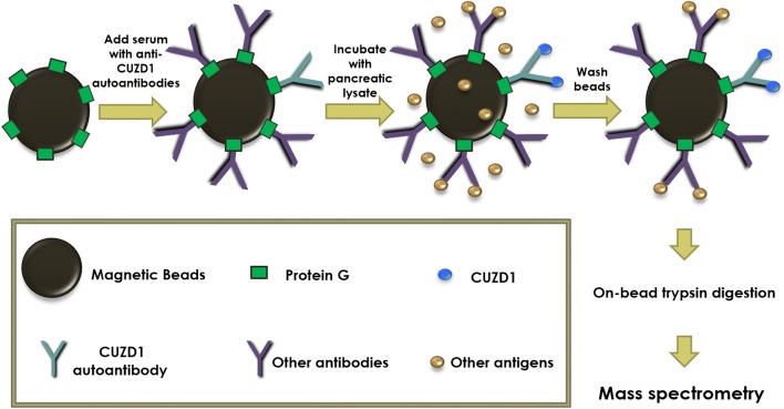
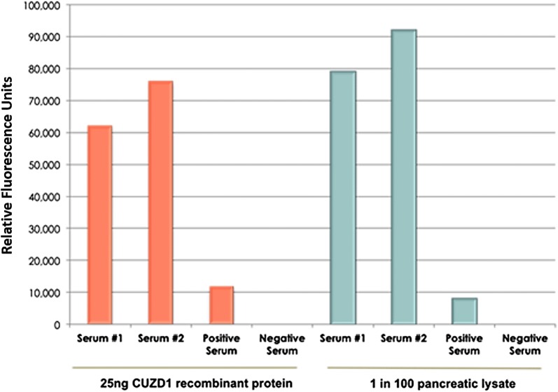
Methods: Serum samples from patients with inflammatory bowel disease (IBD) were analyzed by ELISA for the presence of autoantibodies to CUB and zona pellucida-like domain-containing protein 1 (CUZD1). Protein was extracted from the human pancreas as well as 16 other human tissues to make a complex tissue lysate protein mixture. Antibodies in patient sera were immobilized and purified on protein G magnetic beads and subsequently incubated with pancreatic lysate containing CUZD1 or the aforementioned complex tissue lysate. After extensive washing, antibody-bound protein antigens were trypsin-digested and identified using shotgun mass spectrometry.
Results: The protocol was optimized for the immunoaffinity purification of autoantibody targets from tissue lysate, using CUZD1 from pancreatic lysate and anti-CUZD1 autoantibodies present in IBD patient serum as a proof-of-concept. Pancreatic secretory granule membrane major glycoprotein 2, whose autoantibodies are a known biomarker of Crohn's disease, was also immunoprecipitated from IBD patient serum, as an additional internal positive control.
Conclusions: This study demonstrates the effectiveness of a proteomic approach to identify serum autoantibody targets, using immunoaffinity purification followed by tandem mass spectrometry. Our methodology is applicable for proteome-wide analysis of autoantibody targets in a wide variety of clinical settings.
Keywords: Autoantibodies; Biomarkers; Immuno-MS; Immunoprecipitation; Mass spectrometry; Protein G magnetic beads; Proteomics.
Conflict of interest statement
Competing interestsDr. Eleftherios Diamandis declares that he holds a consultant/advisory role with Abbott Diagnostics.
Figures


Similar articles
-
Novel immunoassays for detection of CUZD1 autoantibodies in serum of patients with inflammatory bowel diseases.Clin Chem Lab Med. 2017 Aug 28;55(10):1574-1581. doi: 10.1515/cclm-2016-1120. Clin Chem Lab Med. 2017. PMID: 28343172
-
Rediscovery of the Anti-Pancreatic Antibodies and Evaluation of their Prognostic Value in a Prospective Clinical Cohort of Crohn's Patients: The Importance of Specific Target Antigens [GP2 and CUZD1].J Crohns Colitis. 2015 Aug;9(8):659-68. doi: 10.1093/ecco-jcc/jjv087. Epub 2015 May 12. J Crohns Colitis. 2015. PMID: 25968583 Clinical Trial.
-
Pancreatic Autoantibodies Against CUZD1 and GP2 Are Associated with Distinct Clinical Phenotypes of Crohn's Disease.Inflamm Bowel Dis. 2015 Dec;21(12):2864-72. doi: 10.1097/MIB.0000000000000564. Inflamm Bowel Dis. 2015. PMID: 26273818
-
CUZD1 and anti-CUZD1 antibodies as markers of cancer and inflammatory bowel diseases.Clin Dev Immunol. 2013;2013:968041. doi: 10.1155/2013/968041. Epub 2013 Apr 22. Clin Dev Immunol. 2013. PMID: 23710207 Free PMC article. Review.
-
Cancer immunomics using autoantibody signatures for biomarker discovery.Mol Cell Proteomics. 2007 Jul;6(7):1115-22. doi: 10.1074/mcp.R600016-MCP200. Epub 2007 Mar 20. Mol Cell Proteomics. 2007. PMID: 17376768 Review.
Cited by
-
Correction to: A proteome-wide immuno-mass spectrometric identification of serum autoantibodies.Clin Proteomics. 2019 Jul 17;16:30. doi: 10.1186/s12014-019-9250-4. eCollection 2019. Clin Proteomics. 2019. PMID: 31346327 Free PMC article.
-
Predicting response and toxicity to PD-1 inhibition using serum autoantibodies identified from immuno-mass spectrometry.F1000Res. 2020 May 7;9:337. doi: 10.12688/f1000research.22715.1. eCollection 2020. F1000Res. 2020. PMID: 33299547 Free PMC article. Clinical Trial.
-
B cell profiles, antibody repertoire and reactivity reveal dysregulated responses with autoimmune features in melanoma.Nat Commun. 2023 Jun 8;14(1):3378. doi: 10.1038/s41467-023-39042-y. Nat Commun. 2023. PMID: 37291228 Free PMC article.
-
Immunotherapy using IgE or CAR T cells for cancers expressing the tumor antigen SLC3A2.J Immunother Cancer. 2021 Jun;9(6):e002140. doi: 10.1136/jitc-2020-002140. J Immunother Cancer. 2021. PMID: 34112739 Free PMC article.
-
Inflammo-immune perspective on the association of eight migraine risk factors with migraine: a multi-omics Mendelian randomization study.Front Neurol. 2024 Aug 7;15:1440995. doi: 10.3389/fneur.2024.1440995. eCollection 2024. Front Neurol. 2024. PMID: 39170074 Free PMC article.
References
-
- Da Gama Duarte J, Parakh S, Andrews MC, et al. Autoantibodies may predict immune-related toxicity: results from a phase I study of intralesional Bacillus Calmette-Guérin followed by ipilimumab in patients with advanced metastatic melanoma. Front Immunol. 2018;9:1–9. doi: 10.3389/fimmu.2018.00001. - DOI - PMC - PubMed
LinkOut - more resources
Full Text Sources
Other Literature Sources
Research Materials