

doi: 10.1016/j.dib.2019.103972.
eCollection 2019 Aug.
Data on SVCT2 transporter expression and localization in cancer cell lines and tissues
Affiliations
- PMID: 31249848
- PMCID: PMC6586595
- DOI: 10.1016/j.dib.2019.103972
Item in Clipboard
Data on SVCT2 transporter expression and localization in cancer cell lines and tissues
Data Brief.
.
Abstract
The data presented in this article are related to the research paper entitled "Increased expression of mitochondrial sodium-coupled ascorbic acid transporter-2 (mitSVCT2) as a central feature in breast cancer", available in Free Radical Biology and Medicine Journal [1]. In this article, we examined the SVCT2 transporter expression in various breast cancer cell lines using RT-PCR and Western blot assays. In addition, we analyzed the subcellular localization of SVCT2 by immunofluorescence colocalization assays and cellular fractionation experiments. Finally, an analysis of different cancer tissue microarrays immunostained for SVCT2 and imaged by The Human Protein Atlas (https://www.proteinatlas.org) is presented.
Figures
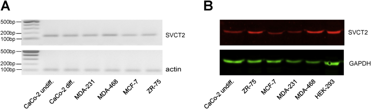
SVCT2 expression in breast cancer cell lines. (A) RT-PCR for SVCT2 expression in breast cancer cells. cDNA was synthesized from total RNA isolated from four breast cancer lines and control cells (undifferentiated and differentiated CaCo-2 cells), followed by amplification by PCR, separation by agarose gel electrophoresis and visualization by ethidium bromide staining. Actin was used as an internal control. (B) Western blot for SVCT2 protein expression in breast cancer cells. Total extracts were prepared from four breast cancer lines and control cells (undifferentiated CaCo-2 and HEK-293 cells) and immunoblotted with anti-SVCT2 antibody. GAPDH was used as a loading control.
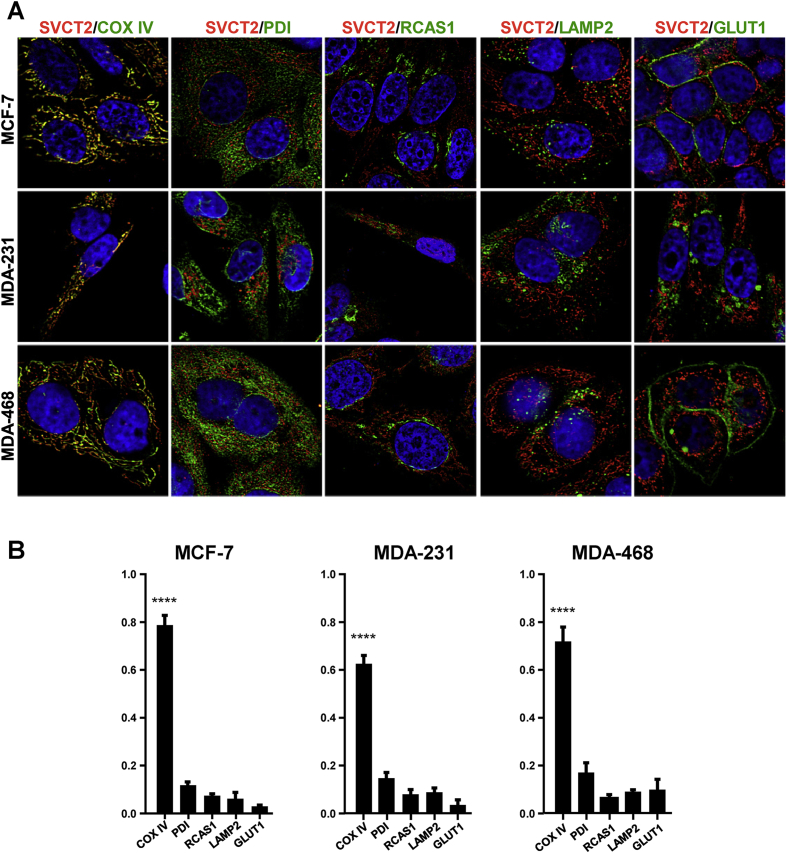
Colocalization of SVCT2 with organelle markers in breast cancer cell lines. (A) Immunofluorescence was performed in MCF-7, MDA-231 and MDA-468 cell lines. Cells were stained with anti-SVCT2 simultaneous with anti-COXIV, anti-LAMP2, anti-PDI, anti-RCAS1 or anti-GLUT1; and stained with Cy3 and FITC secondary antibodies. Samples were observed by confocal microscopy and 0.2-μm successive images were obtained along the z axis for further deconvolution analysis. (B) Pearson colocalization analysis in ImageJ using the JaCop plugin.
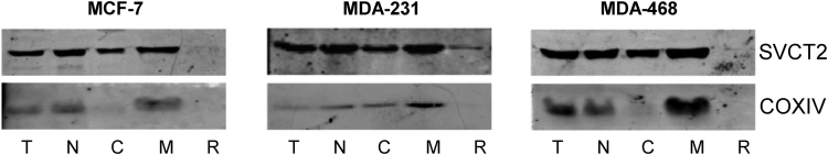
SVCT2 expression in mitochondrial fraction of breast cancer cell lines. MCF-7, MDA-231 and MDA-468 cell lines were homogenized and fractionated by differential centrifugation, and the different fractions (T: total homogenate; N: nuclear fraction; C: cytoplasmic fraction; M: mitochondria; R: endoplasmic reticulum) were immunoblotted with anti-SVCT2 and anti-COXIV antibodies.
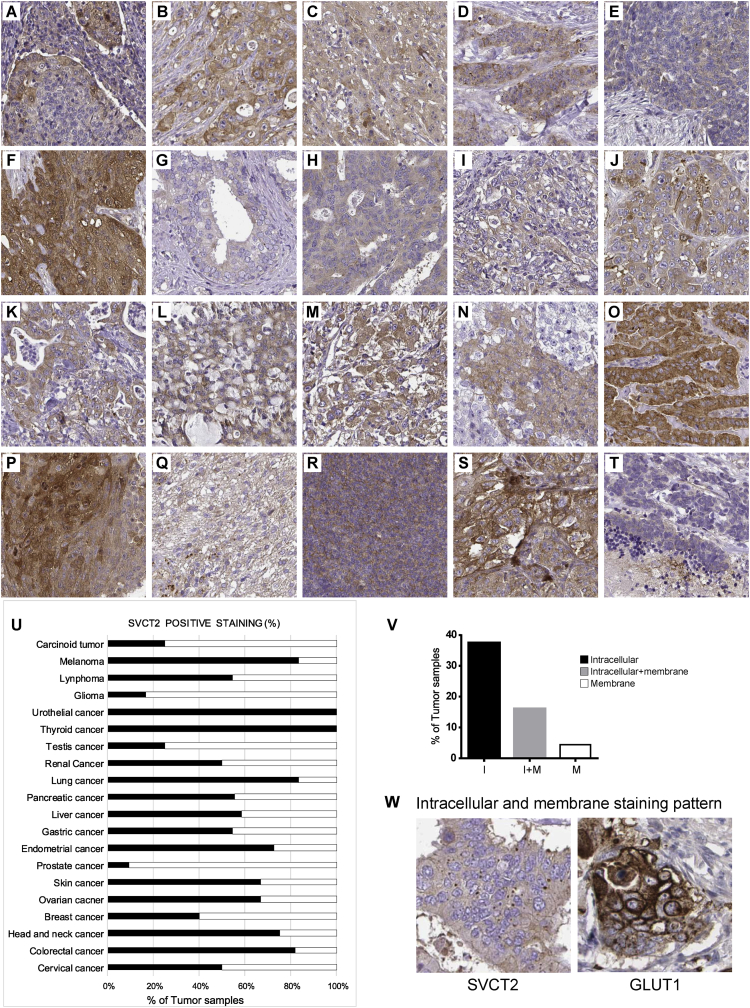
SVCT2 expression in different cancer tissues. Various tissue microarrays processed, immunostained for SVCT2 transporter and imaged by The Human Protein Atlas in different cancer tissues: cervical (A), colorectal (B), head and neck (C), breast (D), ovarian (E), skin (F), prostate (G), endometrial (H), stomach (I), liver (J), pancreatic (K), lung (L), renal (M), testis (N), thyroid (O), urothelial (P), glioma (Q), lymphoma (R), melanoma (S) and carcinoid (T). (U) Percentage of positive SVCT2 samples among different cancer types. (V) Percentage of SVCT2 positive samples with intracellular (I), intracellular plus membrane (I+M) and membrane (M) SVCT2 staining patterns. (W) Staining pattern comparison between membrane staining of GLUT1 vs. intracellular staining of SVCT2. (Images adapted from the protein atlas website https://www.proteinatlas.org/ENSG00000089057-SLC23A2/pathology ).
References
-
- Pena E., Roa F.J., Inostroza E., Sotomayor K., Gonzalez M., Gutierrez-Castro F.A., Maurin M., Sweet K., Labrousse C., Gatica M., Aylwin C.F., Mendoza P., Maldonado M., Delgado C., Madariaga J., Panes J., Silva-Grecchi T., Concha, Moraga-Cid G., Reyes A.M., Munoz-Montesino C., Vera J.C., Rivas C.I. Increased expression of mitochondrial sodium-coupled ascorbic acid transporter-2 (mitSVCT2) as a central feature in breast cancer. Free Radical Biol. Med. 2019;135:283–292. - PubMed
-
- Munoz-Montesino C., Roa F.J., Pena E., Gonzalez M., Sotomayor K., Inostroza E., Munoz C.A., Gonzalez I., Maldonado M., Soliz C., Reyes A.M., Vera J.C., Rivas C.I. Mitochondrial ascorbic acid transport is mediated by a low-affinity form of the sodium-coupled ascorbic acid transporter-2. Free Radical Biol. Med. 2014;70:241–254. - PubMed
-
- Frezza C., Cipolat S., Scorrano L. Organelle isolation: functional mitochondria from mouse liver, muscle and cultured fibroblasts. Nat. Protoc. 2007;2(2):287–295. - PubMed
LinkOut - more resources
Full Text Sources
