. 2017 Dec 15;1(2):e155-e163.
doi: 10.1055/s-0037-1613674. eCollection 2017 Jul.

Release of Prometastatic Platelet-Derived Microparticles Induced by Breast Cancer Cells: A Novel Positive Feedback Mechanism for Metastasis

Affiliations

Release of Prometastatic Platelet-Derived Microparticles Induced by Breast Cancer Cells: A Novel Positive Feedback Mechanism for Metastasis

Marta Zarà et al. TH Open. .

Abstract

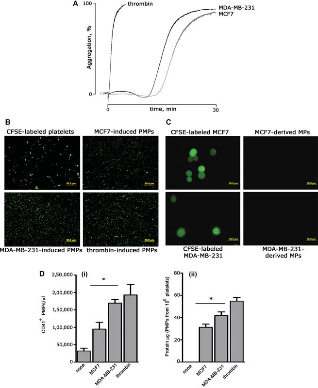
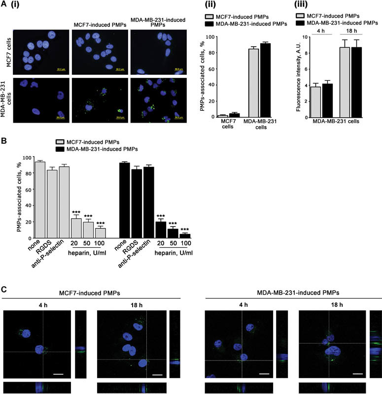
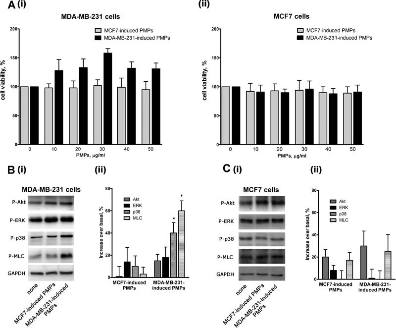
Circulating platelets and platelet-derived microparticles are regulators of cancer metastasis. In this study, we show that breast cancer cells induce platelet aggregation and lead to the release of platelet-derived microparticles. Although able to cause comparable aggregation, the highly aggressive MDA-MB-231 cells were more potent than the poorly aggressive MCF7 cells in inducing platelet-derived microparticles release, which was comparable to that promoted by thrombin. MDA-MB-231 cells were able to bind and internalize both MCF7- and MDA-MB-231-induced platelet-derived microparticles with comparable efficiency. By contrast, MCF7 cells did not interact with either type of platelet-derived microparticles. Upon internalization, only platelet-derived microparticles released by platelet stimulation with MDA-MB-231 cells, but not those released upon stimulation with MCF7 cells, caused activation of MDA-MB-231 cells and promoted the phosphorylation of selected signaling proteins, including p38MAPK and myosin light chain. Accordingly, MDA-MB-231-induced, but not MCF7-induced, platelet-derived microparticles dose-dependently stimulated migration and invasion of targeted MDA-MB-231 cells. These results identify a novel paracrine positive feedback mechanism initiated by aggressive breast cancer cell types to potentiate their invasive phenotype through the release of platelet-derived microparticles.

Keywords: cancer metastasis; cell migration; microparticles; platelet physiology.


Conflict of interest statement

Conflict of Interest None declared.

Figures

Fig. 1
Fig. 1
Breast cancer cells induce the release of platelet-derived microparticles (PMPs). ( A ) Washed human platelets (0.3 × 10 9 platelets/mL) were incubated for 30 minutes at 37°C under constant magnetic stirring with 5 × 10 4 /mL of either MCF7 or MDA-MB-231 cells. Thrombin was used at 0.2 U/mL. Representative aggregation traces are reported. ( B ) PMPs release in the supernatant. Platelets were labeled with 3 µg/mL of carboxyfluorescein succinimidyl ester (CFSE) and co-cultured with either MCF7 or MDA-MB-231 cells or stimulated with thrombin for 30 minutes at 37°C under stirring. Upon centrifugation, to remove platelets and cells, labeled PMPs in the supernatant were visualized by fluorescence microscopy. Representative images of labeled platelets and released PMPs are reported. Scale bars: 50 µm. ( C ) Platelets do not cause the release of breast cancer cell–derived microparticles. Experiments were performed as those in panel B, except that MCF7 and MDA-MB-231 cells were labeled with CFSE before being co-cultured with unlabeled platelets. Scale bars: 50 µm. ( D ) Quantification of PMPs released by platelet stimulation with MCF7, MDA-MB-231, or thrombin. (i) PMPs were quantified using a three laser-equipped BC Navios flow cytometer (Beckman Coulter), upon staining with PE-labeled anti-CD41 antibody (Bio Legend). The instrument was set using Megamix-Plus (BioCytex, Marseille, France) fluorescent calibrated beads (0.1–0.9 μm range). Data were analyzed with the FCS Express 6.0 software (De Novo Software), and are reported as mean ± SEM ( n  = 3). * p  < 0.05. (ii) PMPs in the supernatants were recovered by centrifugation at 20,000 g for 90 minutes, resuspended in HEPES buffer, and analyzed by BCA protein assay. Results report the protein content of PMPs released from the same number of stimulated platelets (10 9 ) and are mean ± SEM ( n  = 7). * p  < 0.05.
Fig. 2
Fig. 2
Interaction of breast cancer cells with platelet-derived microparticles (PMPs). ( A ) MCF7 and MDA-MB-231 cells (5 × 10 4 cells/well) were grown on glass coverslips and then incubated with 30 µg/mL of MCF7-induced or MDA-MB-231-induced PMPs obtained from carboxyfluorescein succinimidyl ester (CFSE)-labeled platelets (green) for 4 or 18 hours. (i) Representative fluorescence microscopy images upon 18 hours of incubation. Cell nuclei were stained with 1 µg/mL of Hoechst (blue); (ii) quantification of the percentage of MCF7 or MDA-MB-231 cells (as indicated on the bottom) associated with fluorescent PMPs, induced by either MCF7- or MDA-MB-231 cells (gray and black bars, respectively, as indicated on the top). Data are the mean ± SEM of three independent experiments. (iii) Comparison of MDA-MB-231 cells' ability to interact with MCF7- or MDA-MB-231-induced PMPs after 4 or 18 hours of incubation. Data are expressed as average green fluorescence intensity associated with each cell and are the mean ± SEM of three independent experiments. ( B ) MDA-MB-231 cells were incubated with MDA-MB-231- or MCF7-induced PMPs obtained from CFSE-labeled platelets (30 μg/mL) in the presence of buffer (none), 0.5 mM RGDS, 10 μg/mL anti-P-selectin antibody CLB/thromb/6, or increasing amount of heparin, as indicated. The percentage of PMPs-associated cells was determined as described in panel A, and results are expressed the mean ± SEM of three independent experiments (*** p  < 0.001). ( C ) Confocal microscopy analysis of MDA-MB-231 cells interacting with MCF7- or MDA-MB-321-induced PMPs for the indicated times of incubation. Representative confocal middle z-sections and orthogonal views are reported. Scale bars: 20 µm.
Fig. 3
Fig. 3
Platelet-derived microparticles (PMPs)-induced activation of MDA-MB-231 cells. ( A ) Viability of MDA-MB-231 (i) or MCF7 (ii) cells incubated with the indicated amounts of PMPs for 24 hours was assessed by a colorimetric MTT assay. Results are reported as the mean ± SEM of three different experiments. ( B and C ) Phosphorylation of selected signaling proteins in MDA-MB-231(panel B) or MCF7 (panel C) cells incubated with MCF7- or MDA-MB-231-induced PMPs for 18 hours, as indicated on the bottom. Representative immunoblot with specific anti-phosphoprotein antibodies directed against the protein indicated on the right is reported in (i), where GAPDH staining is for equal loading control. Quantification of the results by densitometric scanning is reported in (ii), as % of phosphorylation increase over the level of untreated cells. Results are the mean ± SEM of three different experiments. * p  < 0.05.
Fig. 4
Fig. 4
Analysis of cell migration and invasion. Effect of MCF7- or MDA-MB-231-induced PMPs (as indicated on the left) on migration (panels A and B) and invasiveness (panels C and D) of MDA-MB-231 cells (panels A and C) or MCF7 cells (panels B and D), as indicated. Cancer cells were treated with increasing amounts of the two different types of PMPs preparations (0–50 μg/mL, as indicated on the bottom) and then transferred inside cell culture inserts. For the invasion assays (panels C and D), the upper side of the insert was coated with 0.1 mL of Matrigel (50 µg/mL). Incubation was prolonged for 18 hours and the cells that moved through the porous membrane were stained and counted. In all the panels, representative images are reported in (i), while quantification of the results is shown in (ii) as mean ± SEM of three experiments. Statistical significance of the difference was calculated between treated and untreated cells (sample 0). * p  < 0.05; ** p  < 0.01; *** p  < 0.001.

References

    1. Mezouar S, Frère C, Darbousset R et al.Role of platelets in cancer and cancer-associated thrombosis: experimental and clinical evidences. Thromb Res. 2016;139:65–76. - PubMed
    1. Erpenbeck L, Schön M P. Deadly allies: the fatal interplay between platelets and metastasizing cancer cells. Blood. 2010;115(17):3427–3436. - PMC - PubMed
    1. Gay L J, Felding-Habermann B. Contribution of platelets to tumour metastasis. Nat Rev Cancer. 2011;11(02):123–134. - PMC - PubMed
    1. Yan M, Jurasz P. The role of platelets in the tumor microenvironment: From solid tumors to leukemia. Biochim Biophys Acta. 2016;1863(03):392–400. - PubMed
    1. Kopp H G, Placke T, Salih H R. Platelet-derived transforming growth factor-beta down-regulates NKG2D thereby inhibiting natural killer cell antitumor reactivity. Cancer Res. 2009;69(19):7775–7783. - PubMed

LinkOut - more resources