. 2018 Mar 28;2(1):e104-e115.
doi: 10.1055/s-0038-1635572. eCollection 2018 Jan.

Reduced Contraction of Blood Clots in Venous Thromboembolism Is a Potential Thrombogenic and Embologenic Mechanism

Affiliations

Reduced Contraction of Blood Clots in Venous Thromboembolism Is a Potential Thrombogenic and Embologenic Mechanism

Alina D Peshkova et al. TH Open. .

Abstract

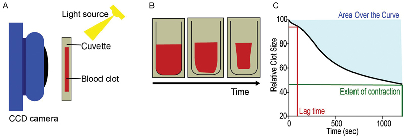
Contraction (retraction) of the blood clot is a part of the clotting process driven by activated platelets attached to fibrin that can potentially modulate the obstructiveness and integrity of thrombi. The aim of this work was to reveal the pathogenic importance of contraction of clots and thrombi in venous thromboembolism (VTE). We investigated the kinetics of clot contraction in the blood of 55 patients with VTE. In addition, we studied the ultrastructure of ex vivo venous thrombi as well as the morphology and functionality of isolated platelets. Thrombi from VTE patients contained compressed polyhedral erythrocytes, a marker for clot contraction in vivo. The extent and rate of contraction were reduced by twofold in clots from the blood of VTE patients compared with healthy controls. The contraction of clots from the blood of patients with pulmonary embolism was significantly impaired compared with that of those with isolated venous thrombosis, suggesting that less compacted thrombi are prone to embolization. The reduced ability of clots to contract correlated with continuous platelet activation followed by their partial refractoriness. Morphologically, 75% of platelets from VTE patients were spontaneously activated (with filopodia) compared with only 21% from healthy controls. At the same time, platelets from VTE patients showed a 1.4-fold reduction in activation markers expressed in response to chemical activation when compared with healthy individuals. The results obtained suggest that the impaired contraction of thrombi is an underappreciated pathogenic mechanism in VTE that may regulate the obstructiveness and embologenicity of venous thrombi.

Keywords: blood clotting; clot contraction; clot retraction; thrombosis; venous thromboembolism.


Conflict of interest statement

Conflict of Interest None declared.

Figures

Fig. 1
Fig. 1
Schematic of experimental methods. ( A ) Recalcified whole blood samples with thrombin were added to a cuvette and allowed to contract. Images were recorded by a charge-coupled device camera every 15 seconds based on the light scattering properties of the clot versus the expelled serum. ( B , C ) Data on relative clot size were recorded over time, where the original kinetic curve could be analyzed for the extent of clot contraction (green) at 20 minutes, the lag time (red) or the time to 5% contraction, or the area over the curve (blue), representing the work done by the contracting clot.
Fig. 2
Fig. 2
Representative scanning electron micrographs of the interior of venous thrombi containing compressed polyhedral erythrocytes (polyhedrocytes) as a morphologic signature of contraction in vivo. The corresponding images were taken from four randomly selected thrombi at lower ( A , C , E , G ) and higher magnification ( B , D , F , H ). Magnification bars = 10 µm.
Fig. 3
Fig. 3
Clot contraction parameters of clots made from the blood of VTE patients ( n  = 55) versus healthy donors ( n  = 60). ( A ) Extent of clot contraction at 20 minutes, ( B ) lag time or time to reach 95% of the initial clot size, ( C ) area over the curve (an integral parameter that characterizes the intensity of the entire process of clot contraction), ( D ) average velocity of contraction.*** p  < 0.001.
Fig. 4
Fig. 4
( A ) Averaged kinetic clot contraction curves obtained from clotting of the blood of VTE patients ( n  = 55) and healthy individuals ( n  = 60). Optical tracking was used to measure the relative changes in clot size over 20 minutes at 15-second intervals. ( BD ) The rate constants of the three phases of clot contraction curves shown in A . The three phases correspond to the initiation of contraction (phase 1), linear contraction (phase 2), and mechanical stabilization (phase 3). Data are shown as mean ± SEM. * p  < 0.5; **** p  < 0.0001.
Fig. 5
Fig. 5
Representative scanning electron micrographs of unstimulated platelets isolated from the blood of a VTE patient ( A ) and a healthy donor ( B ), showing a higher degree of partial activation of the unstimulated (quiescent) platelets in VTE reflected by the shape change, formation of filopodia, and tendency to aggregate. Magnification bars = 2 µm.
Fig. 6
Fig. 6
Proposed pathogenic mechanisms related to the impaired clot contraction in venous thromboembolism.

Similar articles

Cited by

References

    1. Roger V L, Go A S, Lloyd-Jones D M et al.Heart disease and stroke statistics - 2012 update: a report from the American Heart Association. Circulation. 2012;125(01):e2–e220. - PMC - PubMed
    1. Bing R, Chow V, Lau J K, Thomas L, Kritharides L, Ng A CC. Prevalence of echocardiography use in patients hospitalized with confirmed acute pulmonary embolism: a real-world observational multicenter study. PLoS One. 2016;11(12):e0168554. - PMC - PubMed
    1. Lee J S, Moon T, Kim T H et al.Deep vein thrombosis in patients with pulmonary embolism: prevalence, clinical significance and outcome. Vasc Spec Int. 2016;32(04):166–174. - PMC - PubMed
    1. Wolberg A S. Primed to understand fibrinogen in cardiovascular disease. Arterioscler Thromb Vasc Biol. 2016;36(01):4–6. - PMC - PubMed
    1. Geddings J E, Mackman N. Recently identified factors that regulate hemostasis and thrombosis. Thromb Haemost. 2014;111(04):570–574. - PMC - PubMed