

Localization of amyloid beta peptides to locus coeruleus and medial prefrontal cortex in corticotropin releasing factor overexpressing male and female mice
- PMID: 31250157
- PMCID: PMC7371412
- DOI: 10.1007/s00429-019-01915-8
Localization of amyloid beta peptides to locus coeruleus and medial prefrontal cortex in corticotropin releasing factor overexpressing male and female mice
Abstract
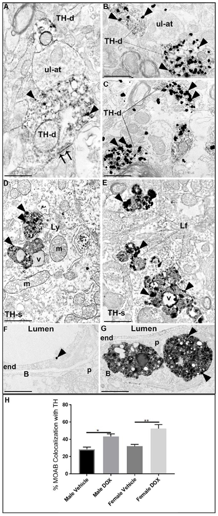
A culmination of evidence from the literature points to the Locus Coeruleus (LC)-Norepinephrine system as an underappreciated and understudied area of research in the context of Alzheimer's Disease (AD). Stress is a risk factor for developing AD, and is supported by multiple clinical and preclinical studies demonstrating that amplification of the stress system disrupts cellular and molecular processes at the synapse, promoting the production and accumulation of the amyloid beta (Aβ42) peptide. Stress-induced activation of the LC is mediated by corticotropin releasing factor (CRF) and CRF receptors exhibit sex-biased stress signaling. Sex differences are evident in the neurochemical, morphological and molecular regulation of LC neurons by CRF, providing a compelling basis for the higher prevalence of stress-related disorders such as AD in females. In the present study, we examined the cellular substrates for interactions between Aβ and tyrosine hydroxylase a marker of noradrenergic somatodendritic processes in the LC, and Dopamine-β-Hydroxylase (DβH) in the infralimbic medial prefrontal cortex (ILmPFC) in mice conditionally overexpressing CRF in the forebrain (CRFOE) under a Doxycycline (DOX) regulated tetO promoter. CRFOE was sufficient to elicit a redistribution of Aβ peptides in the somatodendritic processes of the LC of male and female transgenic mice, without altering total Aβ42 protein expression levels. DOX treated groups exhibited lysosomal compartments with apparent lipofuscin and abnormal morphology, indicating potential dysfunction of these Aβ42-clearing compartments. In female DOX treated groups, swollen microvessels with lipid-laden vacuoles were also observed, a sign of blood-brain-barrier dysfunction. Finally, sex differences were observed in the prefrontal cortex, as females responded to DOX treatment with increased frequency of co-localization of Aβ42 and DβH in noradrenergic axon terminals compared to vehicle treated controls, while male groups showed no significant changes. We hypothesize that the observed sex differences in Aβ42 distribution in this model of CRF hypersignaling is based on increased responsivity of female rodent CRFR1 in the LC. Aβ42 production is enhanced during increased neuronal activation, therefore, the excitation of DOX treated female CRFOE LC neurons projecting to the mPFC may exhibit more frequent co-localization with Aβ due to increased neuronal activity of noradrenergic neurons.
Keywords: Adrenergic receptors; Amyloid; Dopamine-β-hydroxylase; Norepinephrine; Stress.
Conflict of interest statement
Conflict of Interest: The authors declare that they have no conflict of interest.
Figures


References
-
- Abbott NJ, Patabendige AA, Dolman DE, Yusof SR and Begley DJ (2010). “Structure and function of the blood-brain barrier.” Neurobiol Dis 37(1): 13–25. - PubMed
-
- Aliev G, Seyidova D, Neal ML, Shi J, Lamb BT, Siedlak SL, Vinters HV, Head E, Perry G, Lamanna JC, Friedland RP and Cotman CW (2002). “Atherosclerotic lesions and mitochondria DNA deletions in brain microvessels as a central target for the development of human AD and AD-like pathology in aged transgenic mice.” Ann N Y Acad Sci 977: 45–64. - PubMed
-
- Banasr M, Valentine GW, Li XY, Gourley SL, Taylor JR and Duman RS (2007). “Chronic unpredictable stress decreases cell proliferation in the cerebral cortex of the adult rat.” Biol Psychiatry 62(5): 496–504. - PubMed
-
- Bangasser DA, Curtis A, Reyes BA, Bethea TT, Parastatidis I, Ischiropoulos H, Van Bockstaele EJ and Valentino RJ (2010). “Sex differences in corticotropin-releasing factor receptor signaling and trafficking: potential role in female vulnerability to stress-related psychopathology.” Mol Psychiatry 15(9): 877, 896–904. - PMC - PubMed
MeSH terms
Substances
Grants and funding
LinkOut - more resources
Full Text Sources
