. 2020 Mar 1;116(3):721-735.
doi: 10.1093/cvr/cvz164.

Chanzyme TRPM7 protects against cardiovascular inflammation and fibrosis

Affiliations

Chanzyme TRPM7 protects against cardiovascular inflammation and fibrosis

Francisco J Rios et al. Cardiovasc Res. .

Abstract

Aims: Transient Receptor Potential Melastatin 7 (TRPM7) cation channel is a chanzyme (channel + kinase) that influences cellular Mg2+ homeostasis and vascular signalling. However, the pathophysiological significance of TRPM7 in the cardiovascular system is unclear. The aim of this study was to investigate the role of this chanzyme in the cardiovascular system focusing on inflammation and fibrosis.

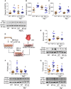
Methods and results: TRPM7-deficient mice with deletion of the kinase domain (TRPM7+/Δkinase) were studied and molecular mechanisms investigated in TRPM7+/Δkinase bone marrow-derived macrophages (BMDM) and co-culture systems with cardiac fibroblasts. TRPM7-deficient mice had significant cardiac hypertrophy, fibrosis, and inflammation. Cardiac collagen and fibronectin content, expression of pro-inflammatory mediators (SMAD3, TGFβ) and cytokines [interleukin (IL)-6, IL-10, IL-12, tumour necrosis factor-α] and phosphorylation of the pro-inflammatory signalling molecule Stat1, were increased in TRPM7+/Δkinase mice. These processes were associated with infiltration of inflammatory cells (F4/80+CD206+ cardiac macrophages) and increased galectin-3 expression. Cardiac [Mg2+]i, but not [Ca2+]i, was reduced in TRPM7+/Δkinase mice. Calpain, a downstream TRPM7 target, was upregulated (increased expression and activation) in TRPM7+/Δkinase hearts. Vascular functional and inflammatory responses, assessed in vivo by intra-vital microscopy, demonstrated impaired neutrophil rolling, increased neutrophil: endothelial attachment and transmigration of leucocytes in TRPM7+/Δkinase mice. TRPM7+/Δkinase BMDMs had increased levels of galectin-3, IL-10, and IL-6. In co-culture systems, TRPM7+/Δkinase macrophages increased expression of fibronectin, proliferating cell nuclear antigen, and TGFβ in cardiac fibroblasts from wild-type mice, effects ameliorated by MgCl2 treatment.

Conclusions: We identify a novel anti-inflammatory and anti-fibrotic role for TRPM7 and suggest that its protective effects are mediated, in part, through Mg2+-sensitive processes.

Keywords: Cardiac hypertrophy; Cations; Magnesium channel; Vascular inflammation.


Figures

Graphical Abstract
Graphical Abstract
Figure 1
Figure 1
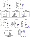
Tissue and cellular Mg2+ and Ca2+ levels in TRPM7+/Δkinase mice. (A) Total Mg2+ concentration in heart and kidneys from WT (n = 9) and TRPM7+/Δkinase (M7+/Δ) (n = 13) mice was assessed by colorimetric assay and normalized by dry weight. Total mononuclear leucocytes were isolated from hearts, kidneys, and blood, and labelled with magnesium green AM (Mg2+ green), followed by specific antibodies: anti-CD45 + anti-F4/80 for macrophage identification in heart and kidney, and anti-CD11b + anti-Ly6C for blood monocytes. (B) Representative flow cytometry histograms and corresponding scatter-plot graphs (C) showing free Mg2+ levels in macrophages isolated from hearts (n = 6/group) and kidneys and blood monocytes (n = 6/group). (D, E) Intracellular free Ca2+ levels in macrophages were investigated using the probe Ca-520 and analysed by flow cytometry (n = 6/group). Data are presented as representative flow cytometry histograms (D) and corresponding scatter-plot graphs (E). Results are mean ± SEM of mean of fluorescence intensity (MFI) of fluorescence-labelled cells assessed by flow cytometry. Statistical significance was determined by a two-tailed unpaired Student’s t-test. *P < 0.05 TRPM7+/Δkinase (M7+/Δ, blue) vs. WT (yellow).
Figure 2
Figure 2
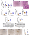
Cardiac dysfunction and fibrosis in TRPM7+/Δkinase mice. (A) Heart weight, normalized to tibia length, in WT and TRPM7+/Δkinase (M7+/Δ) mice (n = 8/group). (B–E) Echocardiography analysis showing ventricular filling velocity assessed by E/A ratio (B), where Early-E and late atrial-A ventricular filling velocity; (C) Left ventricular fractional shortening (FS%); (D) left ventricular AWT (WT n = 7, M7+/Δ n = 8); (E) Representative images by M-mode echocardiography (upper panel) and Doppler (lower panel). (F) Cardiac sections obtained from WT and M7+/Δ were stained with sirius red and collagen content assessed using bright field (scale bar 10 μm) and polarized light (scale bar 200 μm). Images are representative of n = 6/group. Collagen was analysed in bright field (F, H-upper panel) and polarized light (G, H-lower panel) and data expressed as mean ± SEM of % affected area (n = 6/group). Cardiac pro-fibrotic markers were assessed by immunoblotting: (I) fibronectin (n = 5/group), (J) TGFβ (n = 5/group), and (K) phospho-Smad3 (Tyr179) (n = 6/group). Proteins of interest were normalized to α-tubulin. Statistical significance was determined by a two-tailed unpaired Student’s t-test. *P < 0.05 TRPM7+/Δkinase (M7+/Δ, blue) vs. WT (yellow).
Figure 3
Figure 3
Cardiac inflammation in TRPM7+/Δkinase mice. Total RNA was obtained from total cardiac tissues and gene expression for TNFα, IL-12, IL-10, iNOS, Arg-1, and IL-1β was determined by real-time PCR and normalized by GAPDH (A). Data are expressed in 2−ΔΔCt values (WT n = 6, M7+/Δ n = 8). (B) Histological sections from hearts tissues from WT and TRPM7+/Δkinase (M7+/Δ) mice stained with H&E showing an inflammatory infiltrate. The highlighted area in (B) shows the inflammatory infiltrate. Images are representative photomicrographs (n = 8/group), scale bar = 150 μm. Total leucocytes were isolated from hearts by enzymatic digestion and stained for flow cytometry analysis. (C) CD45+ population (total haematopoietic cells), (D) CD45+F4/80+ cells (macrophages); (E) CD45+F4/80+CD206+ (M2 macrophages); (F) CD45+F4/80+CD11c (M1 macrophages) (n = 6/group). Total lysates obtained from cardiac tissues were investigated for the expression of (G) phospho-Stat1 (Tyr701) and (H) phospho-Stat3 (Tyr705) by western blotting and normalized to total Stat1 and Stat3, respectively (n = 6/group). (I, J) Increased galectin-3 expression in the hearts of TRPM7+/Δkinase mice. (I) Representative histological sections obtained from hearts from WT and TRPM7+/Δkinase mice (n = 5/group). Controls for immunoreactivity were assessed using the isotype only. (J) Corresponding scatter-plot graphs indicating galectin-3 expression by immunohistochemistry. Scale bar = 150 μm (n = 5/group). Results are mean ± SEM. Statistical significance was determined by a two-tailed unpaired Student’s t-test. *P < 0.05 TRPM7+/Δkinase (M7+/Δ, blue) vs. WT (yellow).
Figure 4
Figure 4
Cardiac calpain-II and annexin-1, downstream targets of TRPM7, in TRPM7+/Δkinase mice. Cardiac expression of Calpain-II and Annexin-1 (ANXA-1) in total heart lysate (A, D) (n = 6/group), cytosolic (B, E) and membrane fractions (C, F) (n = 7/group) assessed by immunoblotting. Cytosolic and membrane fractions from total cardiac tissues were obtained by ultracentrifugation. Protein expression in total lysate and cytosolic fractions were normalized to α-tubulin and the membrane fraction was normalized to Na-K ATPase content. The ratio of membrane: cytosolic calpain-II (G) and annexin-1 (H) is used as an index of cytosol to membrane translocation. (I) Total lysates from cardiac tissues were used to assess expression of spectrin α II, total (240 kDa) and cleaved forms (110 kDa), by western blotting and further normalized to α-tubulin (n = 6/group). Results are presented as representative immunoblots and corresponding scatter-plot graphs. Data are means ± SEM. Statistical significance was determined by a two-tailed unpaired Student’s t-test *P < 0.05 TRPM7+/Δkinase (M7+/Δ, blue) vs. WT (yellow).
Figure 5
Figure 5
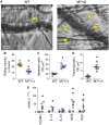
Increased vascular inflammation in TRPM7+/Δkinase mice. (A) Intra-vital images obtained from cremasteric microvessels 3 h after intrascrotal injection of TNFα (20 ng/mL). Images are representative bright field of WT (left panel) and TRPM7+/Δkinase (M7+/Δ) (right panel) animals. Yellow arrows indicate transmigrated cells, scale bars = 25 μm. Images were recorded for 60 s from at least eight vessels from each animal (n = 4/group) and analysed for (B) rolling velocity, (C) number of adherent cells, and (D) number of transmigrated cells and normalized by 100 μm2. Data are presented as mean ± SEM. (E) Total RNA was obtained from aorta and gene expression for VCAM-1, IL-12, IL-10, iNOS, and Arg-1 was determined by real-time PCR and normalized to GAPDH expression. Data are expressed in 2−ΔΔCt values (n = 6–7/group). Data are means ± SEM. Statistical significance was determined by a two-tailed unpaired Student’s t-test. *P < 0.05 TRPM7+/Δkinase (M7+/Δ) (blue) vs. WT (yellow).
Figure 6
Figure 6
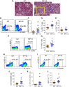
Renal inflammation in TRPM7+/Δkinase mice. (A) Representative photomicrographs of histological kidney sections stained with H&E (n = 8/group). The highlighted area in (A) shows the inflammatory infiltrate. Scale bar = 150 μm. Total leucocytes were isolated from kidneys by enzymatic digestion and stained for flow cytometry analysis. Results are shown as representative plots and as mean ± SEM (WT n = 8, M7+/Δ n = 10) of (B, C) CD45+ population (total haematopoietic cells); (D) CD45+CD3+CD4+ cells (CD4 T lymphocytes); (E) CD45+CD3+CD8+ cells (CD8 T lymphocytes) (n = 6/group); (F, G) CD45+F4/80+ cells (macrophages); (H) CD45+F4/80+CD11c+ (M1 macrophages); (I) CD45+F4/80+CD206+ (M2 macrophages); (J, K) CD11c to CD206 ratio (WT n = 7, M7+/Δ n = 9) and IL-12 to IL-10 mRNA ratio; (L) MCP-1 mRNA gene expression (WT n = 7, M7+/Δ n = 9). Statistical significance was determined by a two-tailed unpaired Student’s t-test. *P < 0.05 TRPM7+/Δkinase (M7+/Δ, blue) vs. WT (yellow). SSC-A, Side-Scatter.
Figure 7
Figure 7
TRPM7+/Δkinase macrophages induce a fibrotic phenotype in cardiac fibroblasts from WT mice: effects of MgCl2 treatment. BMDM were differentiated from TRPM7+/Δkinase (M7+/Δ) and WT mice and treated with MgCl2 (10 mM) for 24 h. (A) Total cell lysate was analysed for Galectin-3 expression by immunoblotting and normalized to GAPDH protein expression. The production of (B) Galectin-3, and (C) IL-10 was analysed in the macrophage supernatant by ELISA (n = 7–8/group). (E) Primary culture cardiac fibroblasts from WT mice were co-cultured in transwell system with macrophage from M7+/Δ and WT animals, treated or not with MgCl2. After 48 h stimulation, the total cell lysate was obtained from cardiac fibroblasts and expression of (F) fibronectin (n = 8/group) (G) PCNA (n = 9/group), and (H) TGFβ (n = 9/group) was analysed by western blotting and normalized to β-actin. Data are presented as representative immunoblots and corresponding scatter-plot graphs. Results are mean ± SEM of M7+/Δ (blue) and WT (yellow). Statistical significance was determined by one-way ANOVA using the Student–Newman–Keuls post-test. *P < 0.05 for TRPM7+/Δkinase (M7+/Δ) compared with WT mice. TRPM7+/Δkinase treated with MgCl2 vs. untreated TRPM7+/Δkinase.

Comment in

References

    1. Abiria SA, Krapivinsky G, Sah R, Santa-Cruz AG, Chaudhuri D, Zhang J, Adstamongkonkul P, DeCaen PG, Clapham DE.. TRPM7 senses oxidative stress to release Zn(2+) from unique intracellular vesicles. Proc Natl Acad Sci USA 2017;114:E6079–E6088. - PMC - PubMed
    1. Nadler MJ, Hermosura MC, Inabe K, Perraud AL, Zhu Q, Stokes AJ, Kurosaki T, Kinet JP, Penner R, Scharenberg AM, Fleig A.. LTRPC7 is a Mg.ATP-regulated divalent cation channel required for cell viability. Nature 2001;411:590–595. - PubMed
    1. Antunes TT, Callera GE, He Y, Yogi A, Ryazanov AG, Ryazanova LV, Zhai A, Stewart DJ, Shrier A, Touyz RM.. Transient receptor potential melastatin 7 cation channel kinase: new player in angiotensin II-induced. Hypertension 2016;67:763–773. - PMC - PubMed
    1. Yogi A, Callera GE, Antunes TT, Tostes RC, Touyz RM.. Transient receptor potential melastatin 7 (TRPM7) cation channels, magnesium and the vascular system in hypertension. Circ J 2011;75:237–245. - PubMed
    1. Wong R, Turlova E, Feng ZP, Rutka JT, Sun HS.. Activation of TRPM7 by naltriben enhances migration and invasion of glioblastoma cells. Oncotarget 2017;8:11239–11248. - PMC - PubMed

Publication types

MeSH terms