. 2019 Jun 28;15(6):e1008214.
doi: 10.1371/journal.pgen.1008214. eCollection 2019 Jun.

TDAG51 is a crucial regulator of maternal care and depressive-like behavior after parturition

Affiliations

TDAG51 is a crucial regulator of maternal care and depressive-like behavior after parturition

Hyeongseok Yun et al. PLoS Genet. .

Abstract

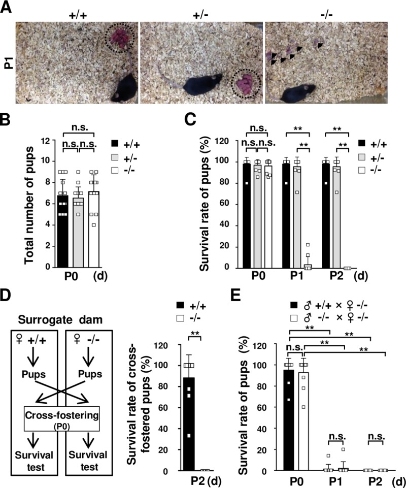
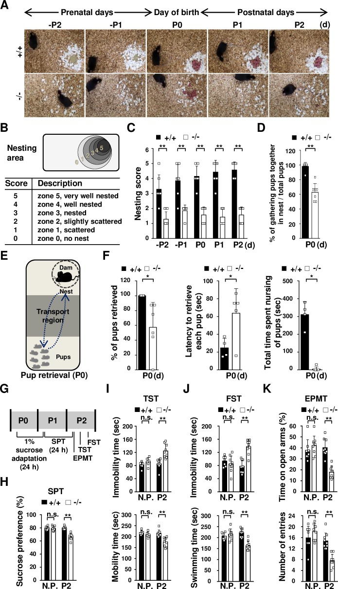
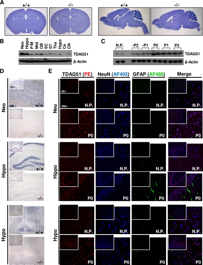
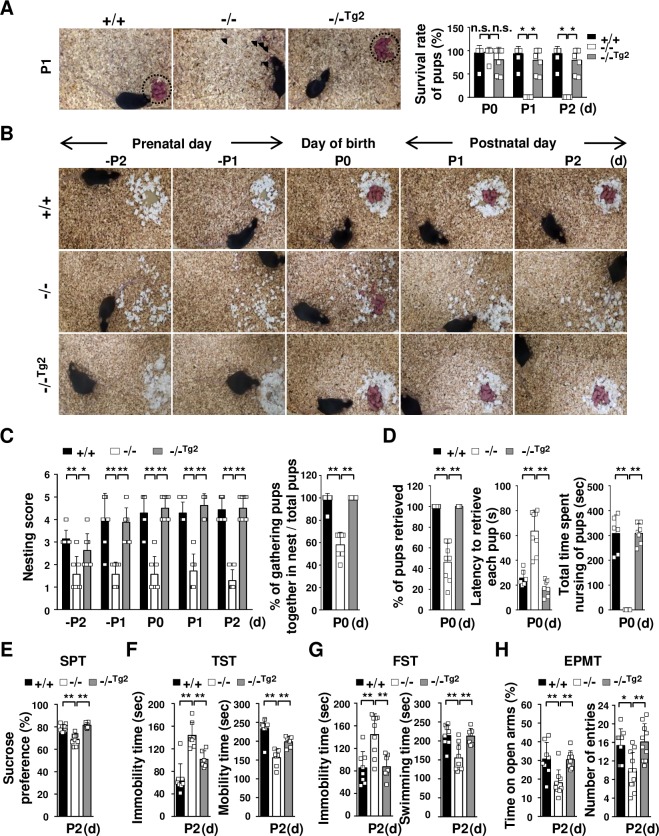
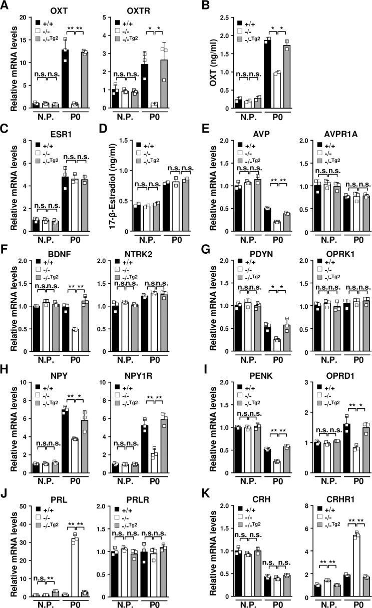
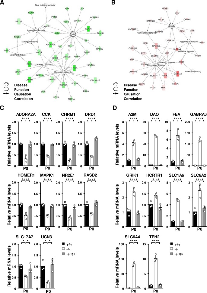
Postpartum depression is a severe emotional and mental disorder that involves maternal care defects and psychiatric illness. Postpartum depression is closely associated with a combination of physical changes and physiological stress during pregnancy or after parturition in stress-sensitive women. Although postpartum depression is relatively well known to have deleterious effects on the developing fetus, the influence of genetic risk factors on the development of postpartum depression remains unclear. In this study, we discovered a novel function of T cell death-associated gene 51 (TDAG51/PHLDA1) in the regulation of maternal and depressive-like behavior. After parturition, TDAG51-deficient dams showed impaired maternal behavior in pup retrieving, nursing and nest building tests. In contrast to the normal dams, the TDAG51-deficient dams also exhibited more sensitive depressive-like behaviors after parturition. Furthermore, changes in the expression levels of various maternal and depressive-like behavior-associated genes regulating neuroendocrine factor and monoamine neurotransmitter levels were observed in TDAG51-deficient postpartum brain tissues. These findings indicate that TDAG51 plays a protective role against maternal care defects and depressive-like behavior after parturition. Thus, TDAG51 is a maternal care-associated gene that functions as a crucial regulator of maternal and depressive-like behavior after parturition.


Conflict of interest statement

The authors have declared that no competing interests exist.

Figures

Fig 1
Fig 1. The survival rate of pups born to TDAG51-/- dams is reduced during the early postpartum period.
(A) Survival of pups born to TDAG51-/- dams. The photographs were obtained on postnatal day 1 (P1). Arrowheads indicate dead pups. Dotted circles indicate pups gathered in a nest. +/+, TDAG51+/+ dams. +/-, TDAG51+/- dams. -/-, TDAG51-/- dams. (B) The average number of pups born to TDAG51-/- dams. The number of pups per dam was analyzed on P0. Black bar (+/+), TDAG51+/+ dams. Gray bar (+/-), TDAG51+/- dams. White bar (-/-), TDAG51-/- dams. (C) The survival rate of pups born to TDAG51-/- dams. Pup survival was measured from P0 to P2 in the absence of male mating partners. (D) The survival rate of pups with surrogate TDAG51-/- dams. A schematic diagram of the cross-fostering experiment between TDAG51+/+ and TDAG51-/- dams is shown in the left panel. Pups born to TDAG51+/+ and TDAG51-/- dams were interchanged on P0, and the survival of the cross-fostered pups with their surrogate dams was analyzed on P2. Black bar (+/+), TDAG51+/+ surrogate dams. White bar (-/-), TDAG51-/- surrogate dams. (E) The effect of male mating partners on the survival of pups born to TDAG51-/- dams. Pup survival was analyzed from P0 to P2 in the presence of the male mating partner. Black bar, TDAG51-/- dams with TDAG51+/+ male mice. White bar, TDAG51-/- dams with TDAG51-/- male mice. **p<0.01. n.s., not significant.
Fig 2
Fig 2. TDAG51 deficiency elicits abnormal maternal behavior and depressive-like behavior after parturition.
(A) Reduced nest building ability in TDAG51-/- female mice. TDAG51+/+ or TDAG51-/- female mice with vaginal plugs were separated from their male mating partners and given nesting material on prenatal day 3 (-P3). Photographs of nest building were obtained from -P2 to P2. +/+, TDAG51+/+ dams. -/-, TDAG51-/- dams. (B) Diagram of the measurement of the nest building score. Nesting scores (0–5) indicate the extent of nest building. (C) TDAG51-/- female mice have impaired nest building abilities. Black bar (+/+), TDAG51+/+ dams. White bar (-/-), TDAG51-/- dams. (D) Percentage of pups gathered in a nest. The number of pups gathered in a nest expressed as a percentage of the total number of neonatal pups measured on P0. (E) Diagram of pup retrieval analysis. (F) Lower pup retrieval by TDAG51-/- dams. Each dam was separated from their pups in a home cage for 5 min. The number of retrieved pups was determined by analyzing a 10-minute video-recording. Left panel, the percentage of retrieved pups per dam. Middle panel, latency to retrieve each pup by the TDAG51-/- dams. Right panel, impaired nursing of the retrieved pups by the TDAG51-/- dams. (G) Diagram of the depressive-like and anxiety-like behavior tests. Nonpregnant (N.P.) female mice or postpartum dams were subjected to 1% sucrose adaptation on P0 for 24 h. Then, 1% sucrose and water were supplied to the mice in their home cages on P1. An SPT was performed on P2; then, the mice were subjected to an EPMT, a TST and an FST in an orderly manner on P2. (H) SPT. Sucrose preference was measured for 24 h. (I) In the TST, the mobility time and immobility time over a 5-min period were analyzed. (J) In the FST, the swimming time and immobility time over a 5-min period were analyzed. (K) In the EPMT, the female mice were exposed to an elevated plus-maze, and the time spent in the open arms and the number of entries were determined over the 10-min test period. *p<0.05. **p<0.01. n.s., not significant.
Fig 3
Fig 3. Expression of TDAG51 in brain tissues.
(A) TDAG51 expression in brain sections from nonpregnant female mice. Histological images of coronal brain sections (top and left panels) and sagittal brain sections (top and right panels) were obtained by hematoxylin staining. +/+, TDAG51+/+. -/-, TDAG51-/-. (B) TDAG51 expression in mouse brain tissues. TDAG51 expression in brain tissues from nonpregnant female mice was analyzed by a western blot analysis with an anti-TDAG51 antibody. β-Actin was used as a loading control. Neo, neocortex. Hippo, hippocampus. P/M, pons/medulla. Mid, midbrain. OB, olfactory bulb. SC, spinal cord. ST, stria terminalis. Tha, thalamus. Hypo, hypothalamus. Ce, cerebellum. CN, cerebral nuclei. (C) Comparison of the TDAG51 expression levels in the brain during pregnancy, parturition and postpartum periods. TDAG51 expression levels in two mice per group were analyzed by a western blot analysis using an anti-TDAG51 antibody. Nonpregnant female mice (N.P.) were used as controls. (D) In situ hybridization analysis of TDAG51 expression in mouse brain tissues. TDAG51 expression was analyzed by an in situ hybridization analysis using DIG-labeled RNA probes on P0. All images were photographed at a 50× or 100× magnification. (E) TDAG51 expression in neuronal cells in mouse brain tissues. Mouse brain tissues were stained with an anti-TDAG51 PE-conjugated, anti-GFAP Alexa Fluor 488 (AF488)-conjugated and anti-NeuN Alexa Fluor 405 (AF405)-conjugated antibodies. All images were photographed at a 60× or 400× magnification. Images from the same observed field were merged.
Fig 4
Fig 4. Effects of the brain-specific expression of the TDAG51 transgene in TDAG51-/- dams.
(A) Rescue effect (as observed in the pup survival test) of brain-specific TDAG51 transgene expression in TDAG51-/- dams. Transgenic lines 2 (TDAG51-/-Tg2) and 3 (TDAG51-/-Tg3) expressing the TDAG51 transgene in the brain of TDAG51-/- female mice were generated. The photographs were obtained on P1 (left panel). Arrowheads indicate dead pups. Dotted circles indicate pups gathered in a nest. Pup survival rate was measured from P0 to P2 (right panel). Black bar (+/+), TDAG51+/+ dams. White bar (-/-), TDAG51-/- dams. Gray bar (-/-Tg2), TDAG51-/-Tg2 dams. (B) Rescue effect on nest building behavior in TDAG51-/-Tg2 pregnant mice. Photographs of the nest were obtained from -P2 to P2. (C) Measurement of nest building abilities. Nesting scores of TDAG51+/+, TDAG51-/- and TDAG51-/-Tg2 pregnant mice were recorded (left panel). The number of pups gathered in a nest expressed as a percentage of the total number of neonatal pups measured on P0 (right panel). (D) Rescue effect on pup retrieval behavior in TDAG51-/-Tg2 dams. Left panel, percentage of retrieved pups per dam. Middle panel, latency to retrieve each pup by TDAG51-/- dams. Right panel, impaired nursing behavior in TDAG51-/- dams. (E) SPT. (F) TST. (G) FST. (H) EPMT. *p<0.05. **p<0.01. n.s., not significant.
Fig 5
Fig 5. Dysregulation of neuroendocrine factors in the brain tissue of TDAG51-/- dams after parturition.
(A) Impaired expression of OXT and its receptor OXTR in TDAG51-/- dams after parturition. Total RNA isolated from brain tissues was analyzed by real-time PCR using specific primers. N.P., nonpregnant mice. Black bar (+/+), TDAG51+/+ dams. White bar (-/-), TDAG51-/- dams. Gray bar (-/-Tg2), TDAG51-/-Tg2 dams. (B) Serum OXT levels in TDAG51-/- dams after parturition. Serum OXT levels were analyzed using ELISA. (C) No difference in ESR1 expression was observed in the TDAG51-/- dams. (D) No difference in the serum estrogen levels was observed in the TDAG51-/- dams. Serum estrogen (17-β-estradiol) levels after parturition were analyzed using ELISA. (E) Downregulation of AVP expression in TDAG51-/- dams after parturition. (F) Downregulation of BDNF expression in TDAG51-/- dams after parturition. (G) Downregulation of PDYN expression in TDAG51-/- dams after parturition. (H) Downregulation of NPY and its receptor NPY1R expression in TDAG51-/- dams after parturition. (I) Downregulation of PENK and its receptor OPRD1 expression in TDAG51-/- dams after parturition. (J) Upregulation of PRL expression in TDAG51-/- dams after parturition. (K) Upregulation of CRHR1 expression in TDAG51-/- dams after parturition. *p<0.05. **p<0.01. n.s., not significant.
Fig 6
Fig 6. Altered gene expression in the brain tissue of TDAG51-/- dams after parturition.
(A) Prediction of the functional networks of the 39 downregulated genes listed in Table 1. Node color intensity indicates the degree of the downregulation of gene expression as follows: a greater intensity of green represents a higher degree of downregulation. Inverted triangle, kinase; dotted square, growth factor; vertical rectangle, G-protein coupled receptor; dotted vertical rectangle, ion channel; horizontal rectangle, ligand-dependent nuclear receptor; vertical diamond, enzyme; trapezoid, transporter; horizontal ellipse, transcription regulator; circle, other. (B) Prediction of the functional networks of the 31 upregulated genes listed in Table 2. Node color intensity indicates the degree of the upregulation of gene expression as follows: a greater intensity of red represents a higher degree of upregulation. (C) Real-time PCR analysis of downregulated genes selected from the list shown in Table 1. Total RNA isolated from brain tissues was analyzed using real-time PCR with specific primers. Black bar (+/+), TDAG51+/+ dams. White bar (-/-), TDAG51-/- dams. Gray bar (-/-Tg2), TDAG51-/-Tg2 dams. (D) Real-time PCR analysis of upregulated genes selected from the list shown in Table 2. *p<0.05. **p<0.01.

Similar articles

Cited by

References

    1. Bridges RS. Neuroendocrine regulation of maternal behavior. Front Neuroendocrinol. 2015;36:178–96. 10.1016/j.yfrne.2014.11.007 - DOI - PMC - PubMed
    1. Pawluski JL, Lonstein JS, Fleming AS. The Neurobiology of Postpartum Anxiety and Depression. Trends Neurosci. 2017;40(2):106–20. 10.1016/j.tins.2016.11.009 - DOI - PubMed
    1. Bergink V, Rasgon N, Wisner KL. Postpartum Psychosis: Madness, Mania, and Melancholia in Motherhood. Am J Psychiatry. 2016;173(12):1179–88. 10.1176/appi.ajp.2016.16040454 - DOI - PubMed
    1. Corwin EJ, Kohen R, Jarrett M, Stafford B. The heritability of postpartum depression. Biol Res Nurs. 2010;12(1):73–83. 10.1177/1099800410362112 - DOI - PMC - PubMed
    1. Josefsson A, Berg G, Nordin C, Sydsjo G. Prevalence of depressive symptoms in late pregnancy and postpartum. Acta Obstet Gynecol Scand. 2001;80(3):251–5. - PubMed

Publication types

LinkOut - more resources