. 2020 Jan;140(1):143-151.e3.
doi: 10.1016/j.jid.2019.06.127. Epub 2019 Jun 26.

IL-20-Receptor Signaling Delimits IL-17 Production in Psoriatic Inflammation

Affiliations

IL-20-Receptor Signaling Delimits IL-17 Production in Psoriatic Inflammation

Hye-Lin Ha et al. J Invest Dermatol. 2020 Jan.

Abstract

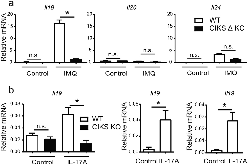
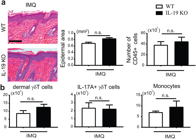
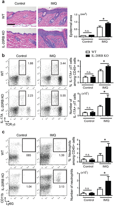
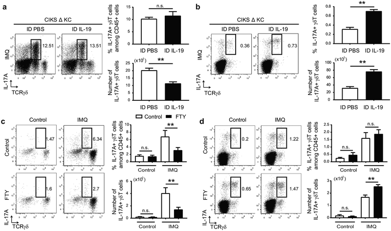
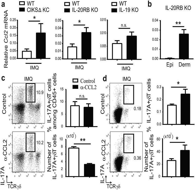
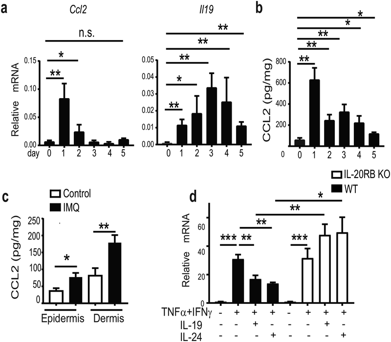
IL-17 cytokines, in particular IL-17A, are critical effectors in psoriasis. Antibodies that block IL-17A are highly efficacious in treating psoriasis. Likewise, disruption of IL-17 cytokines signaling, such as via the loss of the adaptor CIKS/Act1, ameliorates inflammation in mouse models of psoriasis. IL-17A promotes a cascade of effects, including the robust production of IL-19 in both humans and mice. IL-19, along with IL-20 and IL-24, signal via IL-20 receptors and comprise a subgroup within the IL-10 cytokine family. The role of these three cytokines in psoriasis is unresolved. They have been linked to inflammatory processes, including psoriatic pathology, but these cytokines have also been reported to suppress inflammation in other contexts. In this study, we demonstrate that signaling via IL-20 receptors, including in response to IL-19, delimited aspects of imiquimod-induced psoriatic inflammation. IL-20 receptor signaling suppressed the dermal production of the CCL2 chemokine and thereby reduced CCL-2-driven infiltration of inflammatory cells into the dermis, including IL-17A-producing γδT cells. This constitutes a negative feedback, since IL-17A strongly induces IL-19 in keratinocytes. The effects of IL-17 cytokines in this inflammatory setting are dynamic; they are central to the development of both dermal and epidermal hallmarks of psoriasis but also initiate a path to mitigate inflammatory damage.


Conflict of interest statement

CONFLICT OF INTEREST

The authors state no conflict of interest.

Figures

Figure 1.
Figure 1.. IL-17 induces IL-19 in keratinocytes.
(a) Relative mRNA expression of IL-20 family cytokines in epidermis of IMQ- or control-treated ears from wild-type (WT) and CIKS DKC [K5-cre;Traf3ip2/flx] mice. (b) Relative mRNA expression of IL-19 in primary keratinocytes from WT and CIKS KO mice, HaCaT cells and WT ear explants stimulated with IL-17A for 6h (left, middle and right panels, respectively). (*p < 0.05; mean ± SEM; n =5–8, except n=3 for primary CIKS KO keratinocytes).
Figure 2.
Figure 2.. IMQ-induced psoriatic inflammation in IL-19 KO mice.
(a) Representative H&E-stained dorsal sections of IMQ-treated WT and IL-19 KO mice. Left graph: Epidermal thickening quantitated via measurements of epidermal area from sections. Scale bar 100 mm. Right graph: Numbers of CD45+ cells in dorsal skin sections. (b) Numbers of dermal gdT cells (CD45+, TCRγδintermediate), IL-17A+ γδT cells and monocytic cells (CD45+ Ly6C+, CD11b+, excluding MHCII, CD64 cells) based on flow cytometric analyses of IMQ-treated dorsal skin cells from WT and IL-19 KO mice. (for a and b: mean ± SEM; n = 6 mice per group).
Figure 3.
Figure 3.. Exacerbated psoriatic inflammation in IL-20RB KO mice, including a rise in IL-17A+ γδTcells.
(a) Representative H&E-stained dorsal sections of IMQ- or control-treated WT and IL-20RB KO mice. Scale bar 100 μm. Epidermal thickening quantitated via measurement of epidermal area from sections. (b, c) Representative flow cytometric analyses of IMQ- or control-treated dorsal skin cells from WT and IL-20RB KO mice analyzed for expression of markers as shown within the CD45+ gate. (b) Numbers and percentages of IL-17A+ γδT cells and (c) neutrophils generated from flow cytometric analyses. (for a-c: *p < 0.05; mean ± SEM; n = 8–12 mice per group).
Figure 4.
Figure 4.. IL-19 limits accumulation of IL-17+ dermal γδT cells.
(a, b) Representative flow cytometric analyses for markers shown of cells obtained from IMQ-treated ears (a) and earsDLNs (b) of CIKS ΔKC mice i.d. injected with PBS or IL-19. Numbers and percentages of IL-17A+ γδT cells generated from flow cytometric analyses. (**p < 0.01; mean ± SEM; n = 6–8 mice per group). (c, d) Representative flow cytometric analyses for markers shown of cells obtained from IMQ-treated dorsal skin (c) and sDLNs (d) of WT mice injected i.p. with PBS or FTY720. Numbers and percentages of IL-17A+ γδT cells generated from flow cytometric analyses. (**p < 0.01; mean ± SEM; n = 6–9 mice per group).
Figure 5.
Figure 5.. IL-19/IL-20 receptor signaling reduces CCL2 and thereby limits accumulation of IL-17A+ dermal γδT.
(a) Relative mRNA expression for CCL2 in IMQ-treated dorsal skin of CIKS ΔKC, IL-20RB KO, IL-19 KO and WT mice (*p < 0.05; mean ± SEM; n = 6–15 mice per group). (b) Relative mRNA expression for CCL2 in epidermis (Epi) and dermis (Derm) of IMQ-treated ears of IL-20RB KO mice. (**p < 0.01; mean ± SEM; n =7). (c, d) Representative flow cytometric analyses of IMQ-treated ear skin cells (c) and ear sDLNs (d) of CIKS KC mice i.d. injected with α-CCL2 or control antibodies, analyzed for expression of markers shown. Numbers and percentages of IL-17A+ γδT cells generated from flow cytometric analyses. (*p < 0.05, **p< 0.01; mean ± SEM; n = 6–8).
Figure 6.
Figure 6.. IL-19 downregulates expression of CCL2 in dermal fibroblast
(a,b) Relative mRNA expression of CCL2 and IL-19 (a) and CCL2 protein levels (b) in dorsal skin of WT mice during the course of 5 successive IMQ-treatments. (c) Protein levels of CCL2 in one-time IMQ-or control-treated epidermal and dermal layers of ears of WT mice (**p < 0.01; mean ± SEM; n = 6–10 per group). (d) Relative mRNA expression of CCL2 in primary dermal fibroblast cultures of WT and IL-20RB KO mice stimulated (or not) with IFNγ and TNFα in the presence/absence of IL-19 or IL-24, as shown (**p < 0.01; mean ± SEM; n = 10 per group).

References

    1. Autieri MV. IL-19 and Other IL-20 Family Member Cytokines in Vascular Inflammatory Diseases. Front Immunol 2018;9:700. - PMC - PubMed
    1. Bissonnette R, Fuentes-Duculan J, Mashiko S, Li X, Bonifacio KM, Cueto I, et al. Palmoplantar pustular psoriasis (PPPP) is characterized by activation of the IL-17A pathway. J Dermatol Sci 2017;85(1):20–6. - PubMed
    1. Boehncke WH, Schon MP. Psoriasis. Lancet 2015;386(9997):983–94. - PubMed
    1. Cai Y, Shen X, Ding C, Qi C, Li K, Li X, et al. Pivotal role of dermal IL-17-producing gammadelta T cells in skin inflammation. Immunity 2011;35(4):596–610. - PMC - PubMed
    1. Canto E, Garcia Planella E, Zamora-Atenza C, Nieto JC, Gordillo J, Ortiz MA, et al. Interleukin-19 impairment in active Crohn’s disease patients. PLoS One 2014;9(4):e93910. - PMC - PubMed

Publication types

MeSH terms