. 2019 Jun 27;24(13):2370.
doi: 10.3390/molecules24132370.

Computational Investigation of Bisphosphate Inhibitors of 3-Deoxy-d- manno-octulosonate 8-phosphate Synthase

Affiliations

Computational Investigation of Bisphosphate Inhibitors of 3-Deoxy-d- manno-octulosonate 8-phosphate Synthase

Jéssica de Oliveira Araújo et al. Molecules. .

Abstract

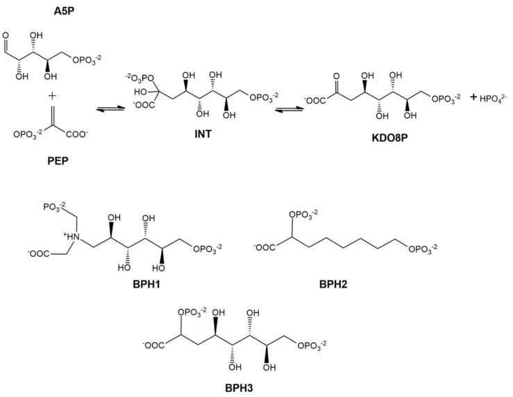
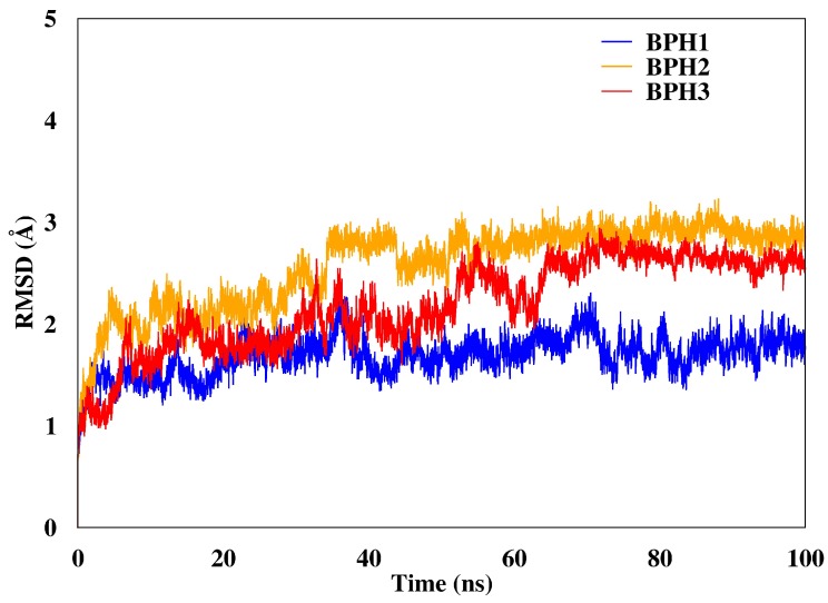
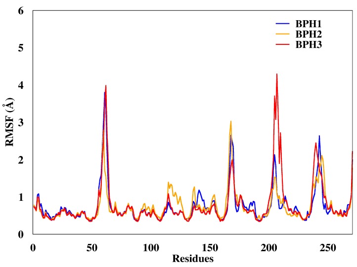
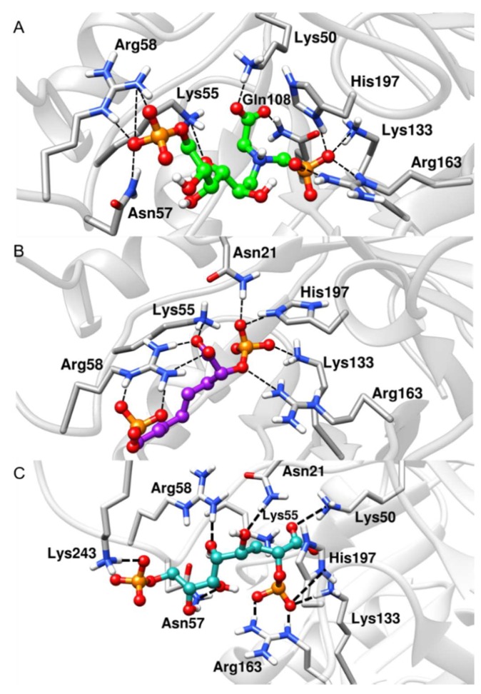
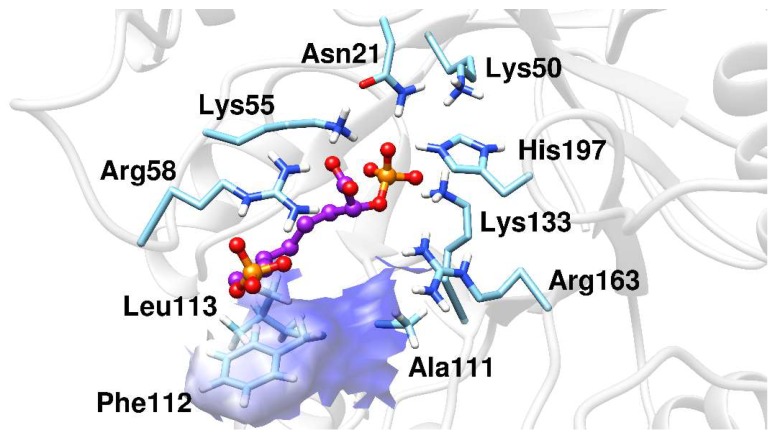
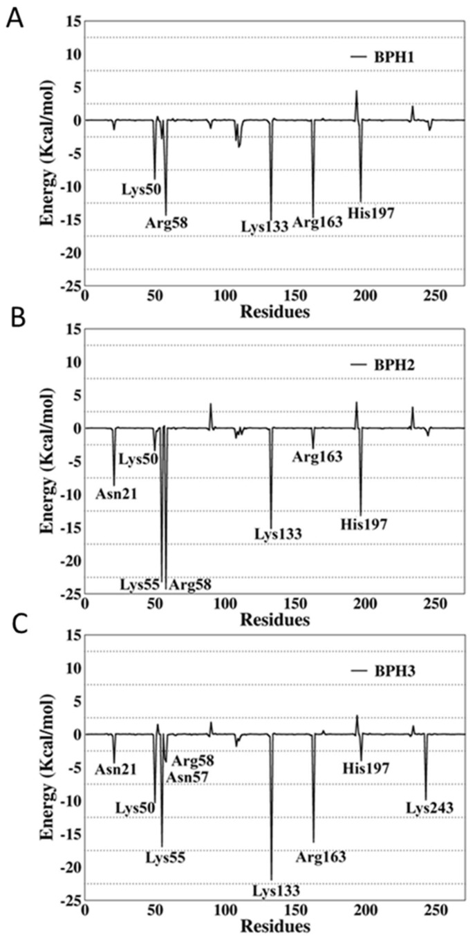
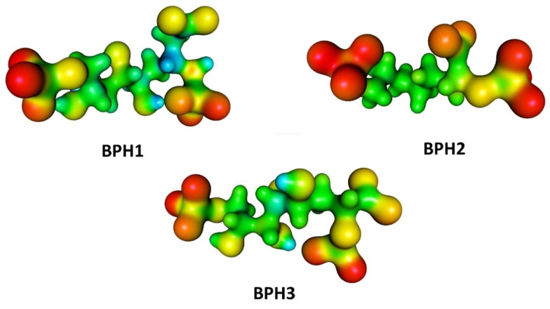
The synthase, 3-deoxy-d-manno-octulosonate 8-phosphate (KDO8P), is a key enzyme for the lipopolysaccharide (LPS) biosynthesis of gram-negative bacteria and a potential target for developing new antimicrobial agents. In this study, computational molecular modeling methods were used to determine the complete structure of the KDO8P synthase from Neisseria meningitidis and to investigate the molecular mechanism of its inhibition by three bisphosphate inhibitors: BPH1, BPH2, and BPH3. Our results showed that BPH1 presented a protein-ligand complex with the highest affinity, which is in agreement with experimental data. Furthermore, molecular dynamics (MD) simulations showed that BPH1 is more active due to the many effective interactions, most of which are derived from its phosphoenolpyruvate moiety. Conversely, BPH2 exhibited few hydrogen interactions during the MD simulations with key residues located at the active sites of the KDO8P synthase. In addition, we hydroxylated BPH2 to create the hypothetical molecule named BPH3, to investigate the influence of the hydroxyl groups on the affinity of the bisphosphate inhibitors toward the KDO8P synthase. Overall, we discuss the main interactions between the KDO8P synthase and the bisphosphate inhibitors that are potential starting points for the design of new molecules with significant antibiotic activities.

Keywords: KDO8P synthase; Neisseria meningitidis; bisphosphate inhibitors; molecular dynamics.


Conflict of interest statement

The authors declare no conflict of interest.

Figures

Scheme 1
Scheme 1
At the top, 2D structures that represent the reaction of d-arabinose 5-phosphate (A5P) and phosphoenolpyruvate (PEP) to form 3-deoxy-d-manno-octulosonate-8-phosphate (KDO8P) and inorganic phosphate (Pi). The doubly phosphorylated intermediate (INT) is used as a reference for the design of the analog inhibitors: BPH1, BPH2, and BPH3.
Figure 1
Figure 1
Structure of the modeled KDO8P synthase from Neisseria meningitidis evidencing the regions of the loops. The loops, L2, L7, and L8, control access to the active site.
Figure 2
Figure 2
Overlapped structures of the bisphosphate inhibitors with PEP and A5P substrates (PDB ID: 1FXQ). The carbon atoms of the substrates are colored yellow. (AC) are the superimpositions with BPH1, BPH2, and BPH3, respectively. It is noted that the phosphate groups derived from the substrates overlap the phosphate groups of the inhibitors.
Figure 3
Figure 3
RMSD of the backbone atoms of the complexes after 100 ns of MD simulations. The RMSD represents the enzyme in the presence of the BPH1 (blue), BPH2 (yellow), and BPH3 (red) inhibitors.
Figure 4
Figure 4
Root mean square deviations (RMSF) of the backbone atoms of the complexes from representative snapshots of MD simulations. Enzyme in the presence of the BPH1, BPH2, and BPH3 inhibitors are colored blue, yellow, and red, respectively.
Figure 5
Figure 5
Representative structures along the MD simulations. (A) BPH1-KDO8P synthase, (B) BPH2-KDO8P synthase, and (C) BPH3-KDO8P synthase complexes. We can see important interactions of phosphate groups of the inhibitors with positively charged groups of KDO8P synthase.
Figure 6
Figure 6
The BPH2 inhibitor having polar contacts at the active site of the KDO8P synthase.
Figure 7
Figure 7
Per-residue decomposition analysis of (A) BPH1, (B) BPH2, and (C) BPH3 in complexes with the KDO8P synthase from N. meningitidis.
Figure 8
Figure 8
Molecular electrostatic potential (MEP) surfaces of biphosphate inhibitors BPH1, BPH2, and the hypothetical BPH3. Calculations were done at the M06-2X/6-31++G (d,p) level.

References

    1. Raetz C.R.H., Whitfield C. Lipopolysaccharide Endotoxins. Annu. Rev. Biochem. 2002;71:635–700. doi: 10.1146/annurev.biochem.71.110601.135414. - DOI - PMC - PubMed
    1. Ray P.H. Purification and characterization of 3-deoxy-d-manno-octulosonate 8-phosphate synthetase from Escherichia coli. J. Bacteriol. 1980;141:635–644. - PMC - PubMed
    1. Brennan P.J., Nikaido H. The Envelope of Mycobacteria. Annu. Rev. Biochem. 1995;64:29–63. doi: 10.1146/annurev.bi.64.070195.000333. - DOI - PubMed
    1. Raetz C. Biochemistry of Endotoxins. Annu. Rev. Biochem. 1990;59:129–170. doi: 10.1146/annurev.bi.59.070190.001021. - DOI - PubMed
    1. Rick P.D., Young D.A. Isolation and characterization of a temperature-sensitive lethal mutant of Salmonella typhimurium that is conditionally defective in 3-deoxy-d-manno-octulosonate-8-phosphate synthesis. J. Bacteriol. 1982;150:447–455. - PMC - PubMed

MeSH terms

LinkOut - more resources