. 2019 Jun 27;11(7):1090.
doi: 10.3390/polym11071090.

Growth on Metallo-Supramolecular Coordination Polyelectrolyte (MEPE) Stimulates Osteogenic Differentiation of Human Osteosarcoma Cells (MG63) and Human Bone Marrow Derived Mesenchymal Stem Cells

Affiliations

Growth on Metallo-Supramolecular Coordination Polyelectrolyte (MEPE) Stimulates Osteogenic Differentiation of Human Osteosarcoma Cells (MG63) and Human Bone Marrow Derived Mesenchymal Stem Cells

Janina Belka et al. Polymers (Basel). .

Abstract

Background: Culturing of cells is typically performed on standard tissue culture plates generating growth conditions, which in general do not reflect the native three-dimensional cellular environment. Recent investigations provide insights in parameters, which strongly affect the general cellular behavior triggering essential processes such as cell differentiation. The physical properties of the used material, such as stiffness, roughness, or topology, as well as the chemical composition of the cell-surface interface are shown to play a key role in the initiation of particular cellular responses.

Methods: We extended our previous research, which identified thin films of metallo-supramolecular coordination polyelectrolytes (MEPEs) as substrate to trigger the differentiation of muscular precursor cells.

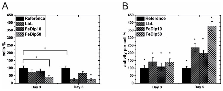
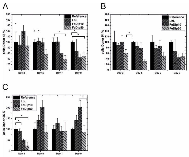
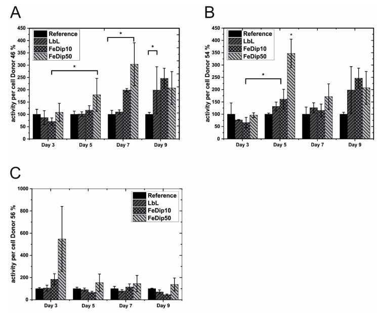
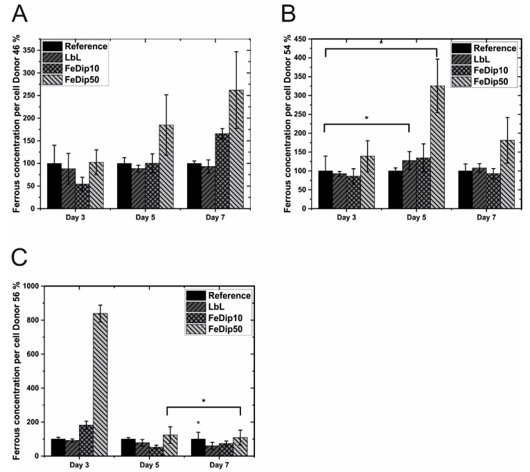
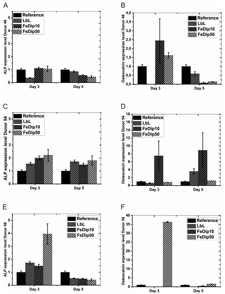
Results: Here, we show that the same MEPEs similarly stimulate the osteogenic differentiation of pre-osteoblasts. Remarkably, MEPE modified surfaces also trigger the differentiation of primary bone derived mesenchymal stem cells (BMSCs) towards the osteogenic lineage.

Conclusion: This result leads to the conclusion that these surfaces individually support the specification of cell differentiation toward lineages that correspond to the natural commitment of the particular cell types. We, therefore, propose that Fe-MEPEs may be used as scaffold for the treatment of defects at least in muscular or bone tissue.

Keywords: cell differentiation; interface; iron metabolism; metallo-supramolecular polymer.


Conflict of interest statement

The authors declare no conflict of interest

Figures

Figure 1
Figure 1
(A) Cell number and (B) metabolic activities of the used MG63 cells was determined at day 3 and 5 for the particular surface modifications. The cell number and the metabolic activity were set in relation to cells grown on tissue culture polystyrene (TCPS) substrates (reference). The experiments were performed at least two times in triplicate. * p < 0.5 (analyzed by one-way ANOVA with Tukey test).
Figure 2
Figure 2
Light microscopy images of Alizarin red stained MG63 cells. (A) Cells grown on Reference substrate, (B) LbL, (C) FeDip10, and (D) FeDip50 at day 3.
Figure 3
Figure 3
Light microscopy images at different magnification of Alizarin red stained MG63 cells grown on (A,B) reference substrates, (C,D) FeDip10, and (E,F) on FeDip50 substrates at day 5.
Figure 4
Figure 4
Cell growth of the used bone derived mesenchymal stem cells (BMSCs) from (A) Donor 46, (B) Donor 54, and (C) Donor 56 relative to that on the reference surface for the particular surface modifications at four different time points. All experiments were performed in triplicate. * p < 0.5 (analyzed by one-way ANOVA with Tukey test).
Figure 5
Figure 5
Relative Metabolic activities of the used hMSC cells from (A) Donor 46, (B) Donor 54, and (C) Donor 56 was determined at four different time points for the particular surface modifications in relation to cells grown on reference substrates. All experiments were performed in triplicate. * p < 0.5 (analyzed by one-way ANOVA with Tukey test).
Figure 6
Figure 6
Examples of light microscopy images of Alizarin red stained BMSCs grown on (A) reference (Donor 409 46), (B) FeDip50 (Donor 46), and (C) FeDip50 (Donor 54) at day 5.
Figure 7
Figure 7
The bar diagrams represent the relative ferrous concentrations per cell for (A) donor 46, (B) donor 54, and (C) donor 56 grown on the indicated substrates. The values obtained for cells grown on TCPS substrates are set to 100% (the values for the references substrates also vary donor dependently. The absolute values for the reference substrates at day 3 are: donor 46 = 68.2 fMol/cell; donor 54 = 31 fMol/cell; donor 56 = 21.6 fMol/cell). All experiments were performed in triplicate. * p < 0.5 (analyzed by one-way ANOVA with Tukey test).
Figure 8
Figure 8
The bar diagrams represent values for relative ALP (A,C,E) and OC (B,D,F) expression levels of cells grown on the indicated substrates of Donor 46 (A,B), Donor 54 (C,D), and Donor 56 (E,F). All values are normalized to GAPDH expression levels. Subsequently the levels of ALP and OC expression determined for cells grown on reference substrates are set to 1. All experiments were performed in triplicate.

References

    1. Belka J., Weigel T., Berninger A.-K., Kurth D.G., Nickel J. Growth and differentiation of myoblastic precursor cells on thin films of metallo-supramolecular coordination polyelectrolyte (mepe) Adv. Mater. Interfaces. 2017;4:1600272. doi: 10.1002/admi.201600272. - DOI
    1. Bains W., Ponte P., Blau H., Kedes L. Cardiac actin is the major actin gene-product in skeletal-muscle cell-differentiation in vitro. Mol. Cell Biol. 1984;4:1449–1453. doi: 10.1128/MCB.4.8.1449. - DOI - PMC - PubMed
    1. Atmani H., Audrain C., Mercier L., Chappard D., Baslé M.F. Phenotypic effects of continuous or discontinuous treatment with dexamethasone and/or calcitriol on osteoblasts differentiated from rat bone marrow stromal cells. J. Cell. Biochem. 2002;85:640–650. doi: 10.1002/jcb.10165. - DOI - PubMed
    1. Carinci F., Pezzetti F., Spina A.M., Palmieri A., Laino G., De Rosa A., Farina E., Illiano F., Stabellini G., Perrotti V., et al. Effect of Vitamin C on pre-osteoblast gene expression. Arch. Oral Boil. 2005;50:481–496. doi: 10.1016/j.archoralbio.2004.11.006. - DOI - PubMed
    1. Giannoudis P.V., Einhorn T.A., Schmidmaier G., Marsh D. The diamond concept—Open questions. Injury. 2008;39:S5–S8. doi: 10.1016/S0020-1383(08)70010-X. - DOI - PubMed

LinkOut - more resources