. 2019 Jun 27;10(7):489.
doi: 10.3390/genes10070489.

Further Insights into the Architecture of the PN Promoter That Controls the Expression of the bzd Genes in Azoarcus

Affiliations

Further Insights into the Architecture of the PN Promoter That Controls the Expression of the bzd Genes in Azoarcus

Gonzalo Durante-Rodríguez et al. Genes (Basel). .

Abstract

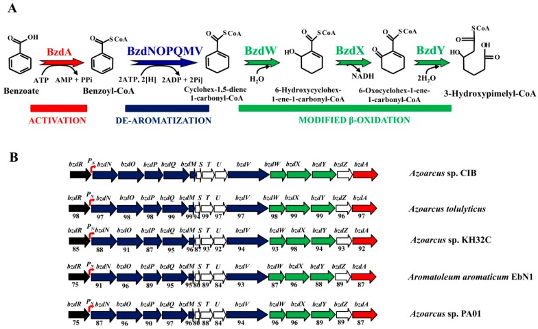
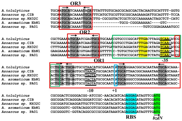
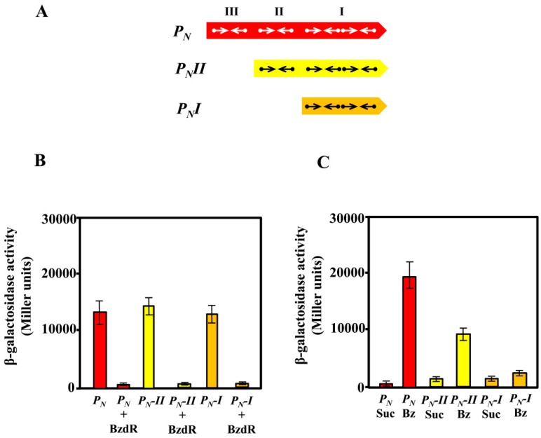
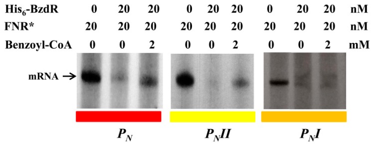
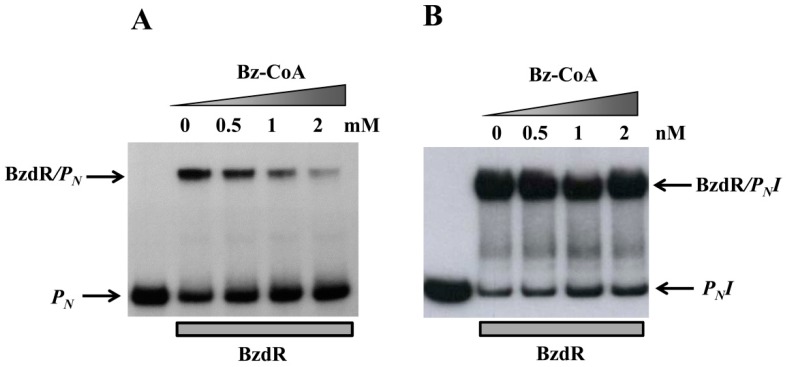
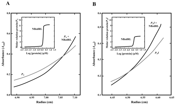
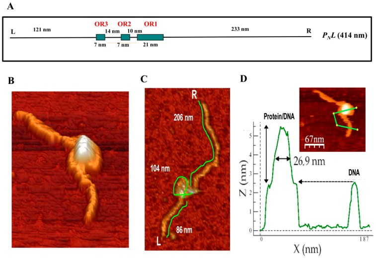
The anaerobic degradation of benzoate in bacteria involves the benzoyl-CoA central pathway. Azoarcus/Aromatoleum strains are a major group of anaerobic benzoate degraders, and the transcriptional regulation of the bzd genes was extensively studied in Azoarcus sp. CIB. In this work, we show that the bzdR regulatory gene and the PN promoter can also be identified upstream of the catabolic bzd operon in all benzoate-degrader Azoarcus/Aromatoleum strains whose genome sequences are currently available. All the PN promoters from Azoarcus/Aromatoleum strains described here show a conserved architecture including three operator regions (ORs), i.e., OR1 to OR3, for binding to the BzdR transcriptional repressor. Here, we demonstrate that, whereas OR1 is sufficient for the BzdR-mediated repression of the PN promoter, the presence of OR2 and OR3 is required for de-repression promoted by the benzoyl-CoA inducer molecule. Our results reveal that BzdR binds to the PN promoter in the form of four dimers, two of them binding to OR1. The BzdR/PN complex formed induces a DNA loop that wraps around the BzdR dimers and generates a superstructure that was observed by atomic force microscopy. This work provides further insights into the existence of a conserved BzdR-dependent mechanism to control the expression of the bzd genes in Azoarcus strains.

Keywords: Azoarcus; anaerobic; benzoate; promoter architecture; regulation.


Conflict of interest statement

Conflicts of interest: The authors declare that there are no competing interests.

Figures

Figure 1
Figure 1
Scheme of anaerobic degradation of benzoate and gene organization of the bzd cluster in different Azoarcus and Aromatoleum strains. (A) Scheme of the anaerobic degradation pathway of benzoate into benzoyl-CoA (red), the de-aromatization of benzoyl-CoA (blue), and the modified β-oxidation that produces 3-hydroxypimelyl-CoA. Enzyme abbreviations: BzdA, benzoate-CoA ligase; BzdNOPQMV, benzoyl-CoA reductase, ferredoxin, and putative reduced nicotinamide adenine dinucleotide phosphate (NADPH)::ferredoxin oxidoreductase; BzdW, cyclohex-1,5-diene-1-carbonyl-CoA hydratase; BzdX, 6-hydroxycyclohex-1-ene-1-carbonyl-CoA dehydrogenase; BzdY, 6-oxocyclohexene-1-ene-carbonyl-CoA hydrolase. (B) Scheme of the bzd cluster in different Azoarcus/Aromatoleum strains. Genes are indicated in the same color code as the corresponding enzymes in panel (A), i.e., genes encoding the activation, de-aromatization, and modified β-oxidation are indicated in red, blue, and green color, respectively. The bzdR regulatory gene is shown in black, and the catabolic PN promoter is shown in red as a curved arrow. Genes of unknown function are colored in white. Below each gene, the percentage of the amino-acid sequence identity to the corresponding Azoarcus sp. CIB ortholog is indicated. The accession numbers of the corresponding genome sequences are as follows: Azoarcus sp. CIB (CP011072), Azoarcus sp. KH32C (AP012304), Aromatoleum aromaticum EbN1 (CR555306), Azoarcus tolulyticus strain ATCC51758 (NZ_FTMD00000000), and Azoarcus sp. PA01 (NZ_LARU00000000).
Figure 2
Figure 2
Sequence comparison analysis between the Azoarcus sp. CIB PN promoter [20] and the PN promoters from other Azoarcus/Aromatoleum strains. The sequence of the PN promoter from strain CIB spanning from position −174 to +79, with respect to the transcription start site (+1, indicated) is shown and compared with that of PN promoters from other Azoarcus strains. The σ70-RNA polymerase (RNAP) recognition sequences −10 and −35 are underlined, the ribosome-binding sequence (RBS) is marked in cyan, and the ATG initiation codon of the bzdN gene is shown in green. The operator regions I, II, and III (OR1, OR2, and OR3), involved in the interaction with the BzdR protein, are boxed in red. The arrows indicate the palindromic TGCA sequences (in gray) present in each of the operator regions. The green box shows the AcpR-binding region (palindromic sequences are indicated in yellow).
Figure 3
Figure 3
Activity of the PN, PNI, and PNII promoters in Escherichia coli and Azoarcus sp. CIB cells. (A) Schematic representation of the PN, PNII, and PNI promoters. Arrows represent the inverted repeat TGCA sequences for BzdR binding at operator regions I, II, and III. (B) The activity of the promoters was analyzed in E. coli MC4100 cells harboring the control plasmid pCK01 or plasmid pCK01-BzdR that expresses the bzdR gene (BzdR), and the plasmids pSJ3-PN, pSJ3-PNI, or pSJ3-PNII that harbor the PN::lacZ (PN), PNI::lacZ (PN-I), or PNII::lacZ (PN-II) translational fusions, respectively. Growth was performed in lysogeny broth (LB) in anaerobic conditions for 16 h as described in Section 2.6. The β-galactosidase activity was measured as detailed in Section 2.6 [34]. Graphed values are the averages from three independent experiments ± SD (error bars). (C) Activity of the PN, PNII, and PNI promoters in Azoarcus sp. CIB cells. Azoarcus sp. CIB cells harboring plasmids pBBR5-PN, pBBR5-PNI, or pBBR5-PNII, which express the PN::lacZ (PN), PNI::lacZ (PN-I), or PNII::lacZ (PN-II) translational fusions, respectively, were anaerobically grown for 72 h in MC medium supplemented with 0.2% succinate (Suc) or 3 mM benzoate (Bz), and the β-galactosidase activity was measured as detailed in Section 2.6. Graphed values are the averages from three independent experiments ± SD (error bars).
Figure 4
Figure 4
In vitro activity of the PN, PNII, and PNI promoters. Multiple-round transcription reactions were carried out as detailed in Section 2.7 by using pJCD-PN, pJCD-PNI, and pJCD-PNII plasmids harboring PN, PNI, and PNII promoter templates, respectively, which produced the corresponding messenger RNA (mRNA). All the in vitro transcription reactions were performed with 100 nM E. coli σ70-RNAP holoenzyme. Purified FNR* was used at 20 nM. When required, His6-BzdR protein was used at 20 nM, and benzoyl-CoA was added at 2 mM.
Figure 5
Figure 5
Gel retardation analysis of BzdR binding to the PN (A) or the PNI promoter (B). Gel retardation analysis was performed as described in Section 2.8 by using 10 nM purified His6-BzdR in the presence of increasing concentrations (from 0 to 2 mM) of benzoyl-CoA (Bz-CoA). The free PN probe and the BzdR/PN complex (A) or PNI probe and the BzdR/PNI complex (B) are indicated by arrows.
Figure 6
Figure 6
Sedimentation equilibrium analysis to study the interaction of purified NBzdRL protein with PN and PNI DNA fragments. (A) Distribution of the gradient in sedimentation equilibrium of the PN fragment (PN, gray line) or PN fragment with His6-NBzdRL protein (black line). The inset graph shows the protein/DNA molar ratio as the concentration of His6-NBzdRL increases. (B) Distribution of the gradient in sedimentation equilibrium of the PNI fragment (gray line) and PNI + His6-NBzdRL complex (black line). The inset graph shows the protein/DNA molar ratio as the concentration of NBzdRL increases.
Figure 7
Figure 7
Analysis of the binding of BzdR to the PN promoter by atomic force microscopy (AFM). (A) Scheme of the PNL DNA fragment used as a template. The DNA fragment extends from position −505 to +720 with respect to the transcription start site of the PN promoter. The operator regions recognized by BzdR are represented by green boxes with their corresponding numbers (OR1, OR2, and OR3). The distances separating each of the indicated regions, the extension of each operator region in nm, and the left (L) and right (R) ends of the PNL fragment are also indicated. (B) Image of a superstructure of the BzdR/PNL complex. (C) Analysis of distances in a BzdR/PNL superstructure (one image, n = 16). The distances of both arms and the loop of the superstructure are detailed. L and R indicate the left and right ends of the PNL DNA fragment, respectively. (D) Image of a BzdR/PNL superstructure and its corresponding height profile. The green line of the image corresponds to the profile outlined in the lower graph. In the graph, the length (in nm) of the line is represented on the x-axis and the height on the y-axis. The first peak corresponds to the BzdR/PNL superstructure and the second peak corresponds to the naked DNA.

References

    1. Pieper D.H., Reineke W. Engineering bacteria for bioremediation. Curr. Opin. Biotechnol. 2000;11:262–270. doi: 10.1016/S0958-1669(00)00094-X. - DOI - PubMed
    1. Fuchs G., Boll M., Heider J. Microbial degradation of aromatic compounds—From one strategy to four. Nat. Rev. Microbiol. 2011;9:803–816. doi: 10.1038/nrmicro2652. - DOI - PubMed
    1. Brzeszcz J., Kaszycki P. Aerobic bacteria degrading both n-alkanes and aromatic hydrocarbons: And undervalued strategy for metabolic diversity and flexibility. Biodegradation. 2018;29:359–407. doi: 10.1007/s10532-018-9837-x. - DOI - PubMed
    1. Díaz E., Jiménez J.I., Nogales J. Aerobic degradation of aromatic compounds. Curr. Opin. Biotechnol. 2013;24:431–442. doi: 10.1016/j.copbio.2012.10.010. - DOI - PubMed
    1. Duarte M., Jauregui R., Vilchez-Vargas R., Junca H., Pieper D.H. AromaDeg, a novel database for phylogenomics of aerobic bacterial degradation of aromatics. Database. 2014;2014:bau118. doi: 10.1093/database/bau118. - DOI - PMC - PubMed

Publication types