

LncRNA MEG3 Inhibits the Degradation of the Extracellular Matrix of Chondrocytes in Osteoarthritis via Targeting miR-93/TGFBR2 Axis
- PMID: 31253047
- PMCID: PMC8804796
- DOI: 10.1177/1947603519855759
LncRNA MEG3 Inhibits the Degradation of the Extracellular Matrix of Chondrocytes in Osteoarthritis via Targeting miR-93/TGFBR2 Axis
Abstract
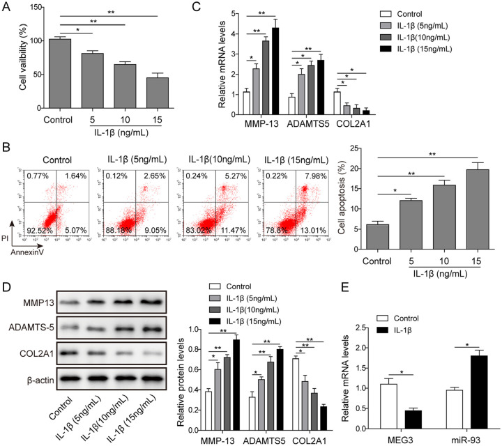
Background: As a degenerative joint disease, osteoarthritis (OA) is characterized by articular cartilage degradation. Long noncoding RNAs (lncRNAs) act critical roles in the regulation of OA development, including affecting the proliferation, apoptosis, extracellular matrix (ECM) degradation, and inflammatory response of chondrocytes. The current study's aim was to investigate the regulatory function and the underlying molecular mechanism of lncRNA MEG3 in ECM degradation of chondrocytes in OA.
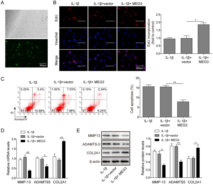
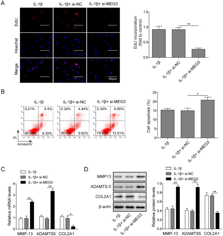
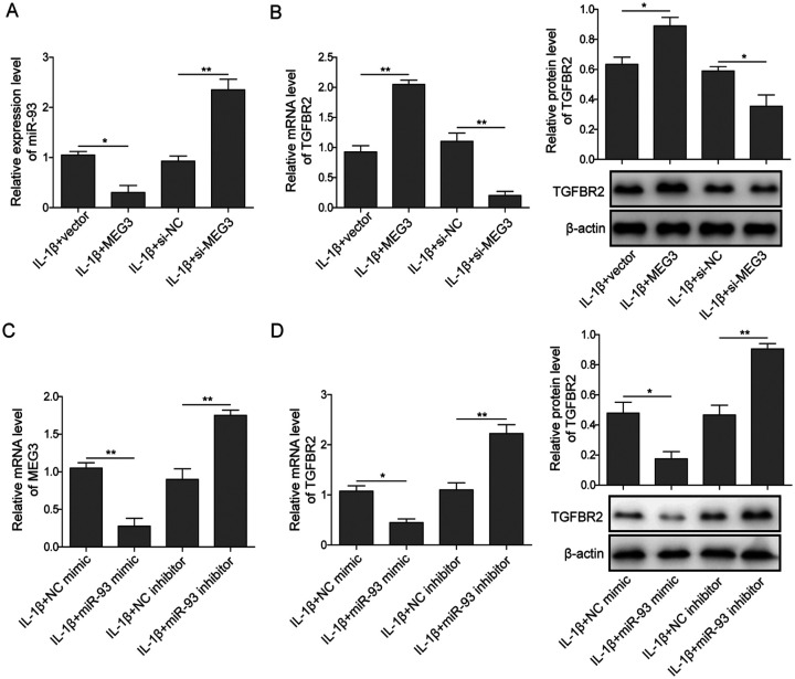
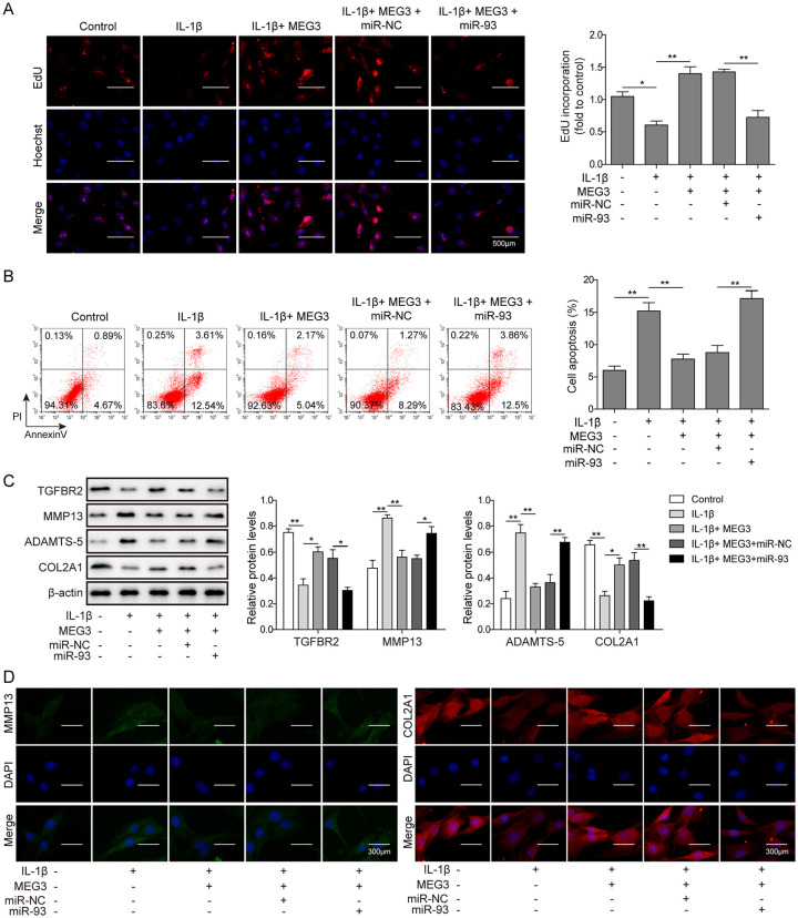
Methods: In the current study, chondrocytes were induced by interleukin-1β (IL-1β) to simulate OA condition, and further assessed cell viability, lncRNA MEG3 and miR-93 expression levels. Overexpression or knockdown of lncRNA MEG3 in chondrocytes treated with IL-1β were performed to investigate the function of MEG3 in regulating cell proliferation, apoptosis and ECM degradation using EdU assay, flow cytometry, quantitative reverse transcription polymerase chain reaction (qRT-PCR), and Western blot. The interaction between MEG3 and miR-93 was assessed using qRT-PCR. Furthermore, overexpression of miR-93 was performed as recovery experiment to explore the functional mechanism of MEG3.
Results: MEG3 was significantly downregulated in chondrocytes treated with IL-1β, whereas miR-93 was upregulated concomitantly. Overexpression of MEG3 induced the proliferation, suppressed the apoptosis, and relieved the degradation of ECM in IL-1β-induced chondrocytes. By contrast, knockdown of MEG3 suppressed the proliferation, promoted the apoptosis, and aggravated ECM degradation in IL-1β induced chondrocytes. In addition, MEG3 was found to relieve the inhibitive expression of TGFBR2 as a competitive endogenous RNA (ceRNA) of miR-93, and then activated transforming growth factor-β (TGF-β) signaling pathway, regulated chondrocytes ECM degradation in IL-1β induced chondrocytes subsequently.
Conclusion: LncRNA MEG3 targeted miR-93/TGFBR2 axis, regulated the proliferation, apoptosis and ECM degradation of chondrocytes in OA.
Keywords: extracellular matrix degradation; lncRNA MEG3; miR-93/TGFBR2; osteoarthritis.
Conflict of interest statement
Figures
Similar articles
-
CircSERPINE2 weakens IL-1β-caused apoptosis and extracellular matrix degradation of chondrocytes by regulating miR-495/TGFBR2 axis.Biosci Rep. 2020 Nov 27;40(11):BSR20201601. doi: 10.1042/BSR20201601. Biosci Rep. 2020. PMID: 33094798 Free PMC article.
-
FGD5-AS1 Inhibits Osteoarthritis Development by Modulating miR-302d-3p/TGFBR2 Axis.Cartilage. 2021 Dec;13(2_suppl):1412S-1420S. doi: 10.1177/19476035211003324. Epub 2021 Apr 9. Cartilage. 2021. PMID: 33834880 Free PMC article.
-
Long noncoding RNA TM1P3 is involved in osteoarthritis by mediating chondrocyte extracellular matrix degradation.J Cell Biochem. 2019 Aug;120(8):12702-12712. doi: 10.1002/jcb.28539. Epub 2019 Mar 19. J Cell Biochem. 2019. PMID: 30887601
-
LncRNA MALAT1 promotes osteoarthritis by modulating miR-150-5p/AKT3 axis.Cell Biosci. 2019 Jul 1;9:54. doi: 10.1186/s13578-019-0302-2. eCollection 2019. Cell Biosci. 2019. PMID: 31304004 Free PMC article. Review.
-
Deciphering the Role of LncRNAs in Osteoarthritis: Inflammatory Pathways Unveiled.J Inflamm Res. 2024 Sep 20;17:6563-6581. doi: 10.2147/JIR.S489682. eCollection 2024. J Inflamm Res. 2024. PMID: 39318993 Free PMC article. Review.
Cited by
-
LncRNA MEG3 suppresses erastin-induced ferroptosis of chondrocytes via regulating miR-885-5p/SLC7A11 axis.Mol Biol Rep. 2024 Jan 18;51(1):139. doi: 10.1007/s11033-023-09095-9. Mol Biol Rep. 2024. PMID: 38236340
-
Knockdown of lncRNA JPX suppresses IL-1β-stimulated injury in chondrocytes through modulating an miR-25-3p/PPID axis.Oncol Lett. 2022 Sep 19;24(5):388. doi: 10.3892/ol.2022.13508. eCollection 2022 Nov. Oncol Lett. 2022. PMID: 36276499 Free PMC article.
-
Dysregulation of lncRNAs in Rheumatoid Arthritis: Biomarkers, Pathogenesis and Potential Therapeutic Targets.Front Pharmacol. 2021 Mar 12;12:652751. doi: 10.3389/fphar.2021.652751. eCollection 2021. Front Pharmacol. 2021. PMID: 33776780 Free PMC article. Review.
-
The Role of Mitochondrial Metabolism, AMPK-SIRT Mediated Pathway, LncRNA and MicroRNA in Osteoarthritis.Biomedicines. 2022 Jun 22;10(7):1477. doi: 10.3390/biomedicines10071477. Biomedicines. 2022. PMID: 35884782 Free PMC article. Review.
-
Interactions Among lncRNA/circRNA, miRNA, and mRNA in Musculoskeletal Degenerative Diseases.Front Cell Dev Biol. 2021 Oct 11;9:753931. doi: 10.3389/fcell.2021.753931. eCollection 2021. Front Cell Dev Biol. 2021. PMID: 34708047 Free PMC article. Review.
References
-
- Liu Q, Zhang X, Dai L, Hu X, Zhu J, Li L, et al.. Long noncoding RNA related to cartilage injury promotes chondrocyte extracellular matrix degradation in osteoarthritis. Arthritis Rheumatol. 2014;66(4):969-78. - PubMed
MeSH terms
Substances
LinkOut - more resources
Full Text Sources
Medical
Research Materials
