. 2019 Jun 28;12(1):62.
doi: 10.1186/s13041-019-0483-y.

Interactions of Rabconnectin-3 with Cav2 calcium channels

Affiliations

Interactions of Rabconnectin-3 with Cav2 calcium channels

Maria A Gandini et al. Mol Brain. .

Abstract

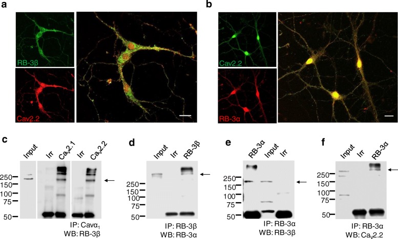
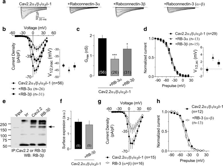
This study describes the interaction between Cav2 calcium channels and Rabconnectin-3, a di-subunit protein that is associated with synaptic vesicles. Immunostaining reveals that both Rabconnectin-3α (RB-3α) and Rabconnectin-3β (RB-3β) are colocalized in mouse hippocampal neurons. Co-immunoprecipitations from brain tissue is consistent with the formation of a protein complex between RB-3α and RB-3β and both Cav2.2 and the related Cav2.1 calcium channel. The coexpression of either RB-3α or RB-3β with Cav2.2 calcium channels in tsA-201 cells led to a reduction in Cav2.2 current density without any effects on the voltage-dependence of activation or inactivation. Coexpression of both Rabconnectin-3 subunits did not cause an additive effect on current densities. Finally, the presence of Rabconnectin-3 did not interfere with μ-opioid receptor mediated Gβγ modulation of Cav2.2 channels. Altogether, our findings show that Rabconnectin-3 has the propensity to regulate calcium entry mediated by Cav2.2 channels.

Keywords: Cav2,2 calcium channels; Hippocampus; N-type channels; Opioid receptor; Rabconnectin-3.


Conflict of interest statement

The authors declare that they have no competing interests.

Figures

Fig. 1
Fig. 1
Colocalization of Rabconnectin-3 and Cav2 channels in hippocampal neurons. Confocal microscopy images from 10 DIV hippocampal neurons showing the distribution of (a) Cav2.2 (red) and RB-3β (green) and (b) Cav2.2 (green) and RB-3α (red). Scale bar 20 μm. Proteins from mouse brain were immunoprecipitated with (c) anti-Cav2.1or Cav2.2, (d) anti-RB-3β, and (e-f) anti-RB-3α, or with control (Irr) antibodies and followed by Western blot analysis using antibodies against the indicated proteins. The experiments are representative of 3 repetitions
Fig. 2
Fig. 2
Functional effects of Rabconnectin-3 on Cav2.2 channels expressed in tsA-201 cells. (a) Representative set of current traces recorded in response to depolarizing steps ranging from − 50 mV to 40 mV from a holding potential of − 80 mV for cells expressing Cav2.2 channels with RB-3α or RB-3β or both. (b) Current density-voltage relationships for cells expressing Cav2.2 channels with RB-3α or RB-3β. Inset. Corresponding mean half activation potential. (c) Corresponding maximal conductance. Numbers shown in the bars reflect numbers of cells (d) Steady-state inactivation curves for cells expressing Cav2.2 channels with RB-3α or RB-3β. Inset. Corresponding mean half activation potential. (e) Proteins from tsA-201 cells transfected with Cav2.2α1 and RB-3β were immunoprecipitated with anti-Cav2.2, anti-RB-3β or control (Irr) antibodies and followed by Western blot analysis using anti-RB-3β. (f) Quantification of biotinylation experiments from tsA-201 cells transfected with Cav2.2/β12δ-1 with and without RB-3β. Biotinylated cell surface protein was isolated and normalized to Na/K-ATPase levels. Numbers shown in the bars reflects numbers of independent experiments. (g) Current density-voltage relationships for cells expressing Cav2.2 channels with RB-3(α + β). (h) Steady-state inactivation curves for cells expressing Cav2.2 channels with RB-3(α + β).
Fig. 3
Fig. 3
Lack of effect of Rabconnectin-3 on G protein modulation of Cav2.2. (a) Representative set of Cav2.2 currents recorded before or after the application of 10 μM DAMGO. As noted in the Methods section, the first current in each trace is evoked by a test depolarization to + 10 mV (P1), the second inward current in a given trace is evoked by a 10 mV test depolarization (P2) that is preceded by a strong depolarizing prepulse (PP, note that the prepulse-evoked outward current has been blanked out). The increase in current amplitude seen during P2 in the presence of DAMGO reflects prepulse relief of Gβγ modulation. (b) Percentage of peak current inhibition (during P1) of Cav2.2 currents after application of 10 μM DAMGO in the presence of absence of RB-3(α + β). (c) Voltage dependent pre-pulse facilitation measured in the presence of DAMGO. The bars reflect the current evoked by test pulse P2 normalized to the current evoked by test pulse P1. This experiment was performed either in the presence or the absence of RB-3(α + β). Numbers in parentheses reflect numbers of cells.

References

    1. Spafford JD, Munno DW, Van Nierop P, Feng ZP, Jarvis SE, Gallin WJ, Smit AB, Zamponi GW, Syed NI. Calcium channel structural determinants of synaptic transmission between identified invertebrate neurons. J Biol Chem. 2003;278:4258–4267. doi: 10.1074/jbc.M211076200. - DOI - PubMed
    1. Catterall WA. Voltage-gated calcium channels. Cold Spring Harb Perspect Biol. 2011;3:a003947. doi: 10.1101/cshperspect.a003947. - DOI - PMC - PubMed
    1. Simms BA, Zamponi GW. Neuronal voltage-gated calcium channels: structure, function, and dysfunction. Neuron. 2014;82:24–45. doi: 10.1016/j.neuron.2014.03.016. - DOI - PubMed
    1. Ackermann F, Waites CL, Garner CC. Presynaptic active zones in invertebrates and vertebrates. EMBO Rep. 2015;16:923–938. doi: 10.15252/embr.201540434. - DOI - PMC - PubMed
    1. Mochida Sumiko. Presynaptic Calcium Channels. International Journal of Molecular Sciences. 2019;20(9):2217. doi: 10.3390/ijms20092217. - DOI - PMC - PubMed

Publication types