. 2019 Aug;123(2):e226-e238.
doi: 10.1016/j.bja.2019.05.027. Epub 2019 Jun 26.

The mitochondrial calcium uniporter contributes to morphine tolerance through pCREB and CPEB1 in rat spinal cord dorsal horn

Affiliations

The mitochondrial calcium uniporter contributes to morphine tolerance through pCREB and CPEB1 in rat spinal cord dorsal horn

K Takahashi et al. Br J Anaesth. 2019 Aug.

Abstract

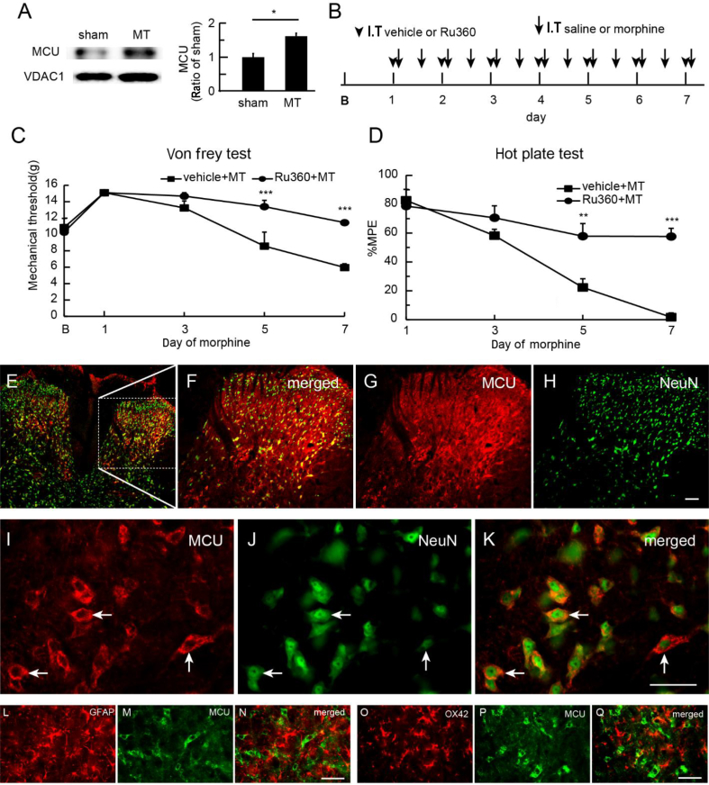
Background: The long-term use of opioid analgesics is limited by the development of unwanted side-effects, such as tolerance. The molecular mechanisms of morphine anti-nociceptive tolerance are still unclear. The mitochondrial calcium uniporter (MCU) is involved in painful hyperalgesia, but the role of MCU in morphine tolerance has not been uncharacterised.

Methods: Rats received intrathecal injection of morphine for 7 days to induce morphine tolerance. The mechanical withdrawal threshold was measured using von Frey filaments, and thermal latency using the hotplate test. The effects of an MCU inhibitor, antisense oligodeoxynucleotide against cyclic adenosine monophosphate response element (CRE)-binding protein (CREB) or cytoplasmic polyadenylation element-binding protein 1 (CPEB1) in morphine tolerance were examined.

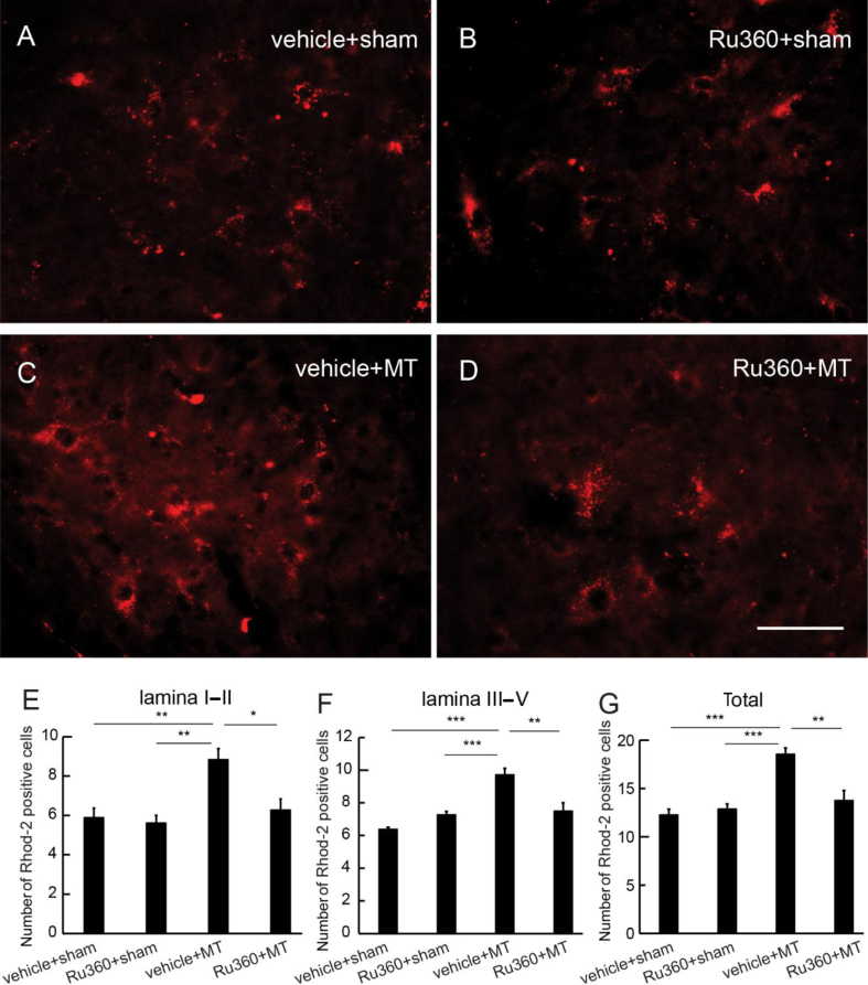
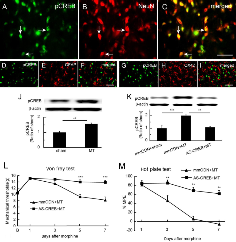
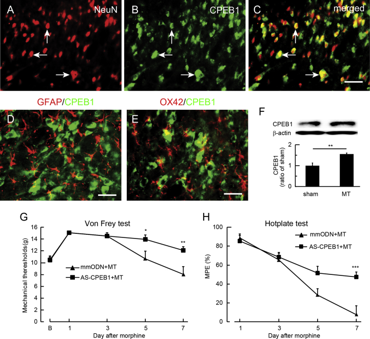
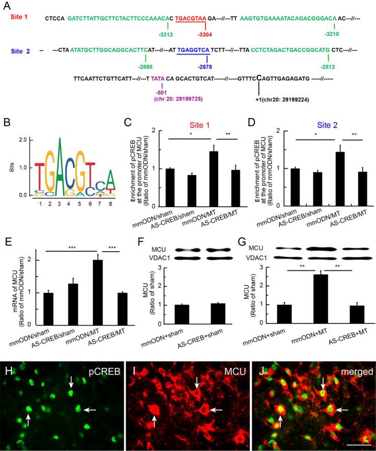
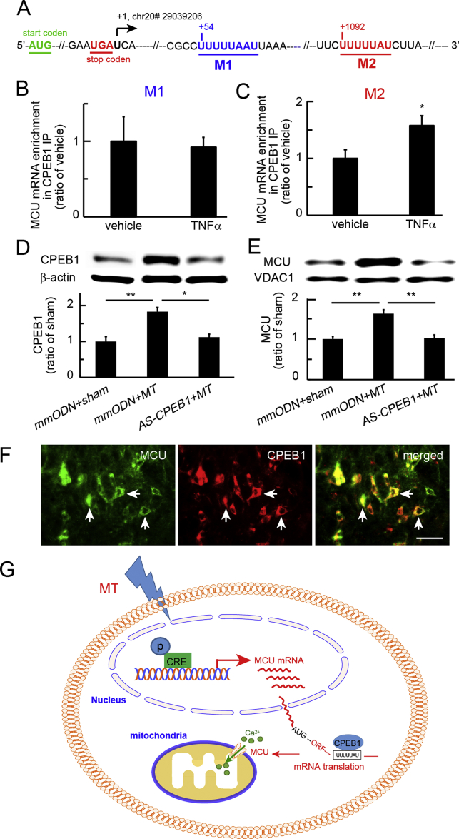
Results: Spinal morphine tolerance was associated with an increased expression of neuronal MCU, phospho-CREB (pCREB), and CPEB1 in the spinal cord dorsal horn. MCU inhibition increased the mechanical threshold and thermal latency, and reduced the accumulation of mitochondrial calcium in morphine tolerance. Intrathecal antisense oligodeoxynucleotide against CREB or CPEB1 restored the anti-nociceptive effects of morphine compared with mismatch oligodeoxynucleotide in von Frey test and hotplate test. Chromatin immunoprecipitation with quantitative PCR assay showed that CREB knockdown reduced the interaction of pCREB with the ccdc109a gene (encoding MCU expression) promoter and decreased the MCU mRNA transcription. RNA immunoprecipitation assay suggested that CPEB1 binds to the MCU mRNA 3' untranslated region. CPEB1 knockdown decreased the expression of MCU protein.

Conclusions: These findings suggest that spinal MCU is regulated by pCREB and CPEB1 in morphine tolerance, and that inhibition of MCU, pCREB, or CPEB1 may be useful in preventing the development of opioid tolerance.

Keywords: CPEB; CREB; calcium; mitochondrial calcium uniporter; morphine; opioid tolerance; opioids.


Conflict of interest statement

The authors declare that they have no conflicts of interest.

Figures

Fig. 1
Fig. 1
Effect of mitochondrial calcium uniporter (MCU) inhibition on morphine tolerance. (a) We harvested spinal cord dorsal horn (SCDH) 1 h after the last morphine dose on Day 7 in morphine-tolerant rats. Western blots demonstrated that morphine tolerance increased the expression of MCU in the SCDH compared with sham (P<0.05; t-test; n=5–6). (b) Scheme for intrathecal administration. Morphine tolerance was induced by intrathecal injection of morphine (arrow; twice a day); sham group was saline. Ruthenium 360 (Ru360) or vehicle (arrowhead) was administered every morning 30 min before morphine or saline (sham). (c) Intrathecal MCU inhibitor, Ru360, significantly up-regulated the mechanical withdrawal threshold compared with vehicle (F(4,40)interaction=6.62, P<0.001; F(4,40)main effect time=23.51, P<0.0001; F(1,10)main effect treatment=12.53, P<0.01; two-way anova repeated measures; n=6). The mechanical withdrawal threshold in the Ru360 group was higher than that in the vehicle group on Days 5 and 7 (***P<0.001 vs vehicle; two-way anova Bonferroni tests). (d) In the hotplate test, there was a significant difference in maximum possible effect (MPE) (%) of thermal latency between Ru360 and saline (F(3,30)interaction=9.47; P<0.001; F(3,30)main effect time=29.45; P<0.0001; F(1,10)main effect treatment=25.47; P<0.001; two-way anova repeated measures; n=6). MPE (%) in the Ru360 group was higher than that for vehicle on Day 5 or 7 (**P<0.01; ***P<0.001 vs vehicle; two-way anova Bonferroni tests). (e–h) Low magnification of MCU immunoreactivity (IR) (MCU-IR) images. (i–k) High-magnification images display that MCU-IR co-localised with NeuN, but not with (l–n) glial fibrillary acidic protein (GFAP) or (o–q) OX42; scale bar: 50 μm. MT, morphine anti-nociceptive tolerance.
Fig. 2
Fig. 2
Effect of mitochondrial calcium uniporter inhibition on mitochondrial calcium in spinal cord dorsal horn (SCDH) neurones. Ruthenium 360 (Ru360) (50 μM) was injected intrathecally every morning 30 min before morphine injection once a day for 7 days. At 30 min after the last morphine, Rhod-2/AM was intrathecally injected for Rhod-2 imaging. (a–d) Representative image of Rhod-2 in the SCDH in (a) saline+sham, (b) Ru360+sham, (c) saline+morphine anti-nociceptive tolerance (MT), and (d) Ru360+MT groups. (e) There was a significant increase in Rhod-2-positive cells at SCDH Laminae I and II in the vehicle+MT group compared with the vehicle+sham or Ru360+sham groups (**P<0.01; one-way anova; n=6); Rhod-2-positive cells in the Ru360+MT group was lower than that in the vehicle+MT group in SCDH Laminae I and II (P<0.05; one-way anova; n=6). Similarly, there was a significant increase in Rhod-2-positive cells in the vehicle+MT group compared with the vehicle+sham or Ru360+sham group in (f) Laminae III–V or (g) Laminae I–V (***P<0.001; one-way anova; n=6). (f) Rhod-2-positive cells in Ru360+MT group were fewer than in the vehicle+MT group in SCDH Laminae III–V (**P<0.01; one-way anova; n=6). (g) Total Rhod-2-positive cells in Laminae I–V in the Ru360+MT group was lower than in the vehicle+MT group (**P<0.01; one-way anova; n=6).
Fig. 3
Fig. 3
Effect of knockdown of transcriptional factor, cyclic adenosine monophosphate response element (CRE)-binding protein (CREB), on spinal morphine tolerance. (a–c) Immunostaining showed that phosphorylated cyclic adenosine monophosphate response element-binding protein (pCREB) co-localised with NeuN, but not with (d–f) glial fibrillary acidic protein (GFAP) or (g–i) OX42 in spinal cord dorsal horn (SCDH) in morphine-tolerant rats, suggesting neuronal pCREB activity in morphine tolerance. (j) Western immunoblots showed that morphine tolerance up-regulated the expression of pCREB in the SCDH compared with the sham group (**P<0.01; t-test; n=6). (k) Expression of spinal pCREB in the mismatch ODN (mmODN)/morphine anti-nociceptive tolerance (MT) group was higher than in the sham group (***P<0.001; one-way analysis of variance [anova] with post hoc Fisher's protected least significant difference [PLSD]; n=4–5); pCREB in the AS-CREB+MT group was significantly lower than that in the mmODN+MT group (**P<0.01; one-way anova with post hoc Fisher's PLSD; n=4–5). (l) Intrathecal AS-CREB caused an elevation of mechanical withdrawal threshold compared with mmODN (F(4,40)main interaction=6.70, P<0.001; F(4,40)main effect time=15.08, P<0.0001; F(1, 10)=22.93, P<0.001; two-way anova repeated measures; n=6). The mechanical withdrawal threshold in the AS-CREB group was higher than in the mmODN group on Days 5 and 7 (***P<0.001 vs mmODN; two-way anova Bonferroni tests; n=6). (m) Intrathecal AS-CREB increased %MPE in thermal latency compared with mmODN (F(3,40)main effect interaction=9.08, P<0.001; F(3,30)main effect time=26.41, P<0.0001; F(1, 10)main effect treatment= 55.29, P<0.0001; two-way anova repeated measures; n=6). Thermal latency in the AS-CREB group was higher than that in the mmODN group on Day 5 or 7 after ODN (**P<0.01 vs mmODN; two-way anova Bonferroni tests; n=6).
Fig. 4
Fig. 4
Effect of spinal cytoplasmic polyadenylation element-binding protein 1 (CPEB1) on morphine tolerance. Double immunostaining showed that the immunoreactivity (IR) of CPEB1-IR co-localised with (a–c) NeuN, but not with (d) glial fibrillary acidic protein (GFAP) or (e) OX42. (f) Western immunoblots showed that morphine tolerance increased the expression of CPEB1 in the spinal cord dorsal horn compared with sham (**P<0.01; t-test; n=4). (g) Intrathecal AS-CPEB1 caused an elevation of mechanical withdrawal threshold compared with mismatch oligodeoxynucleotide (mmODN) (F(4,40)interaction=4.30, P<0.01; F(4,40) main effect time=22.13, P<0.0001; F(1,10) main effect treatment=5.27, P<0.05; two-way analysis of variance [anova] repeated measures). The mechanical withdrawal threshold in the AS-CPEB1+morphine anti-nociceptive tolerance (MT) group was higher than in the mmODN+MT group 5 and 7 days after ODN (*P<0.05, ** P<0.01 vs mmODN+MT; two-way anova Bonferroni tests). (h) Intrathecal AS-CPEB1 increased thermal latency using hotplate test compared with mmODN F(3,30)interaction=5.90, P<0.01; F(3,30) main effect time=43.68, P<0.0001; F(1,10)main effect treatment=6.78, P<0.05; two-way anova repeated measures). The mechanical withdrawal threshold in the AS-CPEB1+MT group was higher than in the mmODN+MT group on Day 7 (***P<0.001 vs mmODN+MT; two-way anova Bonferroni tests).
Fig. 5
Fig. 5
Phosphorylated cyclic adenosine monophosphate response element-binding protein (pCREB) bound mcu gene promoter regions in the spinal cord dorsal horn (SCDH) and mediated mitochondrial calcium uniporter (MCU) transcription. (a) We analysed the alignment of rat MCU gene promoter regions, and found two putative CRE-binding areas, Site 1 (TGACGTAA) and Site 2 (TGAGGTCA),, and chromatin immunoprecipitation with quantitative PCR (ChIP-qPCR) primer areas of the MCU gene at rat chromosome 20 (accession number: NC_005119; NCBI Reference Sequence: NC_005119.4). The TATA box of mcu gene was shown. The first base C (chr 20: 29199224) of the first intron of the mcu gene was referred to as number +1. (b) Motif of the CREB-binding sites based on the JASPAR data sets (http://jaspar.genereg.net/matrix/MA0018.2). (c and d) ChIP-qPCR assay showed that enrichment of pCREB at CRE Site 1 or 2 of the mcu gene promoter region in the mismatch oligodeoxynucleotide (mmODN)+morphine anti-nociceptive tolerance (MT) group was increased compared with the mmODN+sham group (*P<0.05; one-way analysis of variance [anova]; n=5). Enrichment of pCREB at Site 1 or 2 on mcu gene promoter in the AS-CREB+MT group was lower than that in the mmODN+MT group (**P<0.01; one-way anova; post hoc Fisher's protected least significant difference [PLSD]; n=5). (e) RT–PCR showed increased MCU mRNA expression (***P<0.001; one-way anova; post hoc Fisher's PLSD test; n=5). (f and g) Western immunoblots showed no differences in expression of MCU protein between mmODN+sham and AS-CREB+sham groups (f). (g) MCU protein expression increased (**P<0.01; one-way anova). (h–j) Double immunostaining showed that pCREB was co-localised with MCU in the SCDH in morphine-tolerant rats; scale bar: 50 μm.
Fig. 6
Fig. 6
Cytoplasmic polyadenylation element-binding protein 1 (CPEB1) on the mitochondrial calcium uniporter (MCU) mRNA. (a) We analysed the alignment of rat CPEB1 motifs at the MCU mRNA 3′ untranslated region (UTR) and found two putative CPE-binding areas, motif 1, UUUUUAAU (M1), and motif 2, CPE consensus sequence, UUUUUAU, (M2). AUG, translation start codon, and UGA, translation stop codon, are shown. The first base U (chr20# 29039206) following UGA was referred to as number +1. (b) To induce the expression of MCU and CPEB1, we used rat neuronal B35 cells treated with recombinant tumour necrosis factor alpha (rTNFα) for 3 h. Using an RNA immunoprecipitation assay, we observed that MCU mRNA in CPEB1 immunoprecipitation was not increased at the M1 CPE sequence, and that (c) MCU mRNA in CPEB1 immunoprecipitation was increased at the M2 CPE consensus sequence. (d) Morphine-tolerant rats were treated with antisense oligodeoxynucleotide against CPEB1. Western immunoblots showed a significant increase in CPEB1 in the mismatch oligodeoxynucleotide (mmODN)+morphine anti-nociceptive tolerance (MT) group compared with the sham group (**P<0.01; one-way analysis of variance [anova] with post hoc Fisher's protected least significant difference [PLSD] test; n=4–5). Spinal CPEB1 in the AS-CPEB1+MT group was lower than in the mmODN+MT group (*P<0.05; one-way anova with post hoc Fisher's PLSD test; n=4–5). (e) Western immunoblots showed that MCU in the spinal cord dorsal horn (SCDH) was increased in the mmODN+MT group compared with sham rats treated with mmODN; MCU in the SCDH of the AS-CPEB+MT group was lower than in the mmODN+MT group (**P<0.01; one-way anova with post hoc Fisher's PLSD; n=4–5). (f) Double immunostaining showed that CPEB1 immunoreactivity was co-localised with MCU; scale bar: 50 μm. (g) The proposed signalling pathways mediated by phosphorylated cyclic adenosine monophosphate response element-binding protein (pCREB) and CPEB1. Morphine tolerance evoked neuronal activity to induce phosphorylation of CREB. pCREB binds to CRE sites of the mcu gene promoter to induce MCU transcriptional expression. Up-regulated CPEB1 may regulate MCU translation. CPE motifs in MCU mRNA 3′ UTRs reside in a complex containing CPEB1. CPEB1 binds to the MCU mRNA 3′ UTR to regulate MCU mRNA translation. AUG, mRNA translation start codon; ORF, open reading frame of the stretch of codons between AUG and a stop codon.

Similar articles

Cited by

References

    1. O’Brien P.L., Karnell L.H., Gokhale M., Kenneth Pack B.S., Campopiano M., Zur J. Prescribing of benzodiazepines and opioids to individuals with substance use disorders. Drug Alcohol Depend. 2017;178:223–230. - PubMed
    1. Volkow N.D., McLellan A.T. Opioid abuse in chronic pain—misconceptions and mitigation strategies. N Engl J Med. 2016;374:1253–1263. - PubMed
    1. Frenk S.M., Porter K.S., Paulozzi L.J. Prescription opioid analgesic use among adults: United States, 1999–2012. NCHS Data Brief. 2015;1–8 - PubMed
    1. Miller M., Barber C.W., Leatherman S. Prescription opioid duration of action and the risk of unintentional overdose among patients receiving opioid therapy. JAMA Intern Med. 2015;175:608–615. - PubMed
    1. Mao J., Sung B., Ji R.R., Lim G. Neuronal apoptosis associated with morphine tolerance: evidence for an opioid-induced neurotoxic mechanism. J Neurosci. 2002;22:7650–7661. - PMC - PubMed