. 2019 Jul 16;116(29):14661-14670.
doi: 10.1073/pnas.1905179116. Epub 2019 Jun 28.

Unexpected host dependency of Antarctic Nanohaloarchaeota

Affiliations

Unexpected host dependency of Antarctic Nanohaloarchaeota

Joshua N Hamm et al. Proc Natl Acad Sci U S A. .

Abstract

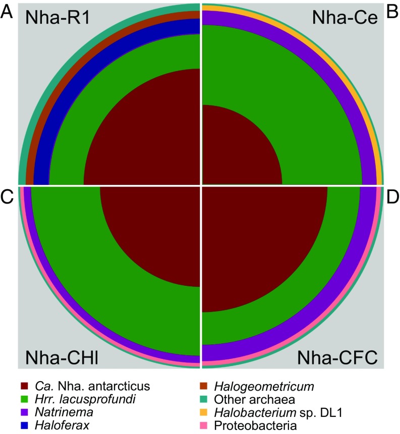
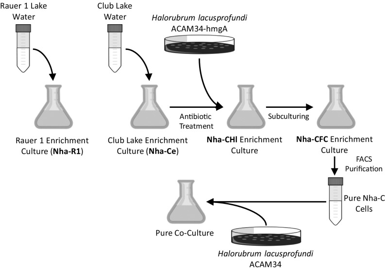
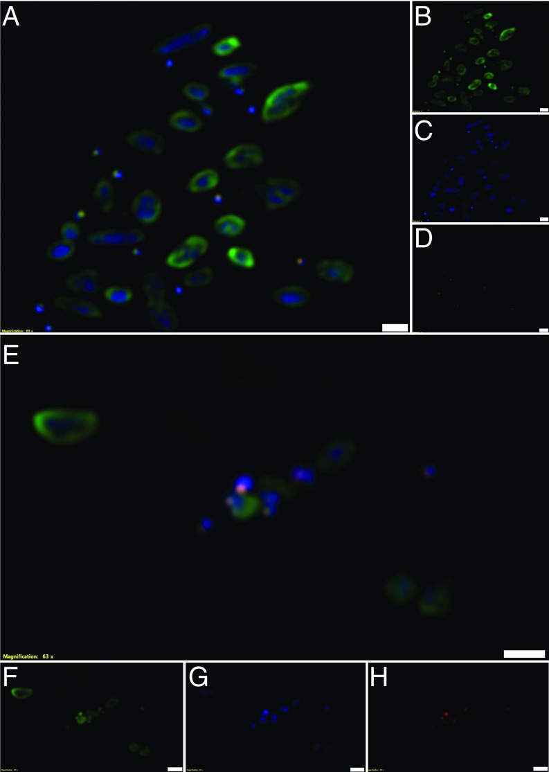
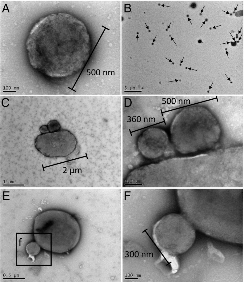
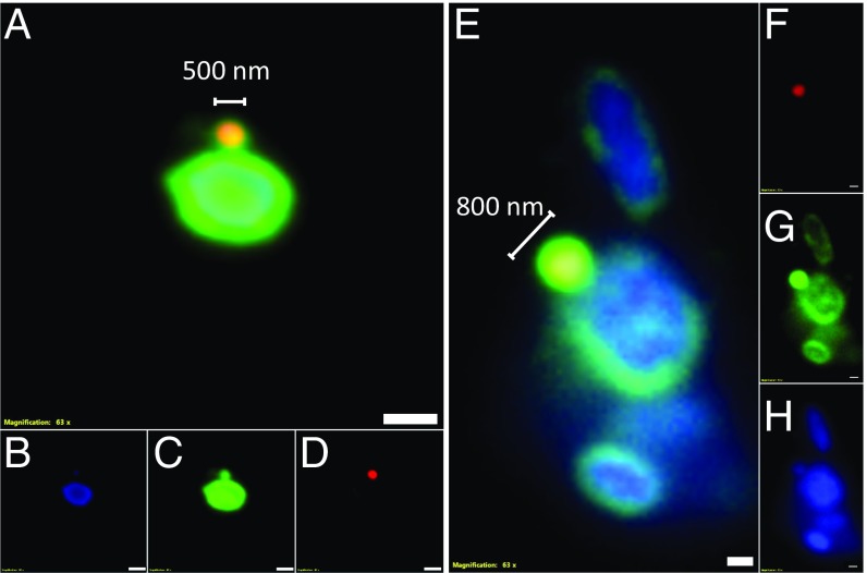
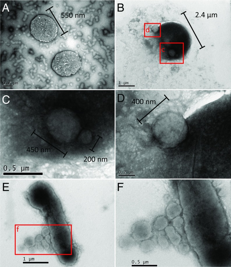
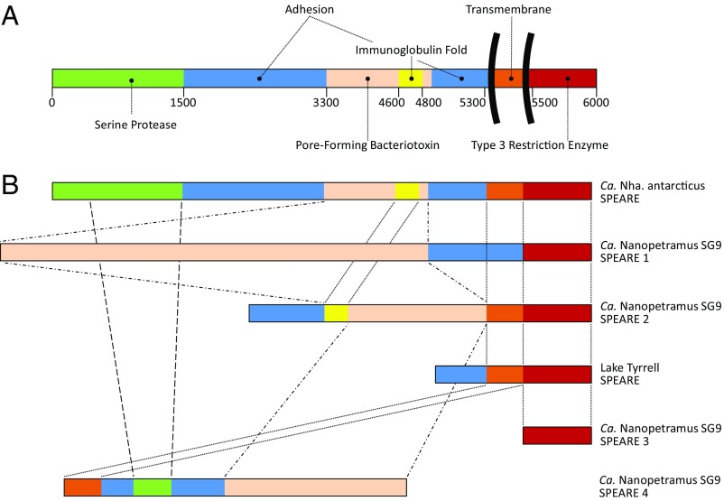
In hypersaline environments, Nanohaloarchaeota (Diapherotrites, Parvarchaeota, Aenigmarchaeota, Nanoarchaeota, Nanohaloarchaeota [DPANN] superphylum) are thought to be free-living microorganisms. We report cultivation of 2 strains of Antarctic Nanohaloarchaeota and show that they require the haloarchaeon Halorubrum lacusprofundi for growth. By performing growth using enrichments and fluorescence-activated cell sorting, we demonstrated successful cultivation of Candidatus Nanohaloarchaeum antarcticus, purification of Ca. Nha. antarcticus away from other species, and growth and verification of Ca. Nha. antarcticus with Hrr. lacusprofundi; these findings are analogous to those required for fulfilling Koch's postulates. We use fluorescent in situ hybridization and transmission electron microscopy to assess cell structures and interactions; metagenomics to characterize enrichment taxa, generate metagenome assembled genomes, and interrogate Antarctic communities; and proteomics to assess metabolic pathways and speculate about the roles of certain proteins. Metagenome analysis indicates the presence of a single species, which is endemic to Antarctic hypersaline systems that support the growth of haloarchaea. The presence of unusually large proteins predicted to function in attachment and invasion of hosts plus the absence of key biosynthetic pathways (e.g., lipids) in metagenome assembled genomes of globally distributed Nanohaloarchaeota indicate that all members of the lineage have evolved as symbionts. Our work expands the range of archaeal symbiotic lifestyles and provides a genetically tractable model system for advancing understanding of the factors controlling microbial symbiotic relationships.

Keywords: DPANN; archaea; symbiont.


Conflict of interest statement

The authors declare no conflict of interest.

Figures

Fig. 1.
Fig. 1.
Relative abundances of the major taxa present in Ca. Nha. antarcticus enrichment cultures. Metagenomes for Nha-R1 (A), Nha-Ce (B), Nha-CHl (C), and Nha-CFC (D). Quadrants show relative abundances of the major taxa identified in each enrichment metagenome. Abundances were calculated using average read depth across a set of universally distributed single-copy genes. Area of the colored sections is directly proportional to the relative abundance of each taxonomic unit. The only taxa identified in all 4 metagenomes were Ca. Nha. antarcticus (Nha-R1: 41%; Nha-Ce: 19%; Nha-CHl: 30%; Nha-CFC: 47%), Hrr. lacusprofundi (Nha-R1: 28%; Nha-Ce: 59%; Nha-CHl: 56%; Nha-CFC: 29%), and Natrinema sp. (Nha-R1: 1%; Nha-Ce: 14%; Nha-CHl: 8%; Nha-CFC: 16%).
Fig. 2.
Fig. 2.
Experimental design for cultivating Ca. Nha. antarcticus in enrichments through to coculture with Hrr. lacusprofundi. The names of the metagenomes associated with the enrichment cultures (Fig. 1) are in bold: Nha-R1, Nha-Ce, Nha-CHl, and Nha-CFC.
Fig. 3.
Fig. 3.
FISH of Nha-C enrichment with Hrr. lacusprofundi ACAM34-hmgA. Fluorescence micrographs show individual Nha-C cells among Hrr. lacusprofundi cells (AD) and Nha-C cells in contact with Hrr. lacusprofundi cells (EH). Nha-C cells are labeled with a Cy5-conjugated (red fluorescence) probe. Hrr. lacusprofundi cells are labeled with a Cy3 (yellow fluorescence; recolored to green to improve contrast) probe; all nucleic acid-containing cells are stained with DAPI (blue fluorescence). Composite image of all 3 filters (A and E). Individual filters for Cy3 (B and F), DAPI (C and G), and Cy5 (D and H). (Scale bars: 2 µm.)
Fig. 4.
Fig. 4.
TEM of Nha-C enrichment with Hrr. lacusprofundi ACAM34-hmgA. Small individual cells consistent with being Nha-C (A and B). Arrows mark putative Nha-C cells (B). Small cells that appear to have intact membranes with minimal visible boundary layers separating them from larger cells, consistent with Nha-C associating with Hrr. lacusprofundi (C and D). Small cell that appears to be attached with a possible membrane collar to a large cell (E and F); the morphology is reminiscent of a site of budding (detachment) or possibly, initial stages of fusing after attachment.
Fig. 5.
Fig. 5.
FISH of Nha-C FACS cells with Hrr. lacusprofundi ACAM34. Fluorescence micrographs show Nha-C cells in contact with Hrr. lacusprofundi cells. The Nha-C cells fluoresced for both the Nha-C and Hrr. lacusprofundi probes, indicating that Hrr. lacusprofundi rRNA transfers to Nha-C cells. Nha-C cells are labeled with a Cy5-conjugated (red fluorescence) probe. Hrr. lacusprofundi cells are labeled with a Cy3 (yellow fluorescence; recolored to green to improve contrast) probe; all nucleic acid-containing cells are stained with DAPI (blue fluorescence). Composite image of all 3 filters (A and E). Individual filters for Cy3 (B and F), DAPI (C and G), and Cy5 (D and H). (Scale bars: 2 µm.)
Fig. 6.
Fig. 6.
TEM of Nha-C FACS cells with Hrr. lacusprofundi ACAM34. Small individual cells consistent with being Nha-C (A). A single and “budded” cell (putative Nha-C) intimately associated with a larger cell (putative Hrr. lacusprofundi) (BD). The budded structure may arise from cell division occurring while Nha-C cells are attached to Hrr. lacusprofundi (D). Multiple small cells (putative Nha-C) seem to be associated with a larger cell (putative Hrr. lacusprofundi) surrounded by possible extracellular material (E and F). The appearance of extracellular material occurred in other images of FACS cultures (SI Appendix, Fig. S14), including in an image of a large cell (putative Hrr. lacusprofundi) that appears to have lysed (SI Appendix, Fig. S14E).
Fig. 7.
Fig. 7.
Domain structure of SPEARE proteins. (A) Protein domains predicted for the 5,998 amino acid Ca. Nha. antarcticus SPEARE protein (IMG gene identification no. 2643306914; locus tag: NAR1_1133). (B) Domain structure comparison of SPEARE proteins from Nha-R1, Ca. Nanopetramus SG9 (8), and Lake Tyrrell metagenome data (2) depicted approximately to scale. Ca. Nanopetramus SG9 SPEARE 1 (8,553 amino acids; hypothetical protein AQV86_04780; accession no. AOV94740.1). Ca. Nanopetramus SG9 SPEARE 2 (4,057 amino acids; hypothetical protein AQV86_02335; accession no. AOV95205.1). Lake Tyrrell SPEARE (717 amino acids; IMG gene identification no. LTJ07AB_218510; locus tag: LTJ07AB_218510). Ca. Nanopetramus SG9 SPEARE 3 (3,315 amino acids; hypothetical protein AQV86_02330; accession no. AOV94739.1).

References

    1. Spang A., Caceres E. F., Ettema T. J. G., Genomic exploration of the diversity, ecology, and evolution of the archaeal domain of life. Science 357, eaaf3883 (2017). - PubMed
    1. Narasingarao P., et al. , De novo metagenomic assembly reveals abundant novel major lineage of Archaea in hypersaline microbial communities. ISME J. 6, 81–93 (2012). - PMC - PubMed
    1. Rinke C., et al. , Insights into the phylogeny and coding potential of microbial dark matter. Nature 499, 431–437 (2013). - PubMed
    1. Andrade K., et al. , Metagenomic and lipid analyses reveal a diel cycle in a hypersaline microbial ecosystem. ISME J. 9, 2697–2711 (2015). - PMC - PubMed
    1. Ghai R., et al. , New abundant microbial groups in aquatic hypersaline environments. Sci. Rep. 1, 135 (2011). - PMC - PubMed

Publication types